
Article Figures & Data
Figures
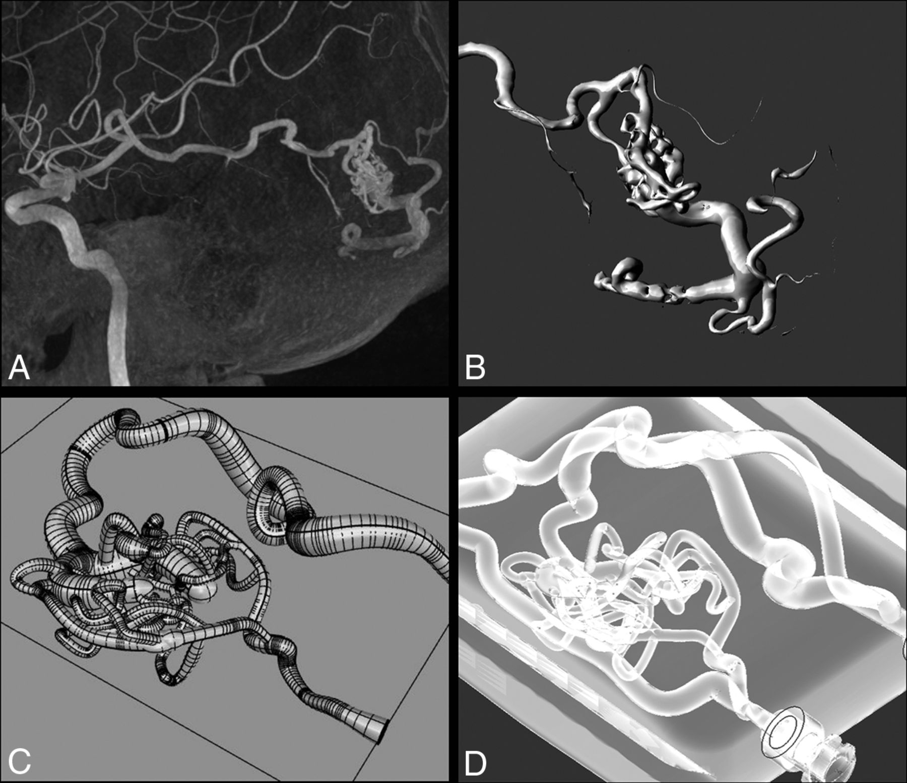
- FIG 1.
The process from the DICOM image to the model. A, MIP 3D reconstruction of the left temporal BAVM. Spetzler-Martin I. B, 3D image of the segmentation manual process by using the region growing technique with 3D Slicer Software. C, CAD representation of the BAVM model design after the main characteristics of the original disease. D, Final in vitro model inside the container, with transparent material and an external “normal” channel. BAVM indicates brain arteriovenous malformation.
- FIG 2.
Contrast media retrograde test. Injections were done by using an Echelon 10 microcatheter and iodinated contrast. A, We tested 10 different input pressures from 10 different flow percentages, and we graded the retrograde advance from 0–3 as follows: 0 = no significant advance; 1 = 1 venous limb complete filling; 2 = 2 venous limbs complete filling; 3 = complete venous filling plus proximal arterial filling. B–E, Retrograde contrast injections with different MAP values. An increase in nidal contrast penetration was seen with lower MAP values, with a statistically significant correlation (P = .001).
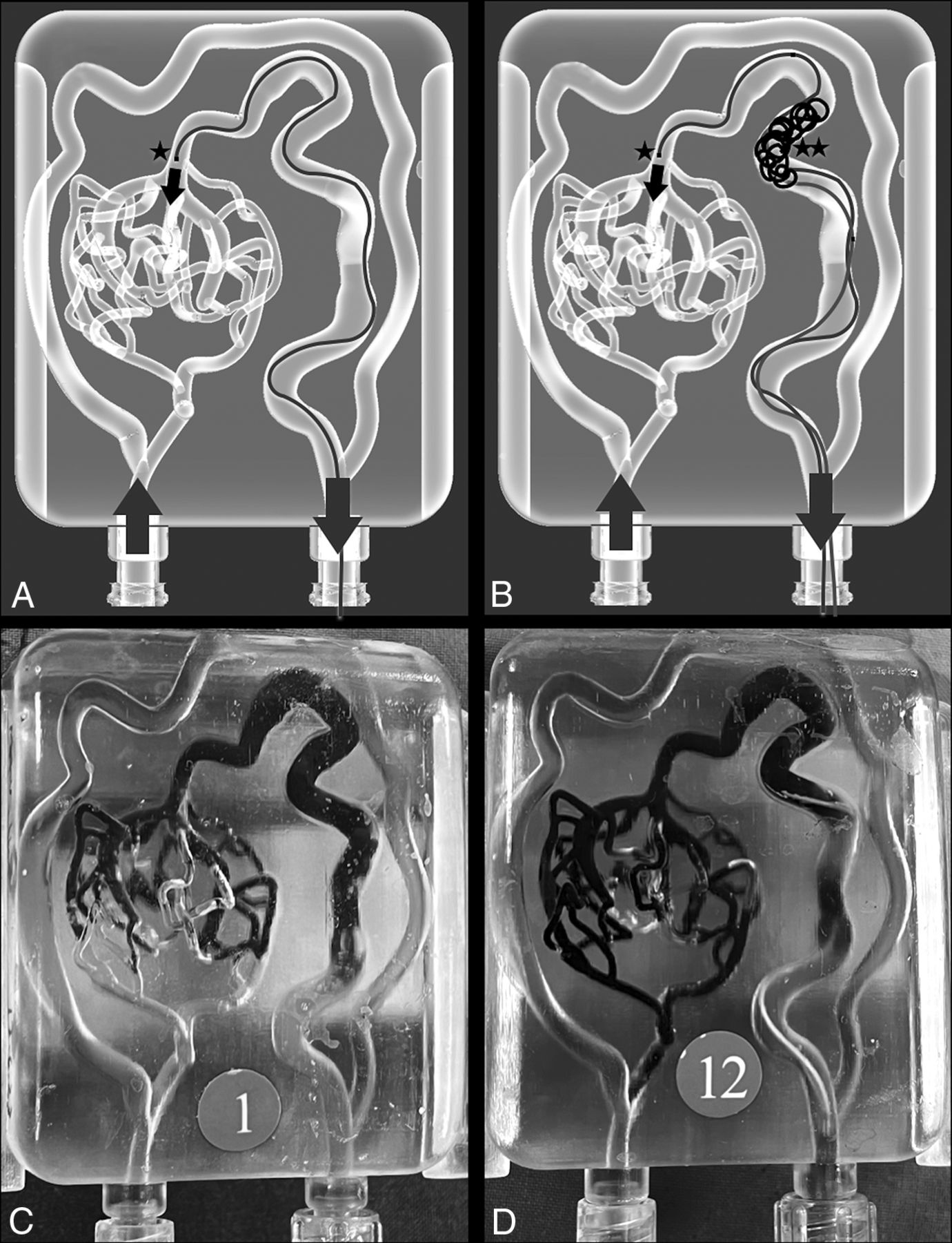
- FIG 3.
Transvenous embolization tests. TVE techniques were performed on 12 identical AVM models, divided into 2 groups (2 × 6). A, Group 1, where retrograde embolization was done by using an Apollo 1.5 30 mm microcatheter (★). B, Group 2, with the same position of the Apollo 1.5 30 mm (★) and a venous coil deployed with a Vasco+ microcatheter in a proximal vein position (★★). C, Real photograph after the EVOH embolization of a group 1 case. D, Real photograph after the EVOH embolization of a group 2 case.


Tables
Transvenous embolization resultsa
Group Group 1 Group 2 Significance Total time (min:sec) 14:40 18:29 NS Number of stops 13 15.5 NS Total LEA (mL) 1.3 mL 1.75 mL NS Nidal LEA volume (mL) 0.46 mL 0.49 mL NS Nidal occlusion (%) 57.6 61.95 NS a Nonparametric test for different variables between groups by using the Mann-Whitney test. NS = nonsignificant.


