
Article Figures & Data
Figures
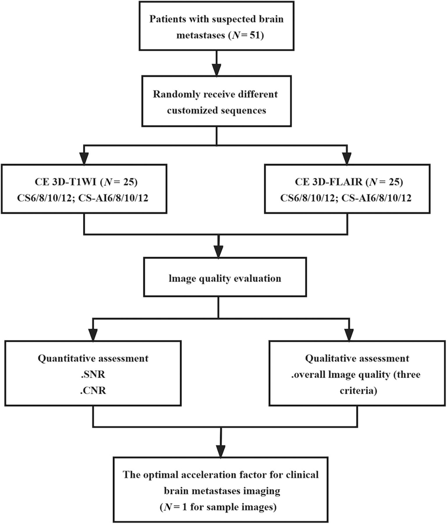
- FIG 1.
Flowchart of the experimental design.
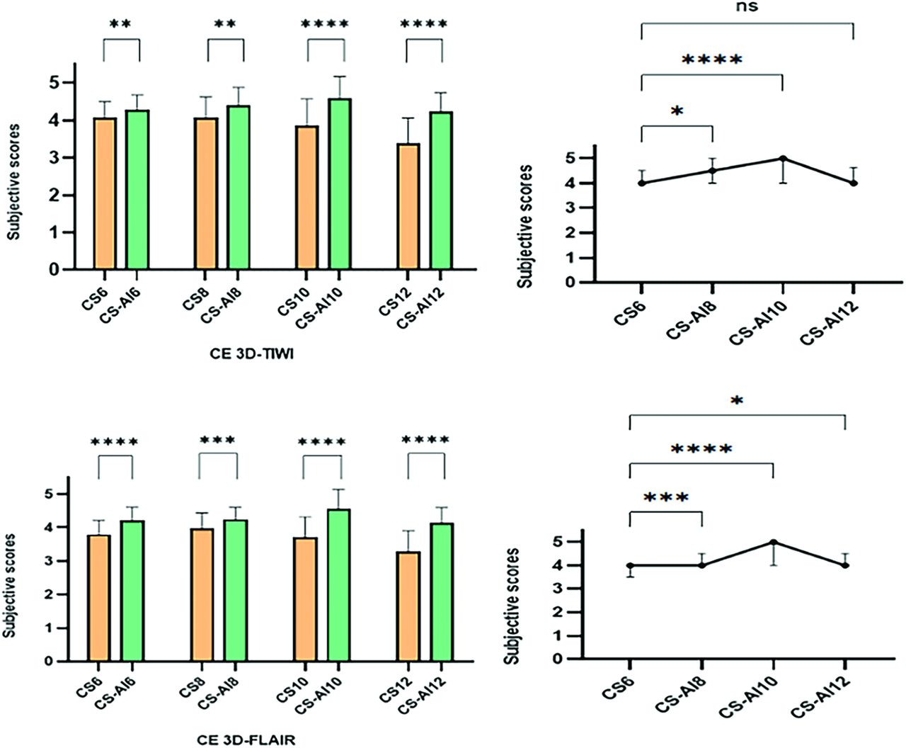
- FIG 2.
Quantitative analysis results of the SNR and CNR of CS- and CS-AI-accelerated CE 3D-T1WI under different AFs. Note: NS indicates not significant; *, P < .05; **, P < .01; ***, P < .001; ****, P < .0001.
- FIG 3.
Quantitative analysis results of the SNR and CNR values of CS- and CS-AI-accelerated CE 3D-FLAIR images under different AFs. Note: NS indicates not significant; *, P < .05; **, P < .01; ***, P < .001; ****, P < .0001.
- FIG 4.
Comparison of the overall image quality score results of different accelerated sequences in patients with BM. Note: NS indicates not significant; *, P < .05; **, P < .01; ***, P < .001; ****, P < .0001.
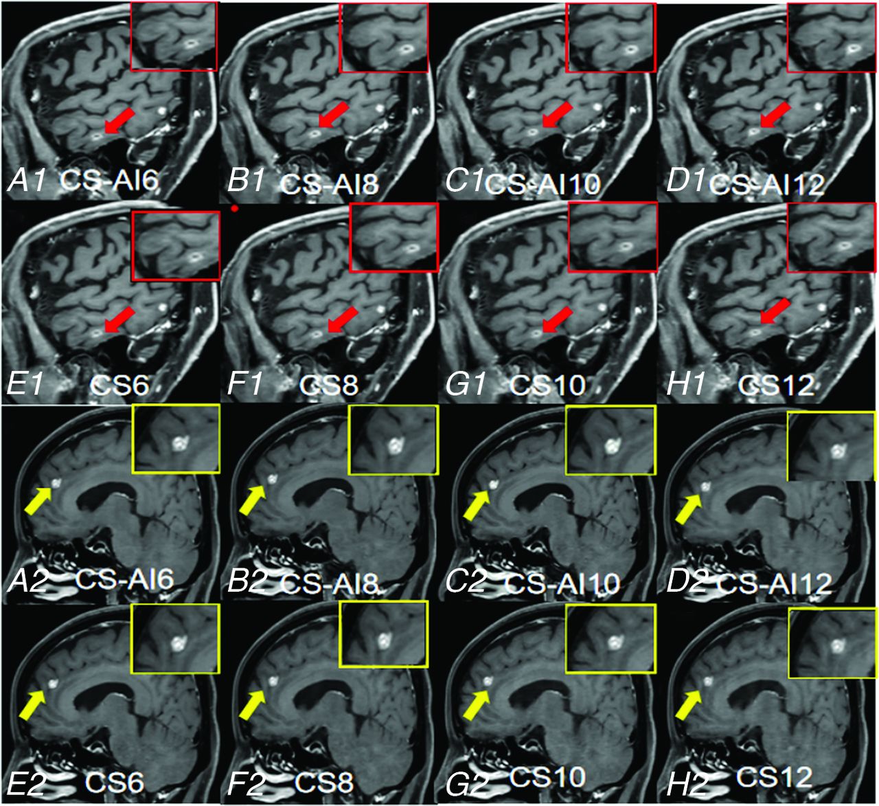
- FIG 5.
MR images of a 68-year-old woman with breast cancer and a 57-year-old man with lung cancer. Reconstructed CE 3D-T1-weighted sagittal images for CS-AI6 (A1, A2), CS-AI8 (B1, B2), CS-AI10 (C1, C2), CS-AI12 (D1, D2), reference CS6 (E1, E2), CS8 (F1, F2), CS10 (G1, G2), and CS12 (H1, H2). The arrows indicate enhanced BM lesions. Notably, the gray-white matter boundaries and lesion edges are clearer in the images accelerated by CS-AI technology, and the image quality is comparable with or better than that of CS6.
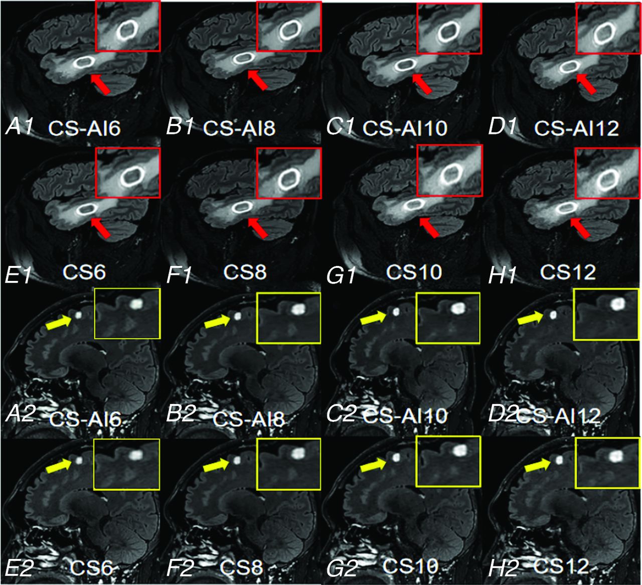
- FIG 6.
MR images of a 55-year-old male patient and a 38-year-old male patient with lung cancer. Reconstructed CE 3D-FLAIR sagittal images for CS-AI6 (A1, A2), CS-AI8 (B1, B2), CS-AI10 (C1, C2), CS-AI12 (D1, D2), reference CS6 (E1, E2), CS8 (F1, F2), CS10 (G1, G2), and CS12 (H1, H2). The arrows indicate enhanced BM. In the CS group, as the AF increases, the images become blurred, and the junctions of gray-white matter and the boundaries of the lesions become less clear.
- FIG 7.
A 60-year-old male patient with lung cancer was scanned with 2 optimized sequences to obtain example images of a good quality in a short time (5:03 minutes). The axial (A), sagittal (B), and coronal (C) images of accelerated CE 3D-T1Wl of CS-AI10. The axial (D), sagittal (E), and coronal (F) images of accelerated CE 3D-FLAIR of CS-AI10.


Tables
Parameter CE 3D-T1WI CS6 (RS) CS 8/10/12 CS-AI 6/8/10/12 TR (ms) 600 600 600 TE (ms) 28 28 28 FOV (mm2) 250 × 250 250 × 250 250 × 250 Voxel size (mm3) 0.99 × 1.00 × 1.10 0.99 × 1.00 × 1.10 0.99 × 1.00 × 1.10 Slices 327 327 327 Acceleration factor 6 8/10/12 6/8/10/12 Scan time (min) 3:34 2:42/2:10/1:49 3:34/2:42/2:10/1:49 Scan time reduction - 24.29%/39.25%/49.06% −/24.29%/39.25%/49.06% Note:—dash indicates no value; RS, reference sequence; FOV, field of view; min, minute.
Parameter CE 3D-FLAIR CS6 (RS) CS 8/10/12 CS-AI 6/8/10/12 TR (ms) 4800 4800 4800 TE (ms) 340 340 340 FOV (mm2) 250 × 250 250 × 250 250 × 250 Voxel size (mm3) 1.12 × 1.12 × 1.12 1.12 × 1.12 × 1.12 1.12 × 1.12 × 1.12 Slices 326 326 326 Acceleration factor 6 8/10/12 6/8/10/12 Scan time (min) 4:48 3:41/2:53/2:24 4:48/3:41/2:53/2:24 Scan time reduction - 23.26%/39.93%/50.00% -/23.26%/39.93%/50.00% Note:—dash indicates no value; RS, reference sequence; FOV, field of view; min, minute.


