
Article Figures & Data
Figures
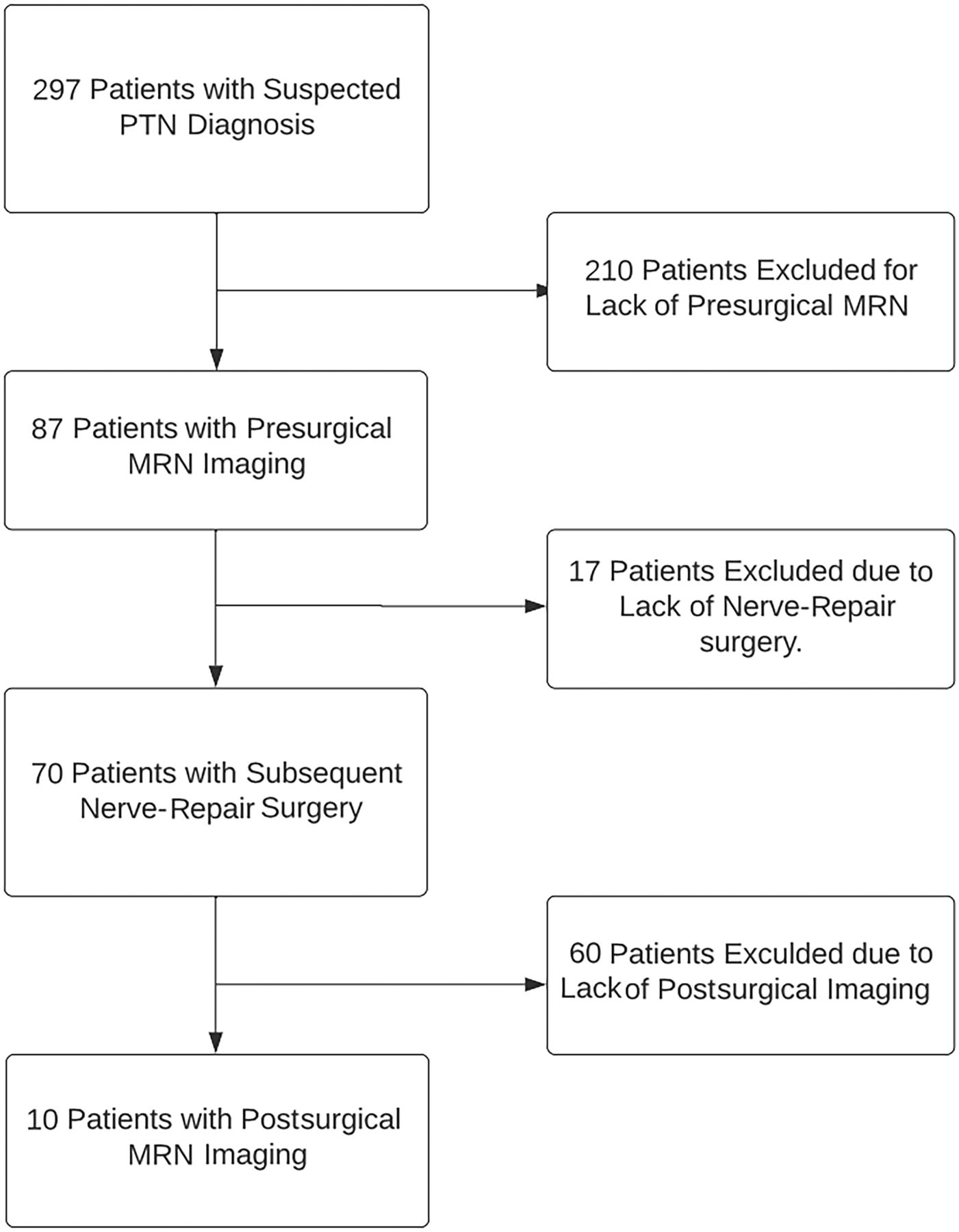
- FIG 1.
Patient population in this study.
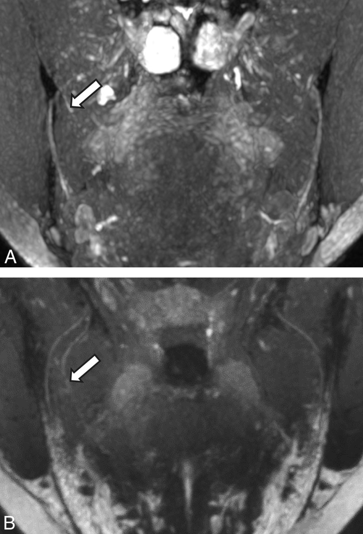
- FIG 2.
NS-RADS PI-1. A 54-year-old woman, status post third molar removal with decreased sensation, burning pain, and dysgeusia. A, Presurgical coronal MRN MIP image 89 days status post inciting event shows a neuroma in continuity of the right lingual nerve (NS- RADS I-4, arrow). B, Postsurgical coronal corresponding MRN 351 days following right lingual nerve neuroma excision and neurorrhaphy with allograft and Axoguard placement demonstrates the expected postsurgical appearance of the nerve (NS-RADS PI-1) with no loss of continuity, neuroma reformation, or substantial nerve-caliber changes (arrow).
- FIG 3.
NS-RADS PI-3. A 64-year-old man with a history of multiple nerve-repair procedures of the inferior alveolar nerve and mental nerve with a history of burning pain, lip biting, and speech difficulties. A and B, Presurgical coronal and sagittal MRN MIP images demonstrate a right mental nerve lateral neuroma in continuity (NS-RADS I-4, arrows). C and D, Postsurgical coronal and sagittal MRN MIP images 353 days following neuroma excision and neurorrhaphy with allograft and Axoguard placement show a recurrent right mental nerve neuroma in continuity (arrows).
- FIG 4.
NS-RADS PI-2. A 27-year-old woman 214 days status post third molar removal with decreased sensation, burning pain, and hypogeusia. A, Presurgical coronal MRN MIP image shows the right lingual nerve demonstrating nerve-caliber focal thickening and increased signal instead of uniformly distally decreasing nerve caliber, consistent with a neuroma in continuity (NS-RADS I-4, arrow). B, Postsurgical coronal MRN MIP image 98 days status post right lingual nerve neuroma excision and neurorrhaphy with allograft and Axoguard placement demonstrates incomplete regeneration, ie, minimal increased residual signal of the nerve without a new neuroma with minor caliber change compared with preoperative MRN, consistent with NS-RADS PI-2 findings (arrow).
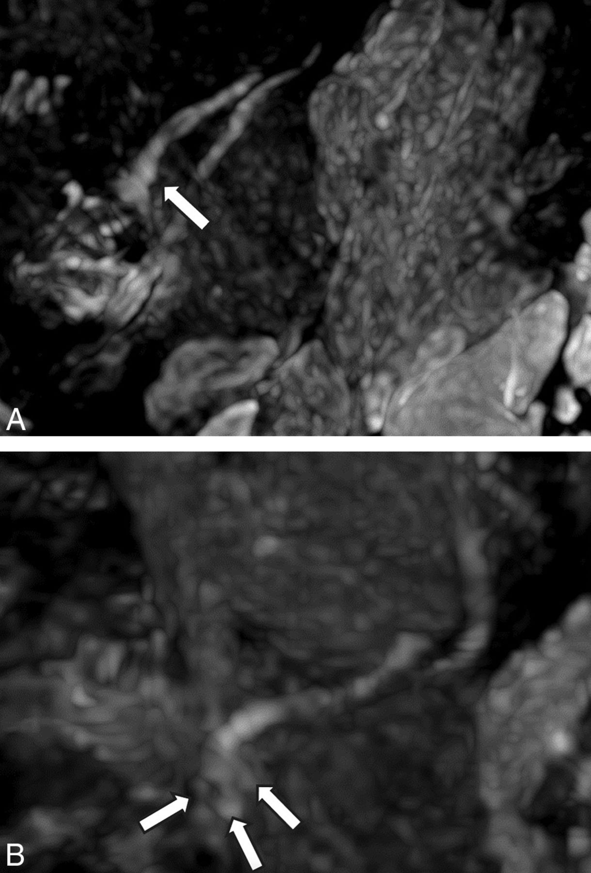
- FIG 5.
NS-RADS PI-3. A 21-year-old woman status post third molar removal experiencing decreased sensation, burning pain, and hypogeusia. A, Presurgical sagittal MRN MIP 55 days status post third molar removal shows right lingual nerve neuroma (NS-RADS I-4, arrow). B, Postsurgical sagittal MRN MIP 166 days status post neuroma excision and neurorrhaphy with allograft and Axoguard placement demonstrates the re-formation of multiple neuromas (arrows).
- FIG 6.
NS-RADS PI-2. A 31-year-old man status post third molar removal experiencing decreased sensation and ageusia. A and B, Presurgical coronal MRN MIP 54 days status post third molar removal shows a right lingual nerve end-bulb neuroma with complete transection with no distal continuity (NS-RADS I-5, arrows). C, Postsurgical coronal MRN MIP 238 days status post neuroma excision and neurorrhaphy with allograft and Axoguard placement demonstrates partial regeneration (arrows).


Tables
Sequence TR/TE (ms) Section Thickness (mm) Matrix FOV (cm) Comments Acquisition Time (min:sec) Axial T2-weighted SPAIR 2000/60 3.0 268 × 248 16 Corpus callosum to chin 5:20 Axial T1-weighted 580/9 3.0 320 × 310 16 Corpus callosum to chin 5:10 Axial 3D balanced FFE 5.32/2.66 0.65 270 × 270 16 Corpus callosum to chin 6:00 Axial DTI 14,000/70 5.0 196 × 192 18 Skull base to chin; b-values = 0 and 600 s/mm2; 12 directions 7:00 Coronal 3D STIR (optional) 1500/78 1.5 (Isotropic Voxel) … 20 Corpus callosum to chin 7:15 Coronal 3D PSIF 12/2.5 0.9 (Isotropic Voxel) … 20 Corpus callosum to chin 7:30 Note:—FFE indicates fast-field echo; SPAIR, spectral attenuated inversion recovery; STIR, short tau inversion recovery; PSIF, diffusion-weighted reversed fast imaging with steady-state precession.
- Table 2:
Postsurgical MRN NS-RADS PI distribution among 10 patients with the number of patients experiencing clinical improvement in specified neuropathic symptoms and overall clinical outcome
NS-RADS PI Patient Count Patients with Postsurgical Clinical Improvement Nerve-Repair PTN Outcome Pain Sensation Taste Lip Biting-Speech Complete Resolution Partial Improvement No Change NS-RADS PI-1 3 3 3 2 3 1 2 0 NS-RADS PI-2 3 0 0 0 2 0 1 2 NS-RADS PI-3 4 0 0 0 3 0 1 3


