
Article Figures & Data
Figures
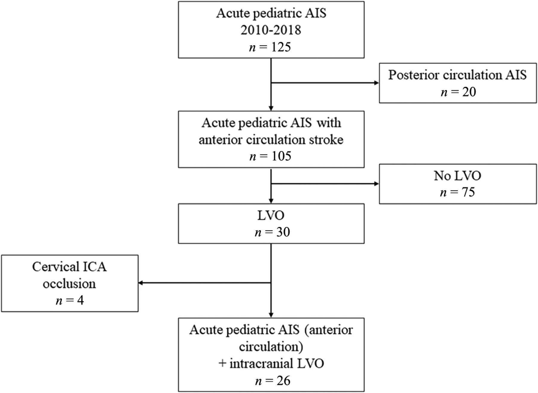
- FIG 1.
Study flow chart.
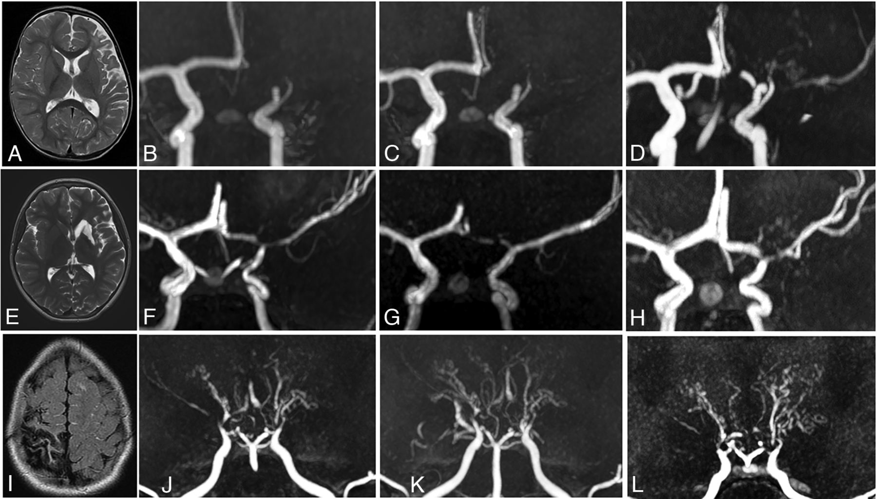
- FIG 2.
U-PS anastomotic bridge development in a patient with AIS and LVO, and unilateral FCA. Upper panel: acute phase MR imaging, showing recent MCA infarction with DWI hypersignal (A), with proximal left MCA occlusion and left A1 stenosis on the time-of-flight MRA, coronal view MIP 15 mm (B) and axial view MIP 10 mm (C). No collateral is visible at the acute phase. Lower panel: MR imaging 12 months after stroke occurrence. Time-of-flight MRA shows strictly unilateral left anomalies. Axial view (D) shows a reverted to normal left A1 segment and a persisting steno-occlusive M1 lesion, favoring the diagnosis of FCA. E, Coronal view shows a U-PS anastomotic bridge bypassing the M1 occlusion, with distal visible flow. F, Closer view of the U-PS anastomotic bridge illustrates the collaterals direction, parallel to the main MCA trunk, without perforating lenticulostriate collaterals.
- FIG 3.
Patterns of angiographic evolution in patients with FCA and U-PS anastomotic bridge development, compared with Moyamoya angiopathy. Upper panel: Patient with left superficial MCA infarction (A, MRI axial T2) and FCA. Angiographic evolution (3D time-of-flight MRA) with persisting occlusion 6 and 12 months after stroke (B and C), and U-PS anastomotic bridge in bypass of the occluded M1 segment with visible downstream MCA segments (D). Middle panel: Patient with left deep and superficial MCA infarction (E, MRI axial T2) and FCA. Angiographic evolution (3D time-of-flight MRA) with initial M1 occlusion (F). Partial improvement of MCA 6 months after stroke (G), with the observation of U-PS anastomotic bridge 12 months after stroke (H). Lower panel: Patient with right superficial MCA infarction (I, MRI axial T2) and Moyamoya angiopathy. Angiographic evolution (3D time-of-flight MRA) with bilateral steno-occlusive lesions of the terminal ICAs, MCAs, and ACAs (J). Bilateral perforating collaterals, present at stroke onset (J) and developing over time with a classical puff of smoke appearance. Progression of the arteriopathy with disappearance of MCAs and ACAs 6 and 24 months after stroke (K and L).
- FIG 4.
Distinctive features of U-PS anastomotic bridge compared with vascular patterns with close appearance. (i)-ICA = (intracranial)-internal carotid artery; ECA = external carotid artery; VA = vertebral artery; BA = basilar artery. Illustration is from Lin et al22 for rete mirabile. Drawings by F.B. and M.K., with courtesy.


