
Article Figures & Data
Figures
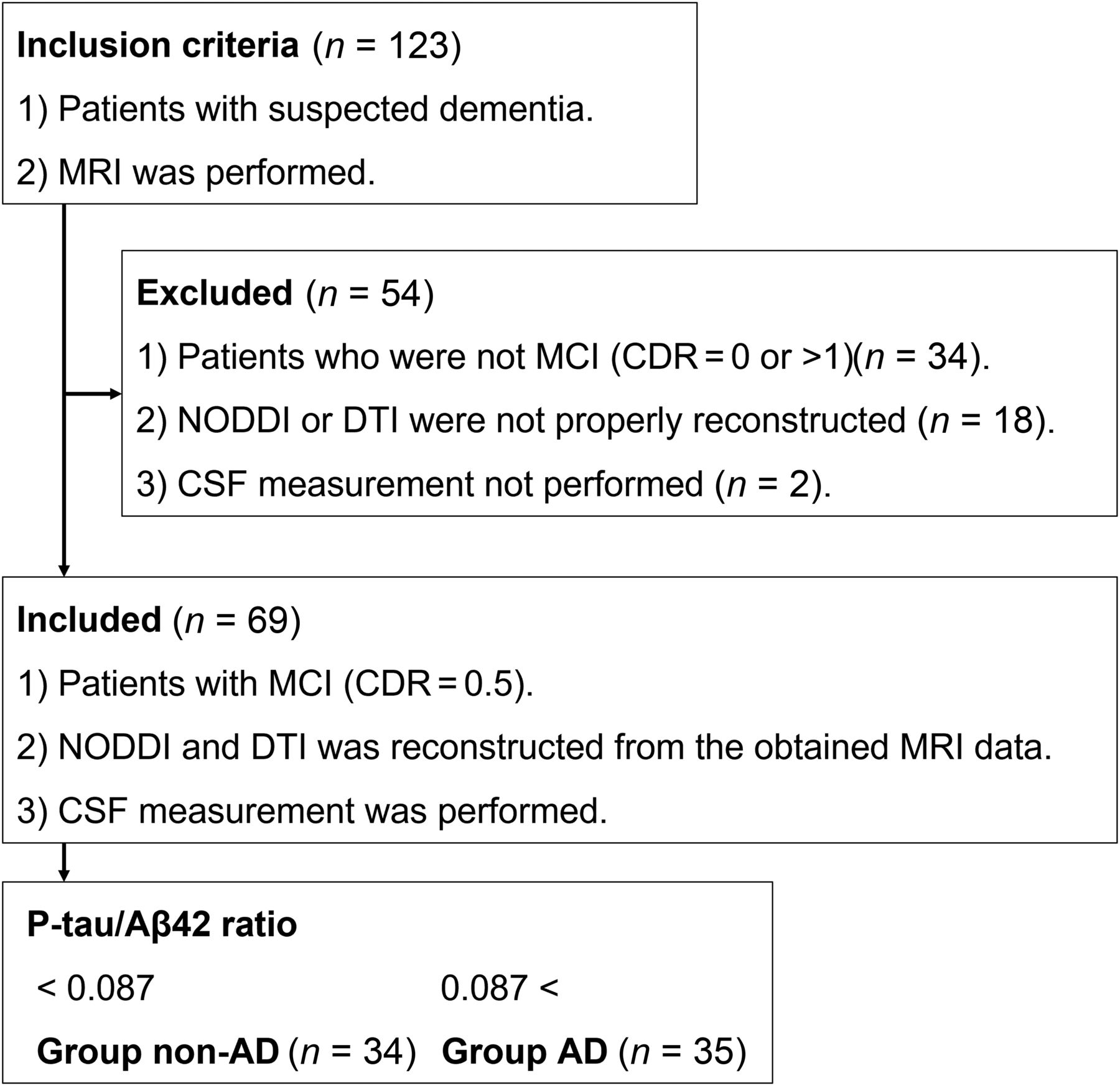
- FIG 1.
Inclusion and exclusion criteria.
- FIG 2.
A b0 image showing the 3-section axial ROI that was created manually for each side of the hippocampus.
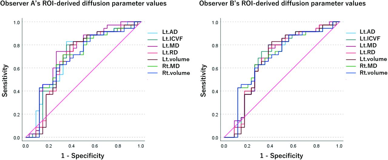
- FIG 3.
Receiver operating characteristic curve of each logistic regression model for each diffusion parameter and volumetry. The diffusion parameter values are from the ROIs of each of observer A and observer B.
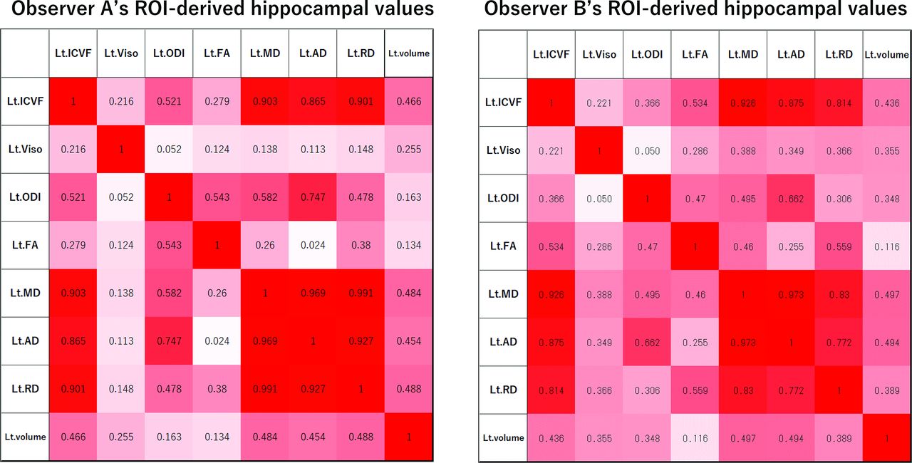
- FIG 4.
R absolute value (|R|) of the left hippocampal values in the AzD group. Heat map of the correlations between the diffusion metrics and the volume. Values of 0.34 < |R| < 0.43 indicate a statistically significant difference with P < .05. Values of 0.43 < |R| indicate a statistically significant difference with P < .01.


Tables
Group non-AzD AzD Number 34 35 Age (Median ± SD) 78 ± 9.15 79 ± 6.58 Sex (M:F) 18:16 17:18 MMSE (Median ± SD) 26 ± 5.09 24 ± 3.81 ADAS-Cog (Median ± SD) 9.40 ± 6.91a 11.60 ± 4.19a RBMT (Median ± SD) 11 ± 6.38a 6.5 ± 4.99a P-tau/Aβ42 ratio 0.05 ± 0.01 0.18 ± 0.07 Aβ42 (pg/mL) 919.38 ± 323.78a 554.94 ± 148.24a P-tau (pg/mL) 46.47 ± 15.36a 91.37 ± 28.69a Note:—MMSE indicates Mini-Mental State Examination; ADAS-Cog, Assessment Scale–Cognitive Subscale; RBMT, Rivermead Behavioral Memory Test; Aβ42, β-amyloid 42; P-tau, phosphorylated tau.
↵a Statistically significant difference between the groups (P < .05).
Left Right NODDI ICC ICC ICVF (mean) 0.87 (0.65−0.94) 0.91 (0.86−0.94) Viso (mean) 0.07 (−0.06−0.23) 0.12 (−0.07−0.31) ODI (mean) 0.70 (0.36−0.85) 0.74 (0.52−0.85) DTI FA (mean) 0.68 (0.53−0.79) 0.59 (0.41−0.72) MD (mean) 0.86 (0.40−0.95) 0.84 (0.69−0.91) AD (mean) 0.82 (0.31−0.93) 0.84 (0.39−0.94) RD (mean) 0.77 (0.53−0.88) 0.84 (0.65−0.92) Note:—ICCs indicates intraclass correlation coefficient.
↵a The data in parentheses are 95% confidence intervals.
- Table 3:
Statistically significant intergroup differences (P < .05) in the left and right hippocampal ROI values
Hippocampal ROI (Observer A) Hippocampal ROI (Observer B) Value (Mean ± Standard Deviation) P Value Value (Mean ± Standard Deviation) P Value non-AzD AzD non-AzD AzD NODDI parameter Left ICVF (dimensionless) Mean 0.377 ± 0.032 0.358 ± 0.031 .008 0.366 ± 0.033 0.349 ± 0.033 .026 90th percentile 0.474 ± 0.041 0.452 ± 0.047 .032 0.469 ± 0.093 0.438 ± 0.049 .029 DTI parameter Right MD (mm2/s) Mean 0.699 ± 0.030 0.715 ± 0.031 .022 0.707 ± 0.031 0.723 ± 0.030 .046 Left MD (mm2/s) Mean 0.706 ± 0.033 0.726 ± 0.034 .004 0.721 ± 0.038 0.739 ± 0.039 .014 10th percentile 0.641 ± 0.032 0.666 ± 0.032 .001 0.658 ± 0.035 0.676 ± 0.039 .011 90th percentile 0.769 ± 0.042 0.787 ± 0.040 .029 0.787 ± 0.058 0.803 ± 0.047 .026 Left AD (mm2/s) Mean 0.790 ± 0.035 0.812 ± 0.036 .002 0.809 ± 0.043 0.829 ± 0.039 .008 10th percentile 0.716 ± 0.033 0.739 ± 0.036 .001 0.733 ± 0.038 0.753 ± 0.040 .003 90th percentile 0.869 ± 0.045 0.887 ± 0.042 .037 0.894 ± 0.069 0.912 ± 0.051 .039 Left RD (mm2/s) Minimum 0.531 ± 0.053 0.556 ± 0.053 .028 0.543 ± 0.076 0.564 ± 0.066 .044 Mean 0.663 ± 0.033 0.682 ± 0.034 .006 0.676 ± 0.036 0.697 ± 0.047 .013 90th percentile 0.730 ± 0.040 0.747 ± 0.038 .038 0.745 ± 0.054 0.771 ± 0.076 .027 Volume (mm3) Left 3148.913 ± 519.884 2872.464 ± 488.542 .006 Right 3236.159 ± 438.547 3015.669 ± 423.202 .017 - Table 4:
The logistic regression model used in the prediction of the intergroup differences
AUCa Odds Ratio Logit (p) Diffusion Parameter Volume Age/Sex MMSE/ADAs-Cog/RBMT Intercept Term NODDI parameter Left ICVF (mean) Observer A 0.69 (0.56−0.82) 0.00 1.00 0.91/1.71 1.19/1.00/0.81 20682.03 Observer B 0.68 (0.55−0.81) 0.00 1.00 0.91/1.79 1.19/1.02/0.82 3487.07 DTI parameter Right MD (mean) Observer A 0.69 (0.56−0.82) 159.68 1.00 0.91/1.73 1.19/1.01/0.82 35.23 Observer B 0.68 (0.55−0.81) 31.01 1.00 0.91/1.69 1.17/1.00/0.82 238.04 Left MD (10th percentile) Observer A 0.73 (0.61−0.85) 199854741945.31 1.00 0.91/2.08 1.22/1.02/0.79 0.00 Observer B 0.68 (0.55−0.81) 126.30 1.00 0.91/1.91 1.19/1.02/0.82 8.02 Left AD (10th percentile) Observer A 0.71 (0.58−0.83) 42437669.81 1.00 0.92/2.05 1.20/1.01/0.80 0.00 Observer B 0.68 (0.55−0.81) 47.98 1.00 0.91/1.93 1.19/1.02/0.82 11.89 Left RD (mean) Observer A 0.68 (0.55−0.81) 128.06 1.00 0.91/1.92 1.18/1.01/0.82 11.57 Observer B 0.68 (0.54−0.81) 46.62 1.00 0.91/1.87 1.18/1.01/0.82 26.58 Volume Left 0.67 (0.54−0.81) NA 1.00 0.91/1.95 1.18/1.01/0.82 543.73 Right 0.68 (0.55−0.81) NA 1.00 0.91/1.72 1.17/1.01/0.82 4849.30 Note:—AUC indicates area under the receiver operating characteristic curve; MMSE, Mini-Mental State Examination; ADAS-Cog, Assessment Scale–Cognitive Subscale; RBMT, Rivermead Behavioral Memory Test; NA, not applicable.
↵a The data in parentheses are 95% confidence intervals.


