
Article Figures & Data
Figures
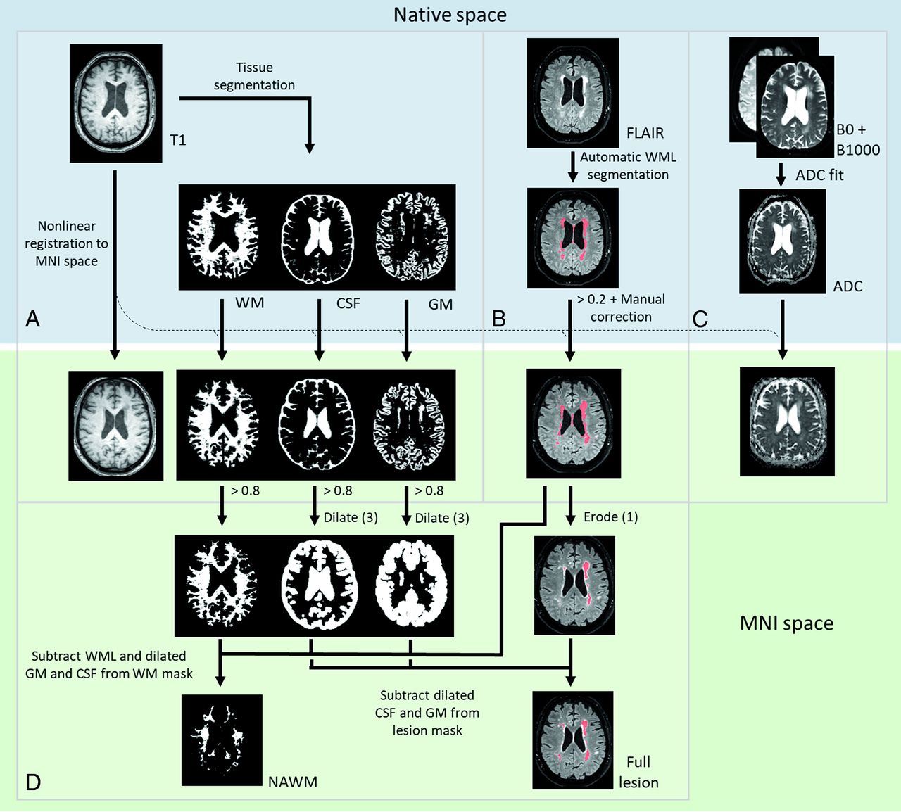
- FIG 1.
Overview of postprocessing steps. T1-weighted scans were segmented in native space and registered to MNI space (A). WMLs were automatically segmented on FLAIR images, thresholded, and manually corrected (B). ADC maps were fitted from DWIs with b-value = 0 and 1000 s/mm2 (C). WML masks and ADC maps were registered to MNI space using the transformation matrix from the T1 registration. NAWM masks were created by subtracting the WML masks from the WM segmentation (D). WML masks were eroded with 2 voxels. Last, GM and CSF masks were dilated with 3 voxels and subtracted from the resulting WML mask and NAWM mask.
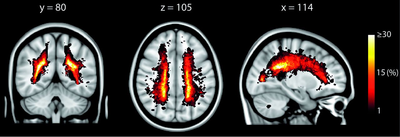
- FIG 2.
Lesion prevalence in patients with Fabry disease. Y, Z, and X coordinates refer to standard MNI space.
- FIG 3.
ADC (A) and ΔADC (B) in new lesion ROIs compared with NAWM. ADC within lesions with the same time (in years) relative to their reference point were combined to calculate mean values (data points) and their SDs (shaded areas). For example, ADCTP1 within the new lesion TP2 and ADCTP2 within new lesion TP3 both have time = −1 but are compared with the HAWM ADC from TP1 and TP2, respectively. Paired t tests showed that the ADC within lesions was significantly higher than the ADC within NAWM and that ΔADC was significantly higher than zero at every time. NAWM indicates normal appearing white matter.
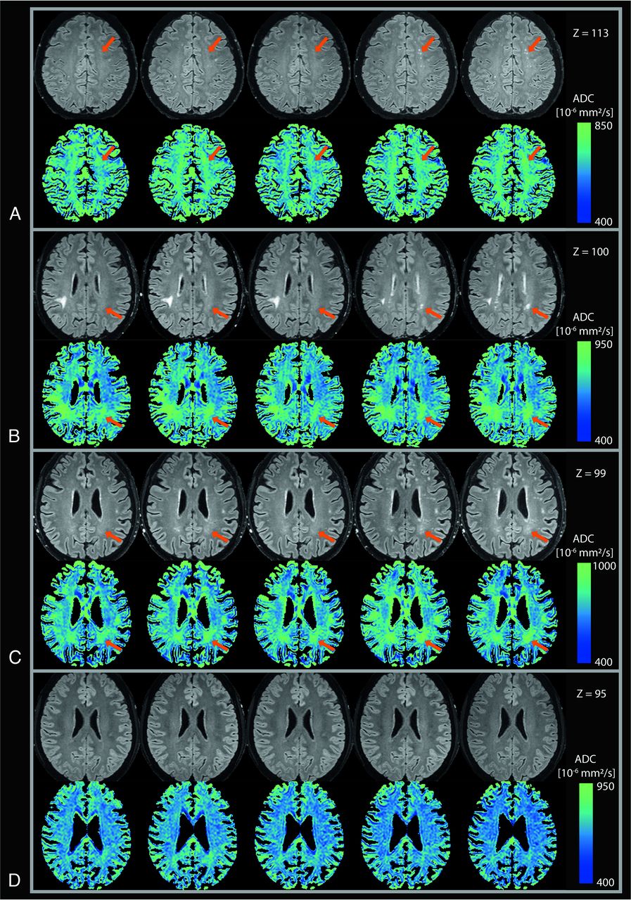
- FIG 4.
Yearly FLAIR scans and ADC maps. Three of these patients developed WMLs during the study (A–C), while 1 patient did not (D). Scans are shown in chronologic order from left to right. New WMLs are indicated by the orange arrows. The patient in B also had a large WM hyperintensity at the right parieto-occipital sulcus, which was diagnosed as a result of infarction before the study. Color scales are optimized for each patient individually. Z values indicate the Z coordinate in standard MNI space.
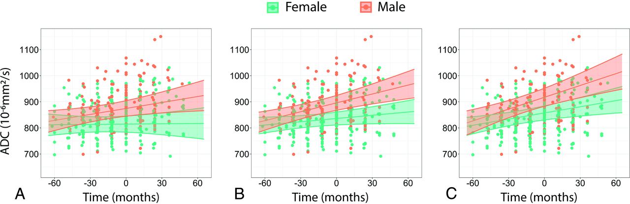
- FIG 5.
Visualization of LME models describing the ADC within new lesions. The graphs show how time and sex would affect the ADC of 36-, 46-, and 56-year-old patients superimposed on the measured data (A–C respectively). ADC is higher and increases faster in men compared with women and increases faster in older patients.


Tables
All Men Women Patient characteristics No. of patients (%) 48 25 (52%) 23 (48%) Classic phenotype (No.) (%) 48 (100%) 25 (100%) 23 (100%) No. of scan data sets (No.) 229 117 112 Age at first MR imaging (median) (range) (yr) 44 (15–69) 31 (15–55) 46 (22–69) Patients <18 yr (No.) (%) 2 (4.2%) 2 (8.0%) 0 (0.0%) Ever ERT (No.) (%) 48 (100%) 25 (100%) 23 (100%) Months treated (mean) 89 (SD, 34) 92 (SD, 39) 86 (SD, 30) Events before first MR imaging Cerebrovascular event (No.) (%) 2 (4.2%) 1 (4.0%) 1 (4.3%) TIA (No.) (%) 1 (2.1%) 1 (4.0%) 0 (0.0%) Kidney function at first MR imaging eGFR in mL/min/1.73 m2 (median) (range) 101 (40–154) 105 (49–154) 100 (40–127) eGFR <60 mL/min/1.73 m2 (No.) (%) 5 (10.4%) 3 (12%) 2 (8.7%) WML scores Fazekas first MR imaging (median) (range) 1 (0–6) 0 (0–6) 1 (0–6) Fazekas first MR imaging (mean) 1.5 (SD, 1.7) 1.4 (SD, 2.0) 1.5 (SD, 1.3) Fazekas > 0 first MR imaging (No.) (%) 29 (60%) 11 (44%) 18 (78%) Fazekas last MR imaging (median) (range)a 1 (0–6) 1 (0–6) 1 (0–6) Fazekas last MR imaging (mean) 1.5 (SD, 1.6) 1.6 (SD, 1.9) 1.4 (SD, 1.3) Fazekas > 0 last MR imaging (No.) (%) 33 (69%) 15 (60%) 18 (78%) Note:—eGFR indicates estimated glomerular filtration rate.
↵a For 32 patients, additional MR imaging after Fazekas scoring was performed. For these patients, the last known Fazekas score was used.
Predictors ADC ΔADC Estimates CI P Estimates CI P (Intercept) 836 810–862 <.01 84.4 63.1–105.7 <.01 Time (mo) 0.41 −0.08–0.91 .10 0.49 0.03–0.95 .04 Male sex 60.1 23.8–96.3 <.01 35.1 6.0–64.2 .02 Age (baseline, mean centered, year) 2.1 0.6–3.7 <.01 1.10 −0.16–2.36 .09 Time:male sex 0.72 −0.05–1.50 .07 0.99 0.27–1.71 <.01 Time:age 0.04 0.00–0.07 .03 0.03 −0.00–0.06 .09 Observations 472 464 Marginal R2 0.227 0.199 Conditional R2 0.689 0.613 - Table 3:
Summary of the obtained LME models describing ADC and ΔADC before lesion detection on FLAIR-weighted images
Predictors ADC before Detection ΔADC before Detection Estimates 95% CI P Estimates 95% CI P (Intercept) 834 806–861 <.01 83.2 59.9–106.5 <.01 Time (mo) 0.44 −0.17–1.04 .15 0.57 0.01–1.14 .047 Male sex 35.4 −3.6–74.5 .08 10.7 −21.8–43.1 .52 Age (baseline, mean centered) (yr) 1.0 −0.8–2.8 .25 0.32 −1.22–1.86 .68 Time:male sex −0.16 −1.10–0.79 .74 0.11 −0.71–0.94 .79 Time:age −0.00 −0.05–0.05 .85 −0.00 −0.05–0.04 .87 Observations 233 225 Marginal R2 0.114 0.035 Conditional R2 0.601 0.452


