Advertisement

Main menu

  • Home
  • Content
    • Current Issue
    • Accepted Manuscripts
    • Article Preview
    • Past Issue Archive
    • Video Articles
    • AJNR Case Collection
    • Case of the Week Archive
    • Case of the Month Archive
    • Classic Case Archive
  • Special Collections
    • AJNR Awards
    • Low-Field MRI
    • Alzheimer Disease
    • ASNR Foundation Special Collection
    • Photon-Counting CT
    • View All
  • Multimedia
    • AJNR Podcasts
    • AJNR SCANtastic
    • Trainee Corner
    • MRI Safety Corner
    • Imaging Protocols
  • For Authors
    • Submit a Manuscript
    • Submit a Video Article
    • Submit an eLetter to the Editor/Response
    • Manuscript Submission Guidelines
    • Statistical Tips
    • Fast Publishing of Accepted Manuscripts
    • Graphical Abstract Preparation
    • Imaging Protocol Submission
    • Author Policies
  • About Us
    • About AJNR
    • Editorial Board
    • Editorial Board Alumni
  • More
    • Become a Reviewer/Academy of Reviewers
    • Subscribers
    • Permissions
    • Alerts
    • Feedback
    • Advertisers
    • ASNR Home

User menu

  • Alerts
  • Log in

Search

  • Advanced search
American Journal of Neuroradiology

American Journal of Neuroradiology

ASHNR American Society of Functional Neuroradiology ASHNR American Society of Pediatric Neuroradiology ASSR
  • Alerts
  • Log in

Advanced Search

  • Home
  • Content
    • Current Issue
    • Accepted Manuscripts
    • Article Preview
    • Past Issue Archive
    • Video Articles
    • AJNR Case Collection
    • Case of the Week Archive
    • Case of the Month Archive
    • Classic Case Archive
  • Special Collections
    • AJNR Awards
    • Low-Field MRI
    • Alzheimer Disease
    • ASNR Foundation Special Collection
    • Photon-Counting CT
    • View All
  • Multimedia
    • AJNR Podcasts
    • AJNR SCANtastic
    • Trainee Corner
    • MRI Safety Corner
    • Imaging Protocols
  • For Authors
    • Submit a Manuscript
    • Submit a Video Article
    • Submit an eLetter to the Editor/Response
    • Manuscript Submission Guidelines
    • Statistical Tips
    • Fast Publishing of Accepted Manuscripts
    • Graphical Abstract Preparation
    • Imaging Protocol Submission
    • Author Policies
  • About Us
    • About AJNR
    • Editorial Board
    • Editorial Board Alumni
  • More
    • Become a Reviewer/Academy of Reviewers
    • Subscribers
    • Permissions
    • Alerts
    • Feedback
    • Advertisers
    • ASNR Home
  • Follow AJNR on Twitter
  • Visit AJNR on Facebook
  • Follow AJNR on Instagram
  • Join AJNR on LinkedIn
  • RSS Feeds

AJNR Awards, New Junior Editors, and more. Read the latest AJNR updates

Research ArticlePediatric Neuroimaging

Acute and Chronic Kernicterus: MR Imaging Evolution of Globus Pallidus Signal Change during Childhood

J. Gburek-Augustat, I. Sorge, M. Stange, J. Kern, A. Merkenschlager, T. Nägele and I. Krägeloh-Mann
American Journal of Neuroradiology August 2023, DOI: https://doi.org/10.3174/ajnr.A7948
J. Gburek-Augustat
aFrom the Division of Neuropaediatrics (J.G.-A., A.M.), Hospital for Children and Adolescents, University Hospital Leipzig, Leipzig, Germany
  • Find this author on Google Scholar
  • Find this author on PubMed
  • Search for this author on this site
  • ORCID record for J. Gburek-Augustat
I. Sorge
bDepartment of Pediatric Radiology (I.S.), University Hospital for Children and Adolescents, University Leipzig, Germany
  • Find this author on Google Scholar
  • Find this author on PubMed
  • Search for this author on this site
  • ORCID record for I. Sorge
M. Stange
cDepartment of Pediatrics (M.S.), University Hospital Halle, Halle, Germany
  • Find this author on Google Scholar
  • Find this author on PubMed
  • Search for this author on this site
  • ORCID record for M. Stange
J. Kern
dDepartment of Paediatric Neurology (J.K., I.K.-M.), University Children's Hospital, Tübingen, Germany
  • Find this author on Google Scholar
  • Find this author on PubMed
  • Search for this author on this site
  • ORCID record for J. Kern
A. Merkenschlager
aFrom the Division of Neuropaediatrics (J.G.-A., A.M.), Hospital for Children and Adolescents, University Hospital Leipzig, Leipzig, Germany
  • Find this author on Google Scholar
  • Find this author on PubMed
  • Search for this author on this site
  • ORCID record for A. Merkenschlager
T. Nägele
eDepartment of Diagnostic and Interventional Neuroradiology (T.N.), Tübingen University Hospital, Tübingen, Germany.
  • Find this author on Google Scholar
  • Find this author on PubMed
  • Search for this author on this site
  • ORCID record for T. Nägele
I. Krägeloh-Mann
dDepartment of Paediatric Neurology (J.K., I.K.-M.), University Children's Hospital, Tübingen, Germany
  • Find this author on Google Scholar
  • Find this author on PubMed
  • Search for this author on this site
  • ORCID record for I. Krägeloh-Mann
  • Article
  • Figures & Data
  • Info & Metrics
  • Responses
  • References
  • PDF
Loading

Article Figures & Data

Figures

  • Tables
  • FIG 1.
    • Download figure
    • Open in new tab
    • Download powerpoint
    FIG 1.

    Examples illustrating the various patterns observed with respect to bilateral signal change in the GP in kernicterus. The upper row displays pattern I, characterized by a diffuse signal increase on T1WI (arrows), while T2WI shows no abnormalities (sample case one, 2 weeks). The middle row represents pattern IIa, where T1WI appears normal, but there is a diffuse signal increase on T2WI (arrows) and FLAIR (arrows), (sample case three, 11 months of age). The lower row shows pattern IIb, featuring normal T1WI findings and a signal increase on T2WI (arrows) and FLAIR (arrows), limited to the borders (sample case two, 2 years 10 months of age).

  • FIG 2.
    • Download figure
    • Open in new tab
    • Download powerpoint
    FIG 2.

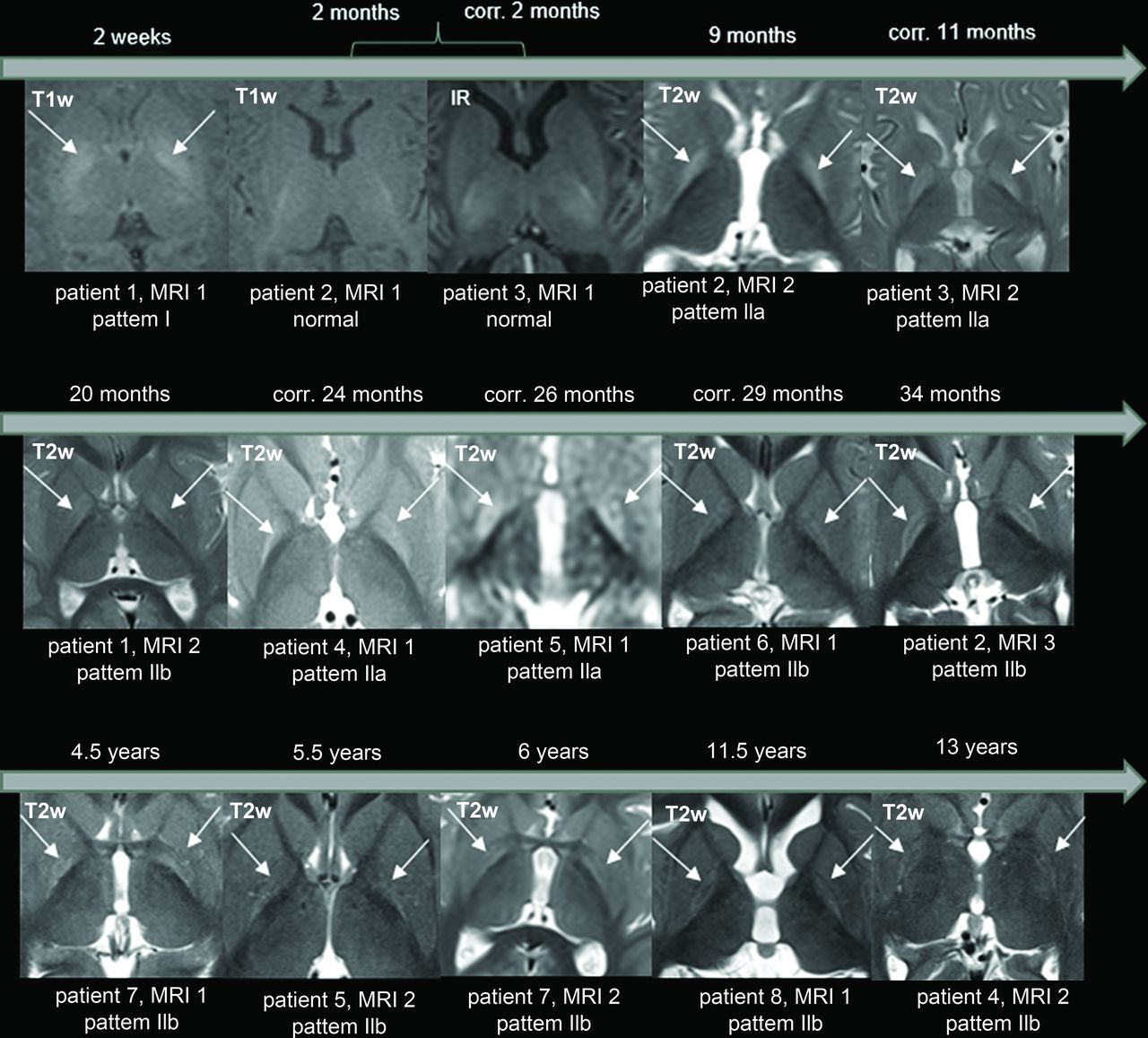
    The temporal progression of the GP signal changes in 8 patients. A bilateral signal change in the GP is the characteristic sign of kernicterus. In the neonatal period, hyperintensity is observed on T1WI (arrows). However, at approximately 2 months of age, a “blind diagnostic window” is encountered, where neither T1WI nor T2WI/FLAIR show abnormal findings. Subsequently, during infancy, there is signal hyperintensity of the entire GP on T2WI/FLAIR (arrows). After around 2 years, the signal hyperintensity on T2/FLAIR is limited to the borders of the GP (arrows). IR indicates inversion recovery; corr., corrected age related to gestational age.

  • FIG 3.
    • Download figure
    • Open in new tab
    • Download powerpoint
    FIG 3.

    Case 2 demonstrates the typical temporal sequence in a single patient (patient 2). The initial MR imaging was performed at approximately 2 months of age, revealing no abnormalities (illustrated on T1WI, left). At 9 months of age (middle), the entire GP exhibited a diffuse signal increase on T2WI (pattern IIa, arrows). At 34 months of age (right), a signal increase on T2WI was observed solely at the borders of the GP (pattern IIb, arrows).

  • FIG 4.
    • Download figure
    • Open in new tab
    • Download powerpoint
    FIG 4.

    Hippocampal volume loss is illustrated in patient 8 at the age of 11.5 years. The coronal T1WI (left) displays the head of the hippocampus only as filiform structures (arrows), resulting in enlarged temporal horns, as also depicted in the axial T2WI (arrows, middle). Both the external and internal spaces are globally enlarged (axial T2WI, right). The right images highlights the bilateral signal changes in the GP (arrows), indicative of pattern IIb.

Tables

  • Figures
    • View popup
    Table 1:

    Absolute inclusion and exclusion criteria

    Inclusion CriteriaExclusion Criteria
    Dyskinetic CPMovement disorder with onset after infancy
    Developmental delay since neonatal periodProgressive disorder
    Hyperbilirubinemia in neonatal periodOther explanatory causes
    • View popup
    Table 2:

    Relative inclusion criteria

    Hearing impairment
    Vertical gaze palsy
    Dental enamel dysplasia
PreviousNext
Back to top
Advertisement
Print
Download PDF
Email Article

Thank you for your interest in spreading the word on American Journal of Neuroradiology.

NOTE: We only request your email address so that the person you are recommending the page to knows that you wanted them to see it, and that it is not junk mail. We do not capture any email address.

Enter multiple addresses on separate lines or separate them with commas.
Acute and Chronic Kernicterus: MR Imaging Evolution of Globus Pallidus Signal Change during Childhood
(Your Name) has sent you a message from American Journal of Neuroradiology
(Your Name) thought you would like to see the American Journal of Neuroradiology web site.
CAPTCHA
This question is for testing whether or not you are a human visitor and to prevent automated spam submissions.
Cite this article
J. Gburek-Augustat, I. Sorge, M. Stange, J. Kern, A. Merkenschlager, T. Nägele, I. Krägeloh-Mann
Acute and Chronic Kernicterus: MR Imaging Evolution of Globus Pallidus Signal Change during Childhood
American Journal of Neuroradiology Aug 2023, DOI: 10.3174/ajnr.A7948

Citation Manager Formats

  • BibTeX
  • Bookends
  • EasyBib
  • EndNote (tagged)
  • EndNote 8 (xml)
  • Medlars
  • Mendeley
  • Papers
  • RefWorks Tagged
  • Ref Manager
  • RIS
  • Zotero
0 Responses
Respond to this article
Share
Bookmark this article
Acute and Chronic Kernicterus: MR Imaging Evolution of Globus Pallidus Signal Change during Childhood
J. Gburek-Augustat, I. Sorge, M. Stange, J. Kern, A. Merkenschlager, T. Nägele, I. Krägeloh-Mann
American Journal of Neuroradiology Aug 2023, DOI: 10.3174/ajnr.A7948
del.icio.us logo Twitter logo Facebook logo Mendeley logo
  • Tweet Widget
  • Facebook Like
  • Google Plus One
Purchase

Jump to section

  • Article
    • Abstract
    • ABBREVIATIONS:
    • MATERIALS AND METHODS
    • RESULTS
    • DISCUSSION
    • CONCLUSIONS
    • Footnotes
    • References
  • Figures & Data
  • Info & Metrics
  • Responses
  • References
  • PDF

Related Articles

  • No related articles found.
  • PubMed
  • Google Scholar

Cited By...

  • No citing articles found.
  • Crossref (6)
  • Google Scholar

This article has been cited by the following articles in journals that are participating in Crossref Cited-by Linking.

  • Predictive value of brain MRI for neurodevelopmental outcome in infants with severe unconjugated hyperbilirubinemia: A systematic review
    Noortje M. van der Meulen, Karin L. Meijers, Jeroen Dudink, Laura A. van de Pol
    European Journal of Paediatric Neurology 2024 53
  • Darstellung eines Kernikterus in der MRT und Änderungen im Verlauf
    Neuroradiologie Scan 2024 14 02
  • Darstellung eines Kernikterus in der MRT und Änderungen im Verlauf
    Radiologie up2date 2024 24 04
  • Hypoxic-Ischemic Encephalopathy and Beyond: A Pictorial Review of Neuroimaging Findings in Neonatal Encephalopathy
    Gleidson Silva, Brandon Simons, Kanika Gupta, Jennifer Kucera, Thomas R VerHage, Manish Bajaj, Tushar Chandra
    Cureus 2025
  • Neonatal Hyperbilirubinemia
    Andrea C. Wickremasinghe, Michael W. Kuzniewicz
    Pediatric Clinics of North America 2025
  • The Importance of Neuroimaging Follow-Up in Bilirubin-Induced Encephalopathy: A Clinical Case Review
    Martina Resaz, Alessia Pepe, Domenico Tortora, Andrea Rossi, Luca Antonio Ramenghi, Andrea Calandrino
    Brain Sciences 2025 15 6

More in this TOC Section

  • FRACTURE MR in Congenital Vertebral Anomalies
  • Neuroimaging Delineation and Progression of SLSMD
  • fetal brain development of 10 weeks gestation
Show more PEDIATRIC NEUROIMAGING

Similar Articles

Advertisement

Indexed Content

  • Current Issue
  • Accepted Manuscripts
  • Article Preview
  • Past Issues
  • Editorials
  • Editor's Choice
  • Fellows' Journal Club
  • Letters to the Editor
  • Video Articles

Cases

  • Case Collection
  • Archive - Case of the Week
  • Archive - Case of the Month
  • Archive - Classic Case

More from AJNR

  • Trainee Corner
  • Imaging Protocols
  • MRI Safety Corner
  • Book Reviews

Multimedia

  • AJNR Podcasts
  • AJNR Scantastics

Resources

  • Turnaround Time
  • Submit a Manuscript
  • Submit a Video Article
  • Submit an eLetter to the Editor/Response
  • Manuscript Submission Guidelines
  • Statistical Tips
  • Fast Publishing of Accepted Manuscripts
  • Graphical Abstract Preparation
  • Imaging Protocol Submission
  • Evidence-Based Medicine Level Guide
  • Publishing Checklists
  • Author Policies
  • Become a Reviewer/Academy of Reviewers
  • News and Updates

About Us

  • About AJNR
  • Editorial Board
  • Editorial Board Alumni
  • Alerts
  • Permissions
  • Not an AJNR Subscriber? Join Now
  • Advertise with Us
  • Librarian Resources
  • Feedback
  • Terms and Conditions
  • AJNR Editorial Board Alumni

American Society of Neuroradiology

  • Not an ASNR Member? Join Now

© 2025 by the American Society of Neuroradiology All rights, including for text and data mining, AI training, and similar technologies, are reserved.
Print ISSN: 0195-6108 Online ISSN: 1936-959X

Powered by HighWire