
Article Figures & Data
Figures
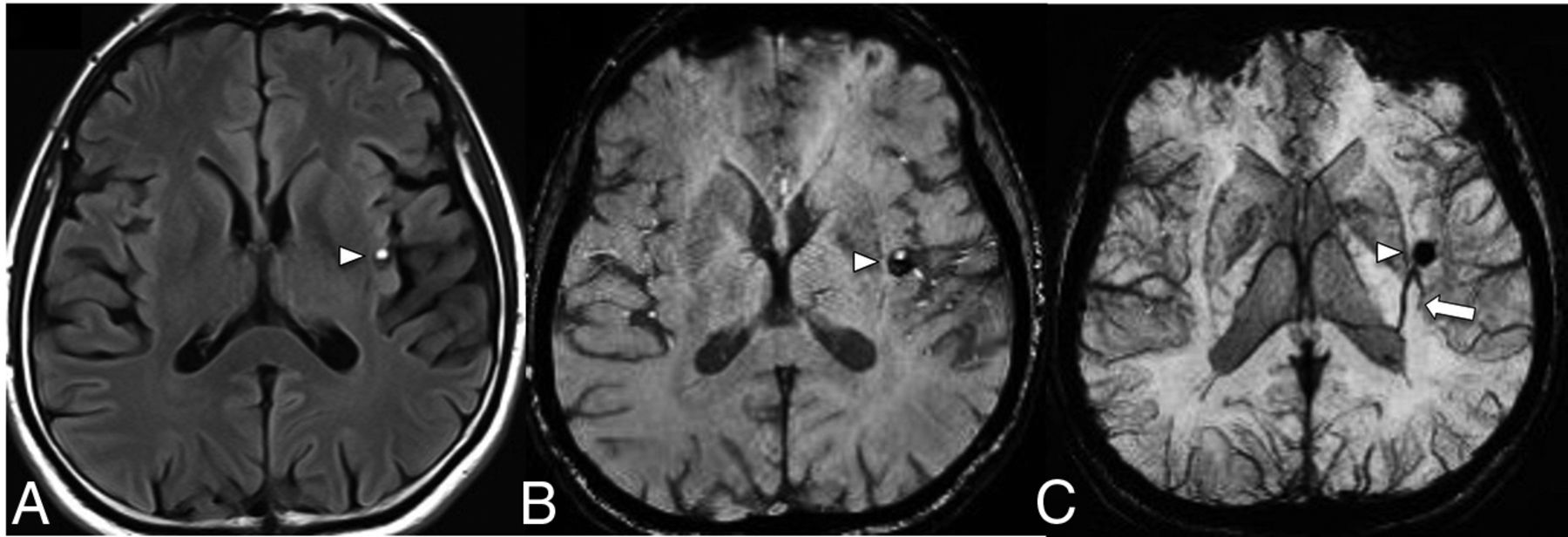
- FIG 1.
FLAIR (A) shows a mixed-signal-intensity CCM (arrowhead) in the left insular cortex with an internal blood-fluid level and no perilesional edema. A collector vein of a DVA (arrow) is seen from the ventricular ependyma to the CCM, which is barely visible on the SWI (B) and becomes more conspicuous on the susceptibility-weighted MIP image (C).
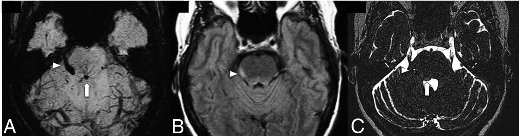
- FIG 2.
A patient with right facial pain and dysesthesia. SWI (A) shows a posterior pontine DVA (arrow) and an associated CCM (arrowhead) involving the intra-axial and cisternal segments of the right trigeminal nerve. FLAIR (B) demonstrates hyperintense edema along the right lateral aspect of the pons (arrowhead). Coregistered T2 sampling perfection with application-optimized contrasts by using different flip angle evolutions (SPACE sequence; Siemens) (C) confirms CCM involvement of the right trigeminal nerve.
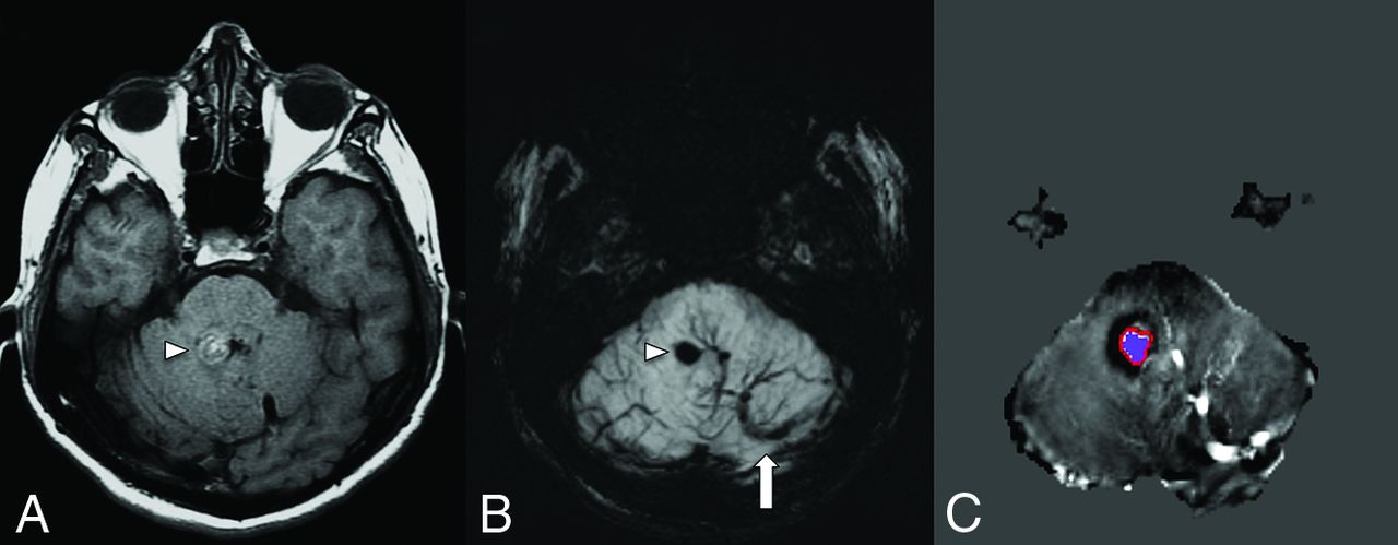
- FIG 3.
A middle-aged patient with new-onset ataxia. T1WI (A) and SWI MIP (B) show a CCM in the right superior cerebellar peduncle (arrowheads) and a large left cerebellar DVA with the collector vein (arrow) draining into the transverse sinus. Quantitative susceptibility mapping (C) analysis of the CCM shows a high mean susceptibility value of 858 parts per billion (with threshold). An ROI with a red boundary represents the exclusive object boundary, and the purple area represents thresholded pixels (150 parts per billion). SWIM (Siemens) parameters: TE = 20.00 ms; TR = 27.00 ms; flip angle = 150; resolution = 0.937 × 0.937 ×2.5 mm. Images courtesy of Dr E. Mark Haacke.
- FIG 4.
Noncontrast CT of the head (A and B) shows dystrophic calcification of the anterior right putamen and pulvinar of the thalamus (arrows). CTV MIP sagittal image (C) shows a right basal ganglia DVA (arrowheads) with the collector vein draining into the ipsilateral internal cerebral vein (arrows).
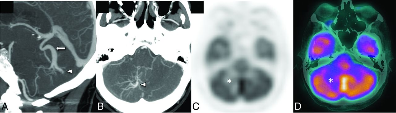
- FIG 5.
A patient with headache and ataxia. CTA sagittal and axial MIP images (A and B) show a right cerebellar hemisphere DVA (arrowheads) with the collector vein (arrow) draining into the vein of Galen. Corresponding [18F] FDG-PET/CT attenuation-corrected image (C) and a fused PET/CT image (D) show moderate reduction of [18F] FDG uptake in the right cerebellar hemisphere in the venous territory of the large DVA (asterisk).
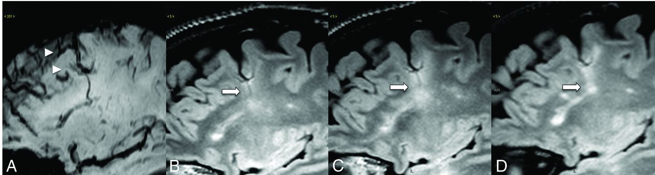
- FIG 6.
A patient with MS with an SWI (A) demonstrating a DVA. FLAIR at 6-month (B), 1-year (C), and 2-year (D) follow-up shows an enlarging demyelinating plaque (arrows) centered around the DVA.
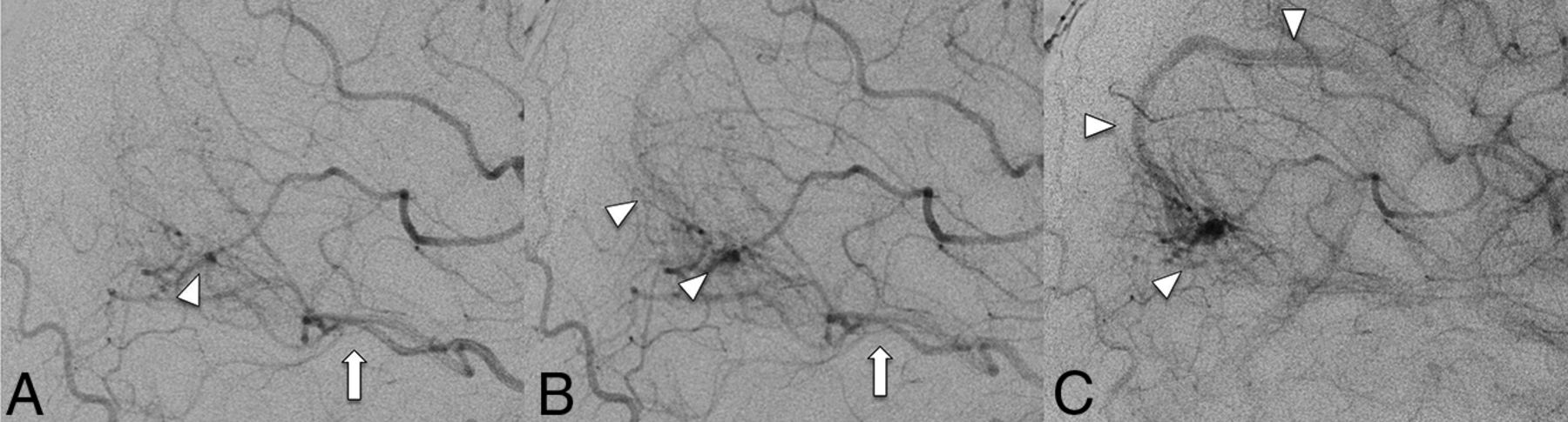
- FIG 7.
DSA cerebral catheter angiogram in the arterial phase image (A) demonstrates a left occipital AVM nidus (arrowhead) supplied by the left posterior cerebral artery (arrow). Subsequent late arterial (B) and early venous (C) images show the AVM nidus (arrowhead) draining into a right occipital DVA (arrowhead), with an early venous filling of the collector vein (arrowhead). The patient underwent stereotactic radiation treatment with successful obliteration of the AVM nidus with preservation of the DVA venous architecture.
- FIG 8.
A patient with headache and ataxia. Contrast-enhanced T1WI (A–C) shows the DVA medusa veins in the cerebellar hemispheres (arrowheads) with a pair of draining veins and ultimately a common collector vein (arrows) draining into the right tentorial venous sinus. DSC MRI perfusion demonstrates a pronounced and asymmetrically increased MTT (D) in the right cerebellar hemisphere, consistent with venous hypertension. CBV (E) and CBF (F) images shows expected increased cerebral blood volume and flow in the DVA medusa veins.
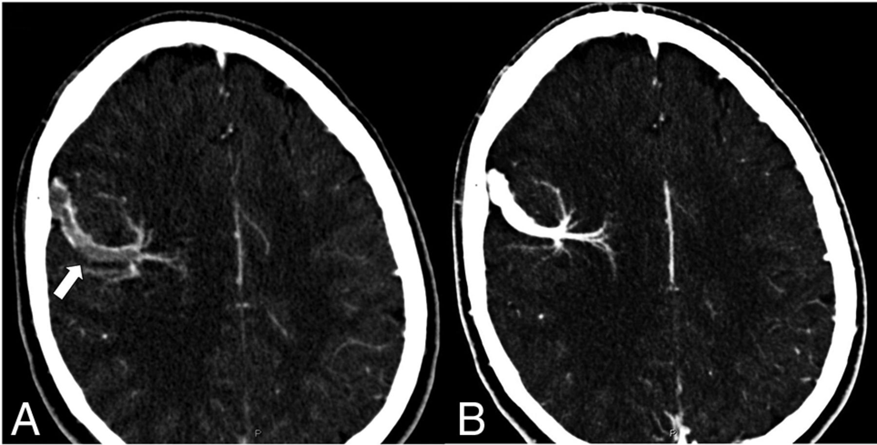
- FIG 9.
A young patient presented with a sudden onset of severe headaches after a marathon race. Presentation CTV (A) shows thrombosis of a DVA collector vein (arrow) overlying the right frontal cerebral convexity. The patient was placed on antiplatelet medication, and a follow-up CTV (B) showed a resolution of the thrombus.
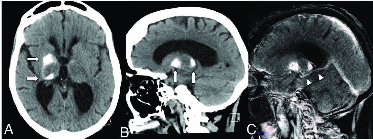
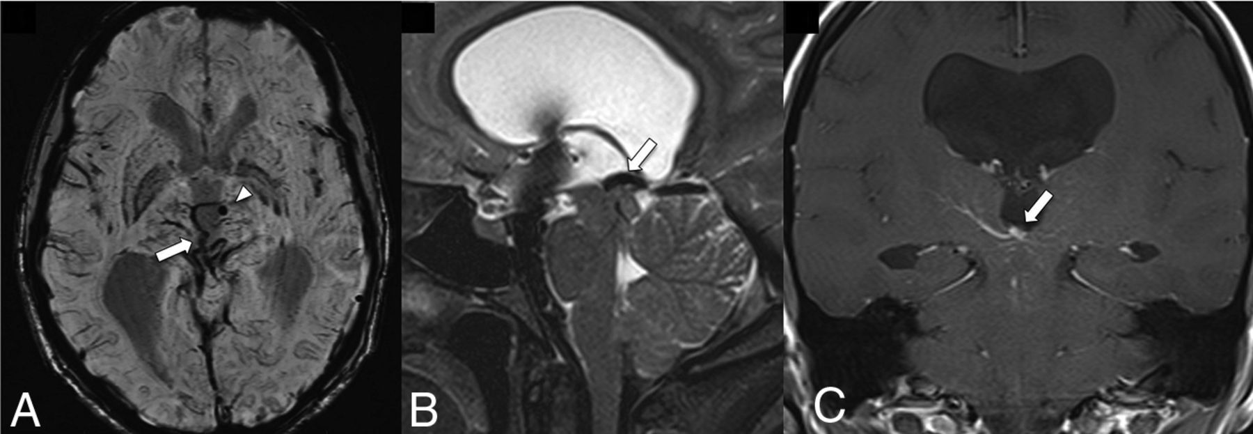
- FIG 10.
A midline midbrain DVA with the collection vein (arrow) obstructs the cerebral aqueduct leading to ventriculomegaly. SWI (A) shows the radially oriented medullary veins in the midbrain and an associated microbleed in the left anterior thalamus (arrowhead). T2-SPACE (B) and gadolinium-enhanced T1-weighted (C) images depict the location of a large collector vein obstructing the entrance into the cerebral aqueduct. Images courtesy of Dr Arjuna Somasundaram and Dr Christian Schwindack.
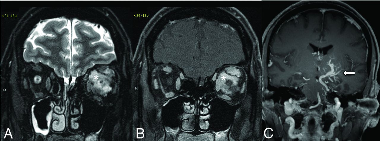
- FIG 11.
CVMS in a patient with a left orbital venous malformation depicted on the coronal T2-weighted fat saturated (A) and coronal post-gadolinium-enhanced T1-weighted fat saturated (B) images, which show an infiltrative T2-weighted hyperintense intraconal lesion with avid contrast enhancement (asterisk). Coronal gadolinium-enhanced T1-weighted image (C) reveals a large left basal ganglia DVA (arrow) with the collector vein draining in the left superior petrosal sinus.


Jump to section
Related Articles
Cited By...
- No citing articles found.