
Article Figures & Data
Figures
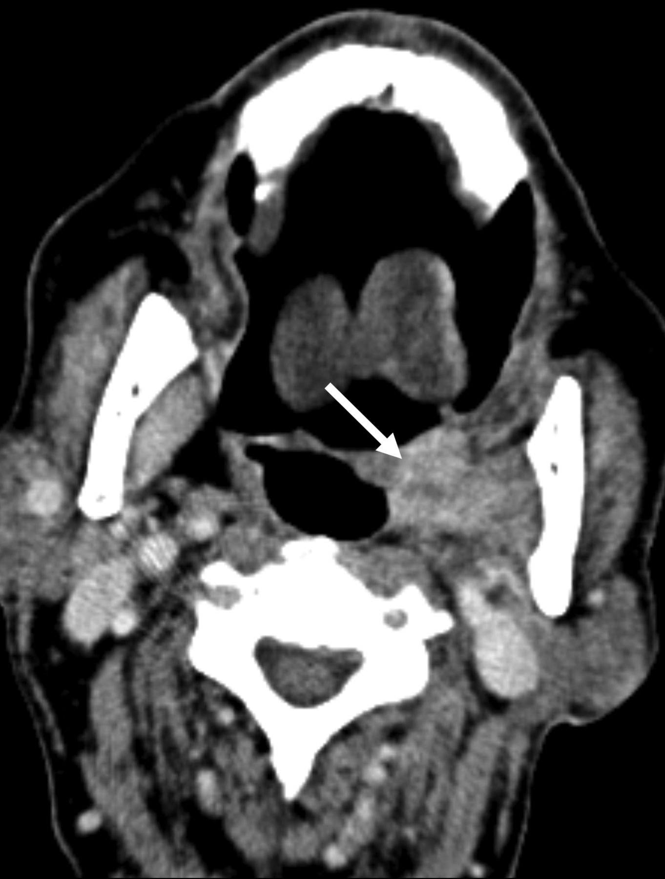
- FIG 1.
SMG flap misdiagnosed as recurrent tumor. Axial contrast-enhanced CT performed as the first posttreatment surveillance scan following resection of an SCC involving the lateral oral tongue, retromolar trigone, and lateral oropharynx. The enhancing nodular masslike lesion of the lateral oropharynx was mistakenly interpreted as recurrent tumor (arrow). After discussion with the otolaryngologist, this was determined to represent an SMG flap reconstruction of the lateral oropharynx.
- FIG 2.
SMG flap of the parotid bed. Axial T2-weighted fat-suppressed (A), T1-weighted (B), and T1-weighted postcontrast fat-suppressed (C) MR images demonstrating facial contour reconstruction with an SMG flap in the parotid bed (arrow), after total parotidectomy for a poorly differentiated carcinoma.
- FIG 3.
Typical SMG flap appearance. A, Axial contrast-enhanced CT of an SCC involving the left lateral oropharynx (dashed arrow). Axial (B) and coronal (C) contrast-enhanced CT images obtained 6 months after resection and reconstruction with an SMG flap show an enhancing, nodular, masslike lesion in the operative bed (solid arrow). The glandular heterogeneous enhancement, the semblance of a preserved hilum, and the absence of the native SMG in its orthotopic location in the submandibular space are useful in differentiating a normal SMG flap from recurrent tumor.
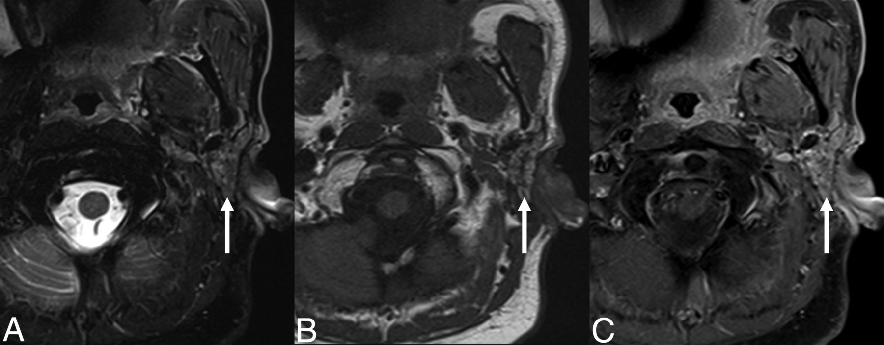
- FIG 4.
T2 signal intensity of an SMG flap. Coronal T2-weighted (A) and T2-weighted fat-suppressed (B) MR images of an SMG flap within the parapharyngeal space (solid arrow) for reconstruction following resection of a synovial cell sarcoma. MR imaging characteristics of SMG flaps are variable compared with the contralateral gland. Due to atrophy in this case, the mobilized gland is increased in signal intensity relative to the contralateral gland in the orthotopic location (dashed arrow) in the first image, but decreased in signal intensity in the fat-suppressed image.
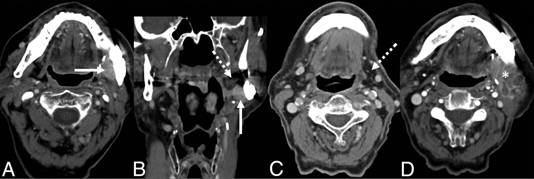
- FIG 5.
A potential interpretation pitfall of SMG flaps. Axial (A) and coronal (B) contrast-enhanced CT images of a patient undergoing a first postoperative surveillance scan following marginal mandibulectomy and internal fixation for resection of an SCC of the retromolar trigone. The patient declined adjuvant chemoradiation. After reviewing the operative report, an enhancing masslike lesion medial to the mandible was presumed to represent the SMG flap (solid arrow). However, this enhancing tissue was later retrospectively revealed to be recurrent tumor adjacent to the SMG flap with an atrophic, fatty gland (dashed arrow). C, Axial contrast-enhanced CT from the preoperative staging study shows that the gland was originally low in attenuation due to fat content (dashed arrow). D, Axial contrast-enhanced CT of the second postoperative surveillance scan shows heterogeneous enhancement of progressive tumor (asterisk).
- FIG 6.
Distortion of the SMG flap into a fusiform shape. Axial contrast-enhanced CT following marginal mandibulectomy for resection of an ameloblastoma. The SMG flap appears as a fusiform enhancing nodule inferior to the operative bed (arrow). While similar in imaging appearance to a SMG transfer, this gland was mobilized for reconstruction of the ipsilateral surgical defect rather than to shield the contralateral gland away from the high-dose radiation treatment field.
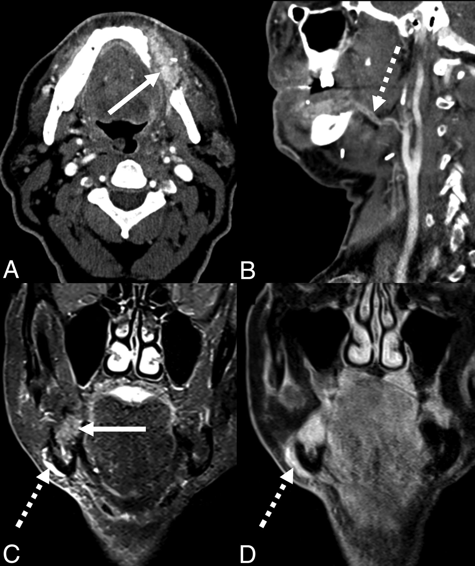
- FIG 7.
SMG flaps and vascular pedicles in 2 patients following marginal mandibulectomy and SMG flap reconstruction for primary resection of an SCC of the oral cavity. Axial (A) and sagittal (B) contrast-enhanced CT images reveal the hyperenhancing, heterogeneous, glandular texture of the SMG flap (solid arrow) with a vascular pedicle including the facial artery coursing medial to the mandibular ramus (dashed arrow). Coronal STIR (C) and coronal T1-weighted fat-suppressed (D) postcontrast MR images show an SMG flap in the mandibulectomy defect (solid arrow) and vascular pedicle, including the facial vein coursing lateral to the mandibular body (dashed arrows).
- FIG 8.
SMG flap glandular texture. Axial T2-weighted fat-suppressed (A), T1-weighted (B), and T1-weighted postcontrast fat-suppressed (C) MR images in a patient with a pleomorphic adenoma of the parapharyngeal space following resection and reconstruction with an SMG flap (arrow). The gland conforms to the triangular shape of the parapharyngeal space, while maintaining a glandular heterogeneity similar to that in the adjacent parotid gland.


Jump to section
Related Articles
Cited By...
- No citing articles found.