
Article Figures & Data
Figures
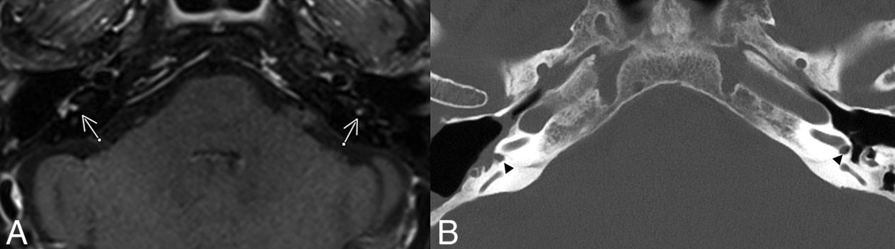
- FIG 1.
Example of enhancing granulation tissue in the RW. A, Axial postcontrast FS T1WI demonstrates enhancement present bilaterally in the RWs (arrows). B, Axial head CT with thin-section bone window reconstructions demonstrates a right-sided canal wall-down mastoidectomy for resection of a cholesteatoma. Opacification of the round windows can be seen bilaterally, correlating to the area of enhancement (arrowheads).
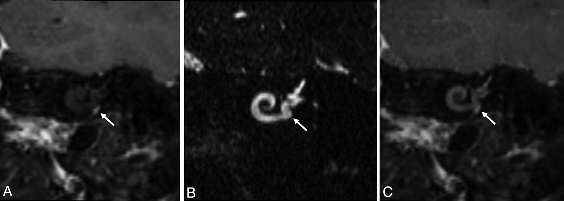
- FIG 2.
A, Postcontrast FS T1WI reconstructed in the Stenvers view demonstrates focal contrast enhancement. B, Thin-section 3D SPACE T2WI reconstructed in the Stenvers view demonstrates fluid signal throughout the basal turn of the cochlea as well as hyperintense signal lateral to the RW membrane (white arrow). Specifically, there is no filling defect within the basal turn of the cochlea. C, Coregistered overlay of the 3D SPACE T2WI and postcontrast FS T1WI illustrate that the enhancement is located lateral to the basal turn and entirely within the RWN (white arrow). Without the aid of the T2 SPACE and coregistration, it would be possible to misdiagnose a small intralabyrinthine schwannoma in the basal turn or a glomus tympanicum. The white arrow in A is the focal enhancement.


Tables
- Table 1:
Summary of patient demographics, reasons for initial ENT visit, and relevant medical history relating to temporal bone
Patient information Demographics No. of patients 95 Age at MR imaging (average) (range) (yr) 58 (18−84) Sex (No.) Female 53 Male 42 Reason for ENT visit Hearing loss 51 Tinnitus 11 Vertigo/dizziness/ataxia 13 Schwannoma/cholesteatoma/other tumor 13 Headache 2 Other 5 No. of patients with a mastoid effusiona 24 No. of patients with prior surgery on temporal bone 8 Tympanomastoidectomy 6 Tympanotomy 1 Myringotomy 1 ↵a No. of patients with clinical charts indicating the presence of granulation tissue in RW.
Findings Patients with MR imaging 95 Enhancement in RW 15 Right 6 Left 7 Bilateral 2 Patients with both MR imaging and CT 27 CT–/MR imaging– 22 CT+/MR imaging+ 4 CT–/MR imaging+ 1 Patients with CT 27 Soft tissue in RW 4 Right 0 Left 3 Bilateral 1 Note:—CT+ indicates soft tissue found, MR imaging+ indicates enhancement found, CT– indicates no soft tissue found, and MR imaging indicates no enhancement found.


Jump to section
Related Articles
Cited By...
- No citing articles found.