
Article Figures & Data
Figures
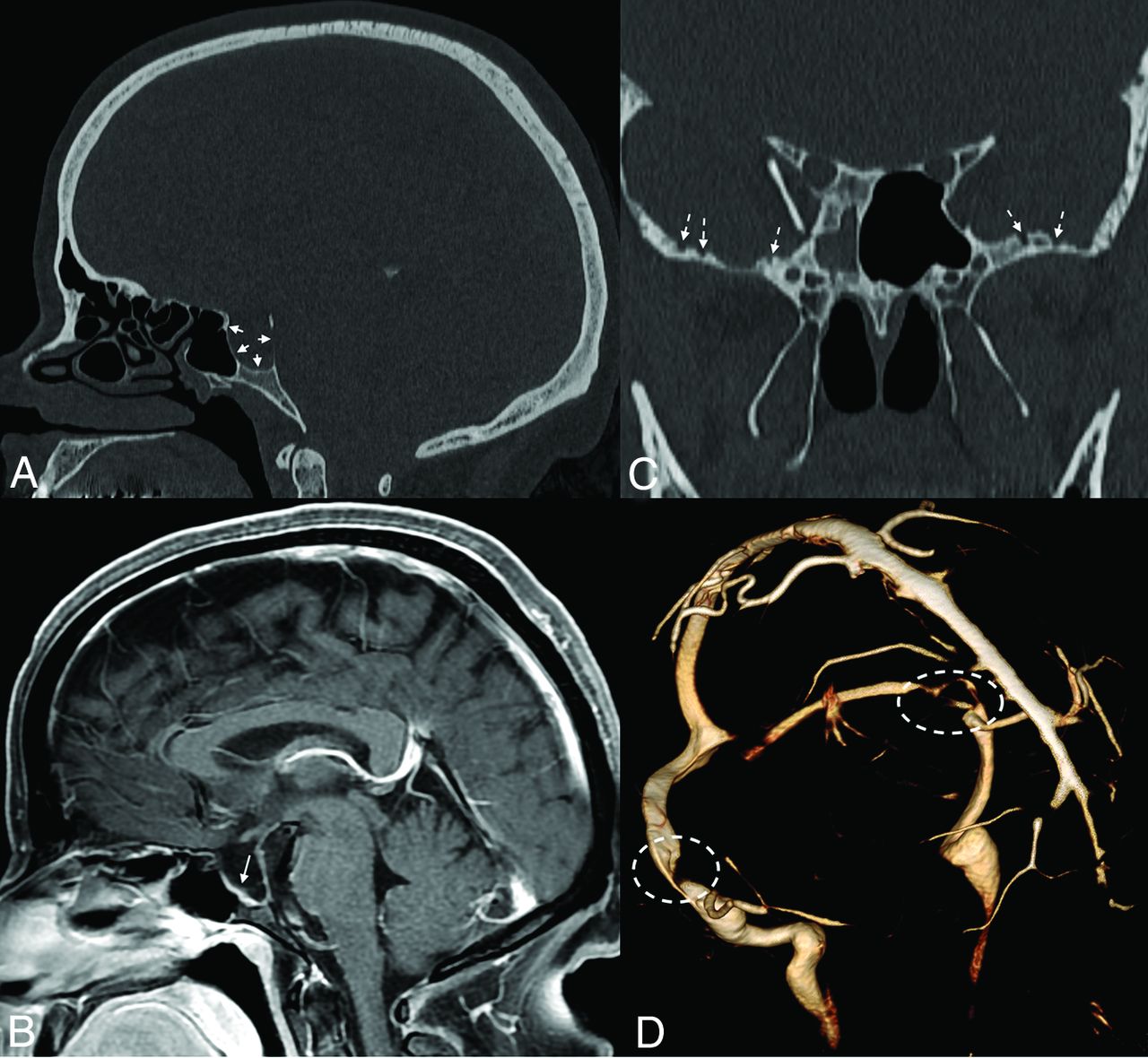
- FIG 1.
Sella expansion and skull base pitting in IIH. Sagittal CT image (A) in a 38-year-old woman with pseudotumor cerebri demonstrates marked expansion of the osseous walls of the sella (short solid arrows), with frank dehiscence posteriorly. Corresponding sagittal MR image (B) shows flattening of the pituitary tissue along the floor of the sella (long solid arrow). Prominent pitting is also noted along the anterior margins of both middle cranial fossae (dashed arrows, C). 3D reconstruction image of an MRV (D) demonstrates smooth tapering of the bilateral transverse sinuses (dashed ovals), compatible with IIH.
- FIG 2.
Skull pitting, meningoceles, and CSF leak in a patient who presented with rhinorrhea. Axial and coronal CT images (A and B) demonstrate substantial pitting along the anteromedial aspect of the left middle cranial fossa (dashed ovals, A and B), with complete opacification of the left sphenoid sinus (asterisks), concerning for a CSF leak. Milder pitting is noted on the right (solid arrows). Corresponding MR imaging (C and D) confirms a meningocele protruding into the left sphenoid sinus (dashed ovals, C and D). MRV (not shown) noted smooth tapered stenoses involving both transverse sinuses, and the patient was ultimately diagnosed with pseudotumor cerebri.
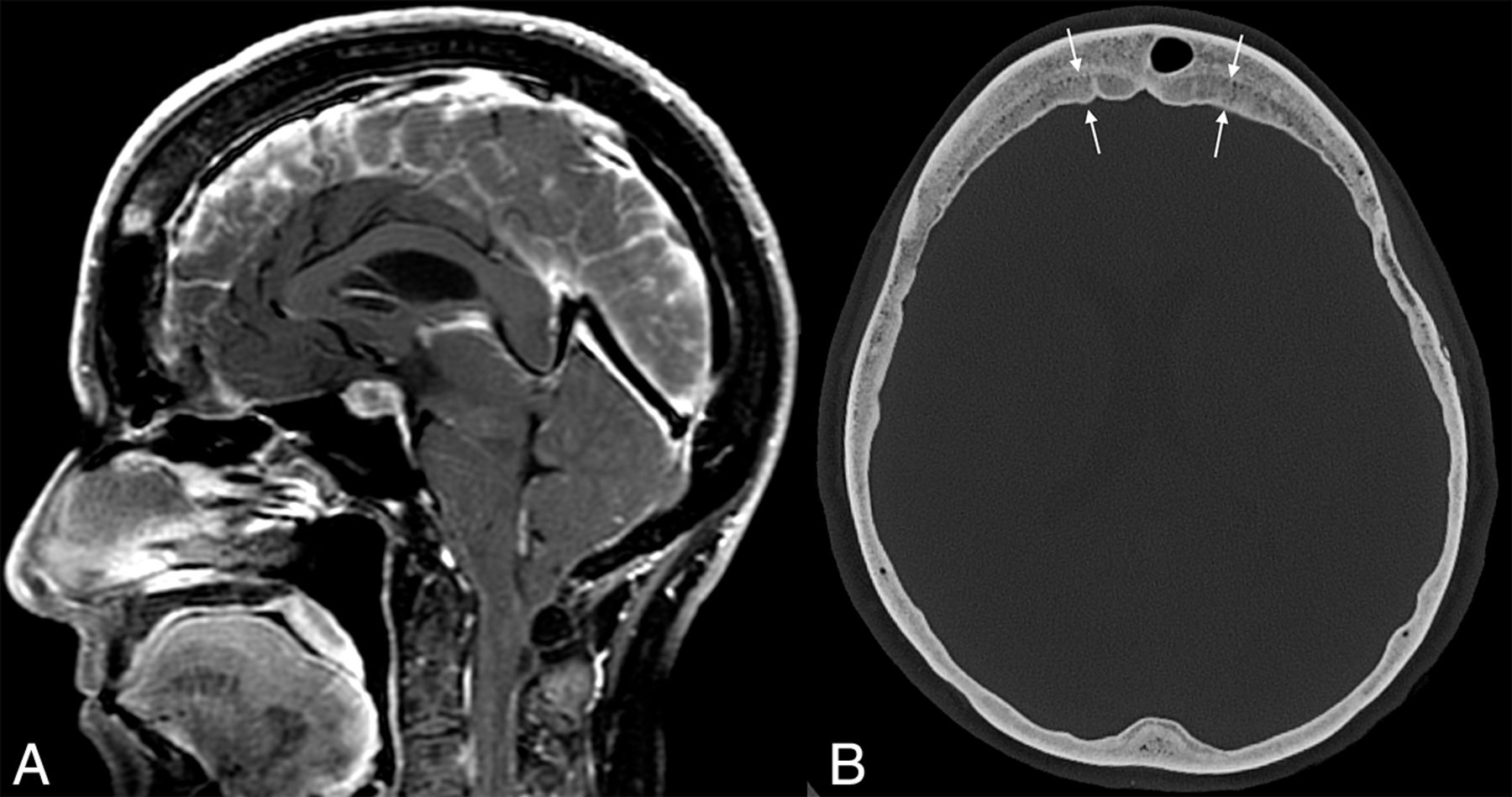
- FIG 3.
Example of layered calvarial thickening in a patient with SIH. Sagittal MR images (A) demonstrate classic findings of SIH, including pituitary enlargement, brain sag, and dural venous sinus engorgement. Axial CT image (B) in the same patient shows thickening of the bifrontal calvaria with characteristic layering related to preferential growth along the inner table of the skull (between straight arrows).
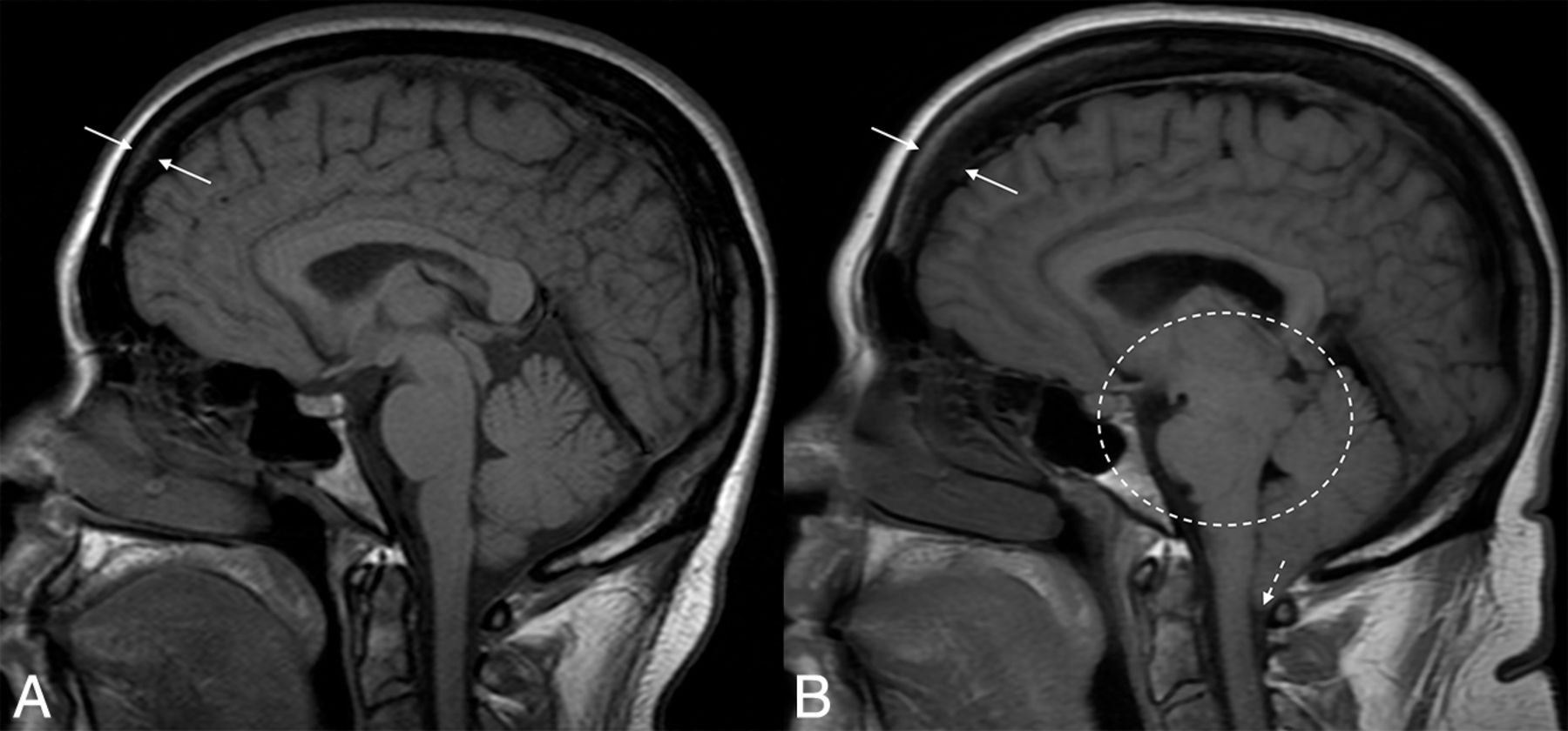
- FIG 4.
Hyperostosis related to a CSF leak. Sagittal T1-weight images before (A) and after (B) the development and diagnosis of SIH, with 16 years between examinations. Multiple classic findings of SIH are seen, including brain sag (dashed oval) and herniation of the cerebellar tonsils through the foramen magnum (dashed arrow). The patient also developed substantial frontal-predominant calvarial hyperostosis (between solid arrows), particularly along the inner table of the skull.


Jump to section
Related Articles
Cited By...
- Spinal CSF Leaks: The Neuroradiologist Transforming Care
- Skull Base CSF Leaks: Potential Underlying Pathophysiology and Evaluation of Brain MR Imaging Findings Associated with Spontaneous Intracranial Hypotension
- Investigating Sea-Level Brain Predictors for Acute Mountain Sickness: A Multimodal MRI Study before and after High-Altitude Exposure
- Identifying Patients with CSF-Venous Fistula Using Brain MRI: A Deep Learning Approach
- Likelihood of Discovering a CSF Leak Based on Intracranial MRI Findings in Patients without a Spinal Longitudinal Extradural Collection: A New Probabilistic Scoring System