Advertisement

Main menu

  • Home
  • Content
    • Current Issue
    • Accepted Manuscripts
    • Article Preview
    • Past Issue Archive
    • Video Articles
    • AJNR Case Collection
    • Case of the Week Archive
    • Case of the Month Archive
    • Classic Case Archive
  • Special Collections
    • AJNR Awards
    • Low-Field MRI
    • Alzheimer Disease
    • ASNR Foundation Special Collection
    • Photon-Counting CT
    • View All
  • Multimedia
    • AJNR Podcasts
    • AJNR SCANtastic
    • Trainee Corner
    • MRI Safety Corner
    • Imaging Protocols
  • For Authors
    • Submit a Manuscript
    • Submit a Video Article
    • Submit an eLetter to the Editor/Response
    • Manuscript Submission Guidelines
    • Statistical Tips
    • Fast Publishing of Accepted Manuscripts
    • Graphical Abstract Preparation
    • Imaging Protocol Submission
    • Author Policies
  • About Us
    • About AJNR
    • Editorial Board
    • Editorial Board Alumni
  • More
    • Become a Reviewer/Academy of Reviewers
    • Subscribers
    • Permissions
    • Alerts
    • Feedback
    • Advertisers
    • ASNR Home

User menu

  • Alerts
  • Log in

Search

  • Advanced search
American Journal of Neuroradiology

American Journal of Neuroradiology

ASHNR American Society of Functional Neuroradiology ASHNR American Society of Pediatric Neuroradiology ASSR
  • Alerts
  • Log in

Advanced Search

  • Home
  • Content
    • Current Issue
    • Accepted Manuscripts
    • Article Preview
    • Past Issue Archive
    • Video Articles
    • AJNR Case Collection
    • Case of the Week Archive
    • Case of the Month Archive
    • Classic Case Archive
  • Special Collections
    • AJNR Awards
    • Low-Field MRI
    • Alzheimer Disease
    • ASNR Foundation Special Collection
    • Photon-Counting CT
    • View All
  • Multimedia
    • AJNR Podcasts
    • AJNR SCANtastic
    • Trainee Corner
    • MRI Safety Corner
    • Imaging Protocols
  • For Authors
    • Submit a Manuscript
    • Submit a Video Article
    • Submit an eLetter to the Editor/Response
    • Manuscript Submission Guidelines
    • Statistical Tips
    • Fast Publishing of Accepted Manuscripts
    • Graphical Abstract Preparation
    • Imaging Protocol Submission
    • Author Policies
  • About Us
    • About AJNR
    • Editorial Board
    • Editorial Board Alumni
  • More
    • Become a Reviewer/Academy of Reviewers
    • Subscribers
    • Permissions
    • Alerts
    • Feedback
    • Advertisers
    • ASNR Home
  • Follow AJNR on Twitter
  • Visit AJNR on Facebook
  • Follow AJNR on Instagram
  • Join AJNR on LinkedIn
  • RSS Feeds

AJNR Awards, New Junior Editors, and more. Read the latest AJNR updates

Review ArticleReview Articles
Open Access

The Monro-Kellie Doctrine: A Review and Call for Revision

J.C. Benson, A.A. Madhavan, J.K. Cutsforth-Gregory, D.R. Johnson and C.M. Carr
American Journal of Neuroradiology December 2022, DOI: https://doi.org/10.3174/ajnr.A7721
J.C. Benson
aFrom the Departments of Radiology (J.C.B., A.A.M., D.R.J., C.M.C.)
  • Find this author on Google Scholar
  • Find this author on PubMed
  • Search for this author on this site
  • ORCID record for J.C. Benson
A.A. Madhavan
aFrom the Departments of Radiology (J.C.B., A.A.M., D.R.J., C.M.C.)
  • Find this author on Google Scholar
  • Find this author on PubMed
  • Search for this author on this site
  • ORCID record for A.A. Madhavan
J.K. Cutsforth-Gregory
bNeurology (J.K.C.-G.), Mayo Clinic, Rochester, Minnesota
  • Find this author on Google Scholar
  • Find this author on PubMed
  • Search for this author on this site
  • ORCID record for J.K. Cutsforth-Gregory
D.R. Johnson
aFrom the Departments of Radiology (J.C.B., A.A.M., D.R.J., C.M.C.)
  • Find this author on Google Scholar
  • Find this author on PubMed
  • Search for this author on this site
  • ORCID record for D.R. Johnson
C.M. Carr
aFrom the Departments of Radiology (J.C.B., A.A.M., D.R.J., C.M.C.)
  • Find this author on Google Scholar
  • Find this author on PubMed
  • Search for this author on this site
  • ORCID record for C.M. Carr
  • Article
  • Figures & Data
  • Info & Metrics
  • Responses
  • References
  • PDF
Loading

Abstract

SUMMARY: The Monro-Kellie doctrine is a well-accepted principle of intracranial hemodynamics. It has undergone few consequential revisions since it was established. Its principle is straightforward: The combined volume of neuronal tissue, blood, and CSF is constant. To maintain homeostatic intracranial pressure, any increase or decrease in one of these elements leads to a reciprocal and opposite change in the others. The Monro-Kellie doctrine assumes a rigid, unadaptable calvaria. Recent studies have disproven this assumption. The skull expands and grows in response to pathologic changes in intracranial pressure. In this review, we outline what is known about calvarial changes in the setting of pressure dysregulation and suggest a revision to the Monro-Kellie doctrine that includes an adaptable skull as a fourth component.

ABBREVIATIONS:

IIH
idiopathic intracranial hypertension
SIH
spontaneous intracranial hypotension

The Monro-Kellie doctrine has been a fundamental principle of neurophysiology for >200 years. Its hypothesis is based on simple reasoning: Within a rigid calvarial vault, the total volume of brain, CSF, and blood is constant. Any change in one of these elements results in an opposing compensatory response by the other 2 components.1 On neuroimaging, the effects of this doctrine are ubiquitous: Encephalomalacia leads to ex vacuo dilation of an adjacent ventricle, an edematous and enlarged brain compresses ventricles and vessels, and CSF hypovolemia results in pituitary and venous engorgement.

Multiple studies, however, now suggest that the Monro-Kellie doctrine is due for further examination, particularly in the setting of abnormal intracranial pressure. Both increased and decreased intracranial pressure exerts downstream effects on the calvaria. Increased pressure leads to calvarial thinning and pitting,2 while decreased pressure leads to development of layering hyperostosis along the inner table of the skull.3 These findings have substantial implications, both in terms of how we think of the Monro-Kellie doctrine and potentially how pathologies of intracranial pressure are identified and treated. In light of these findings, we review the history of the Monro-Kellie hypothesis and describe mounting evidence of a nonrigid, adaptable calvaria in the setting of pathologically elevated or reduced CSF pressure.

BACKGROUND

The Monro-Kellie doctrine underwent many early changes before achieving its current form. Alexander Monro, a Scottish doctor of impressive medical lineage, first proposed that a rigid skull contained an incompressible brain and a constant amount of blood; ie, a steady input of arterial blood led to a compensatory output in venous blood.4 In 1824, his student George Killie de Leith provided postmortem evidence of this theory. Neither, however, mentioned CSF.5 Around the same time, John Ambercrombie made similar observations in animals; some early citations of this theorem were entitled the “Monro-Abercrombie doctrine.”6

George Burrows first included CSF in this model, and it was not until 1926 that Harvey Cushing7 neatly summarized the Monro-Kellie doctrine as it is generally known today. According to Cushing, the “three elements” (blood, nervous tissue, and fluid) in the skull “must always remain the same in bulk….any increase in blood volume, for example, can only take place at the expense of one of the other elements.” Cushing considered the responsiveness of any reciprocal action to be prompt, “These changes all take place rapidly—a matter of minutes.”7

Of course, self-evident exceptions to this rule abound. A foreign body may enter the calvarial vault, displacing or decreasing all 3 native intracranial components. Many types of intracranial masses are not composed of brain, CSF, or blood (eg, abscesses, metastases, and granulation tissue). These exceptions do not require a revision of the doctrine because they neither challenge the underlying mechanism on which the Monro-Kellie doctrine is built nor change the primary assumption of a fixed-volume skull.

Our understanding of intracranial pressure dynamics has become more nuanced since the inception of the doctrine. It is now known, for example, that blood plays a much greater role in dictating intracranial pressure than CSF. The total intracranial in- and outflow of blood is approximately 700 mL/min, occupying 100–130 mL of intracranial volume at any time. CSF, conversely, is produced at only 0.35 mL/min, with a total volume of about 75 mL.5 The cardiac cycle also dynamically influences intracranial pressure, with variations in arterial input during systolic and diastolic flow resulting in pulsatile brain movement, brain expansion and contraction, and intraventricular CSF flow.8

Although our understanding of intracranial pressure has become more refined, the Monro-Kellie doctrine has notably not been adjusted or revised. A few prior studies have proposed minor updates regarding concepts such as tissue elasticity and compliance9 and the dynamic nature of intracranial arterial and venous vascularity.5 None of these proposals have strayed far from the tenets of the doctrine: 1) The 3 major constituents that play a role in intracranial pressure homeostasis are blood, CSF, and brain parenchyma, and 2) the calvaria is rigid and has a fixed volume.

The closest proposed revision came from Mascarenhas et al,6 who performed a set of postmortem experiments in which they expanded a rubber balloon in a skull while measuring skull deformation. The results indicated that deformation was noted during increases and decreases in internal pressure. The authors consequently contended that the Monro-Kellie doctrine was, therefore, invalid because it failed to acknowledge such deformational changes in the skull.

Now there is convincing evidence of a more chronic type of elasticity of the calvaria in response to increases and decreases in intracranial pressure. Unlike the experiments of Mascarenhas et al,6 however, this elasticity manifests as changes in the thickness and shape of the calvarium, rather than transient deformations. Both types of changes represent adaptivity of the skull to expand or contract the calvarial vault in response to CSF pressure abnormalities. Below, we review in detail what is currently known about such calvarial changes.

Intracranial Hypertension

Intracranial hypertension was first described in 1893 by Heinrich Quincke, a disease he labeled “meningitis serosa” attributed to alterations in CSF secretions.10 In 1904, Max Nonne first used the term pseudotumor cerebri (“false brain tumor”) to describe this syndrome. Nonne’s intent was to propose a state of elevated intracranial pressure that was distinct from that related to cerebral tumors: intercranial hypertension that followed a less sinister course.11 John Foley12 later offered the term “benign intracranial hypertension.”

These labels have become outdated. First, several other etiologies of increased intracranial pressure have become known. Such cases are referred to as secondary intracranial hypertension, reserving the term “primary pseudotumor cerebri” (also known an idiopathic intracranial hypertension [IIH]) for cases of cryptogenic elevated intracranial pressure. Next, the vision loss associated with IIH has made the label “benign” inappropriate.13

The etiologic mechanism for IIH remains unknown. Theories generally fall into the categories of altered CSF hydrodynamics or hindrances to venous outflow.14 The disorder represents a dysfunction in the homeostatic regulatory role of the Monro-Kellie doctrine; the increase in ≥1 intracranial component is insufficiently balanced by a corresponding decrease in the others.15

On imaging, numerous sequelae of IIH have been well-established. The pituitary gland is typically flattened along the floor of the sella, thought to be related to downward herniation of an arachnocele through the diaphragma sella. Ventricles are often slitlike.16 Many findings have been reported in the orbits: flattening of the posterior sclera, distention of the perioptic subarachnoid space, tortuosity of the optic nerves, and intraocular protrusion of the optic nerve.17 Most important, lateral transverse sinus stenosis is also common, likely mediated by and contributing to increased intracranial pressure.18

Recent studies have demonstrated that remodeling of the calvaria is common in the setting of chronic pseudotumor cerebri. In such patients, the elevated intracranial pressure can lead to enlargement of the sella turcica (Fig 1),19 as well as prominent arachnoid pits and/or arachnoid granulations.2 Meningoceles are also common, typically forming in the temporal bone (Fig 2).20 As Bialer et al20 noted, these findings are all biomechanically similar in that they increase the volume of the subarachnoid space, thereby at least conceptually decreasing intracranial pressure. Skull base erosion and meningocele formation can lead to the development of CSF leaks.21 In some cases, encephaloceles in these locations may also lead to epilepsy.22

FIG 1.
  • Download figure
  • Open in new tab
  • Download powerpoint
FIG 1.

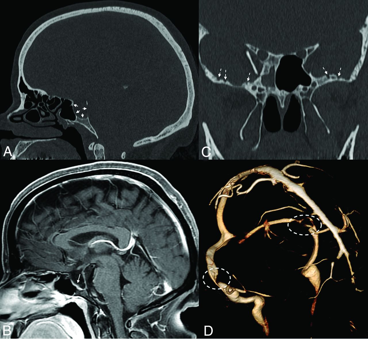
Sella expansion and skull base pitting in IIH. Sagittal CT image (A) in a 38-year-old woman with pseudotumor cerebri demonstrates marked expansion of the osseous walls of the sella (short solid arrows), with frank dehiscence posteriorly. Corresponding sagittal MR image (B) shows flattening of the pituitary tissue along the floor of the sella (long solid arrow). Prominent pitting is also noted along the anterior margins of both middle cranial fossae (dashed arrows, C). 3D reconstruction image of an MRV (D) demonstrates smooth tapering of the bilateral transverse sinuses (dashed ovals), compatible with IIH.

FIG 2.
  • Download figure
  • Open in new tab
  • Download powerpoint
FIG 2.

Skull pitting, meningoceles, and CSF leak in a patient who presented with rhinorrhea. Axial and coronal CT images (A and B) demonstrate substantial pitting along the anteromedial aspect of the left middle cranial fossa (dashed ovals, A and B), with complete opacification of the left sphenoid sinus (asterisks), concerning for a CSF leak. Milder pitting is noted on the right (solid arrows). Corresponding MR imaging (C and D) confirms a meningocele protruding into the left sphenoid sinus (dashed ovals, C and D). MRV (not shown) noted smooth tapered stenoses involving both transverse sinuses, and the patient was ultimately diagnosed with pseudotumor cerebri.

Beyond distinct areas of pitting, the skull itself becomes thinned in IIH. A case-control study by Barke et al23 found both skull base thickness (P < .001) and calvarial width (P = .024) to be significantly smaller in patients with IIH than in controls. Rabbani et al24 confirmed such findings, noting that while advancing age was typically associated with increased calvarial thickness, increased age portended calvarial thinning in patients with IIH. These calvarial changes are in opposition to the conventional understanding of the Monro-Kellie doctrine.

Except for CSF leaks and rare cases of epilepsy related to encephaloceles, little remains known about the clinical relevance of calvarial expansion and/or thinning in the setting of IIH. To date, no studies have shown that these calvarial changes affect patient outcomes. Still, IIH remains an evolving field. As our knowledge of this disorder continues to grow, it is possible that calvarial changes among some patients with IIH could affect treatment strategies or could be used as a prognostic marker.

Intracranial Hypotension

Spontaneous intracranial hypotension (SIH) is related to CSF volume depletion. Although CSF leaks may be secondary to trauma and iatrogenic causes such as surgery or lumbar punctures, intracranial hypotension by fiat is due to spontaneous spinal CSF leaks.25 Like IIH, the intracranial sequelae of intracranial hypotension have been well-described on imaging.

Even more so than intracranial hypertension, most of the intracranial sequelae of SIH can be explained by the conventional understanding of the Monro-Kellie doctrine. For example, the relative dearth of CSF results in pituitary enlargement (ie, opposite to IIH) and expansion of the dural venous sinuses.26,27 The dura engorges, leading to diffuse dural thickening and enhancement.28 In some patients, subdural fluid collections, either hygromas or hematomas, develop over the cerebral convexities.29 On the basis of the presence or absence of these findings, one can reliably predict the likelihood of a patient having SIH.29 Ultimately, SIH represents a disorder in which autoregulatory mechanisms are insufficient to compensate for the CSF loss, and other imaging abnormalities reflect this breakdown. Specifically, the brain begins to sag inferiorly, with effacement of the suprasellar and prepontine cisterns and decreased mamillopontine distance.29

Despite the number of changes observed, most of the compensatory changes in SIH are related to hyperemia; other than the pituitary gland, the brain parenchyma is relatively unable to expand in response to CSF depletion.30 The same is true following treatment. Once a leak has been repaired, intracranial CSF volume has been shown to increase significantly over its hypotensive baseline. The brain volume in the posttreatment setting, in contrast, remains the same.17,31 Thus, the main compensatory action following CSF leak repair is a reciprocal reduction of intracranial blood volume.

It is only recently that we have become aware of calvarial changes in the setting of SIH. Johnson et al32 first showed that patients with SIH were often observed to have a thickened calvaria, often with a characteristic layered hyperostotic growth pattern along the inner table of the skull. In the setting of long-standing SIH, the authors opined, calvarial growth served as an additional compensatory mechanism for the depleted intracranial CSF volume. Babcock et al3 confirmed such findings in a case-control study, noting that layered hyperostosis was present in 32% of patients with SIH but in only 5% of controls (P < .001; OR = 11.58) (Fig 3). Most important, the study of Babcock et al found no significant difference in the prevalence of diffuse (nonlayered) hyperostosis between groups (P = .465). Thus, the “layer cake” type of hyperostosis is much more indicative of intracranial hypotension (Fig 4). Although the expected location of this layer cake appearance was not discussed in detail in either publication, it is possible that these findings tend to be frontal-predominant, similar to benign hyperostosis frontalis interna. Specifically, the representative examples in the article by Babcock et al seem to suggest that such findings are at least prone to more substantial development along the frontal bone.

FIG 3.
  • Download figure
  • Open in new tab
  • Download powerpoint
FIG 3.

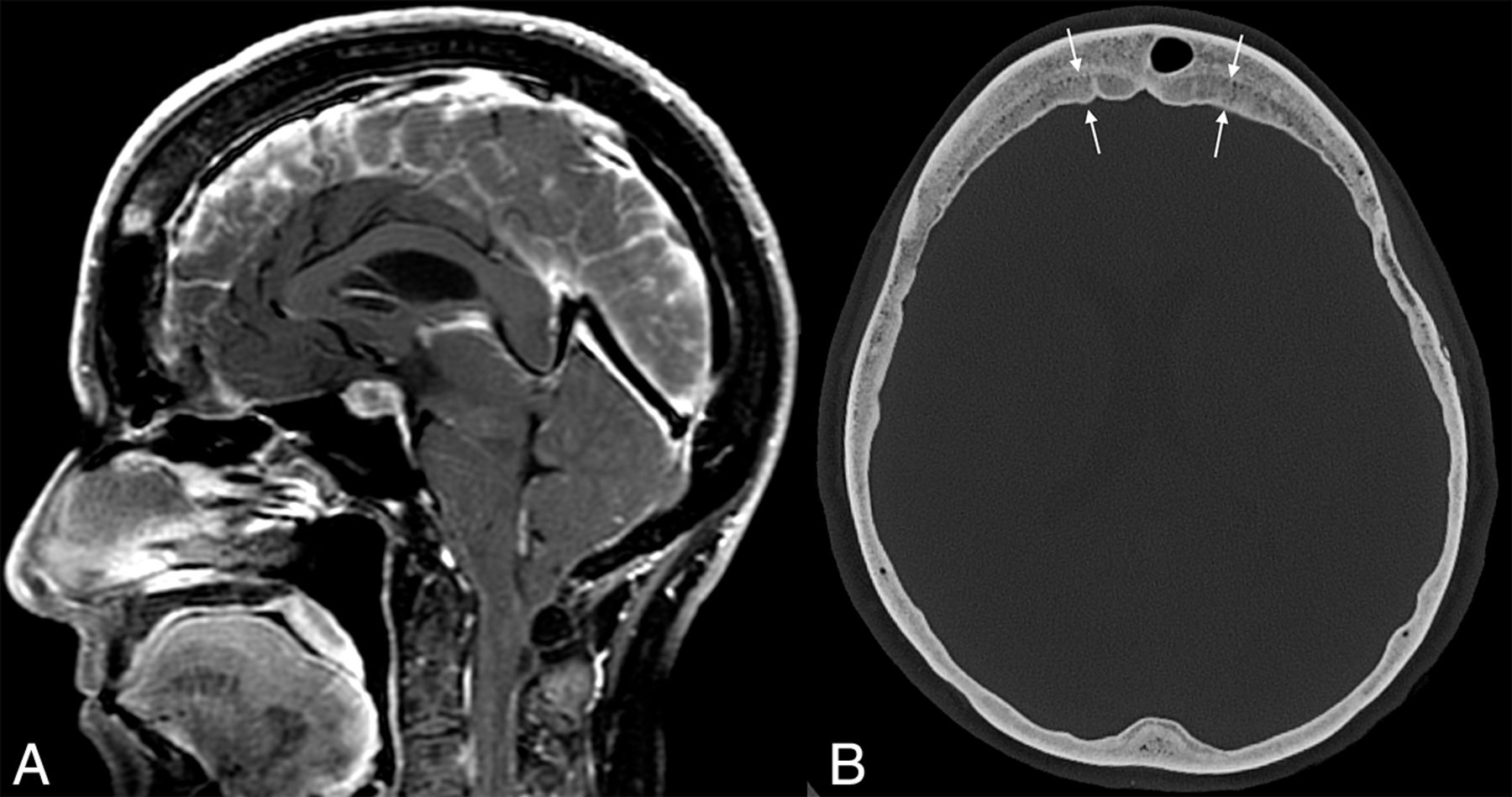
Example of layered calvarial thickening in a patient with SIH. Sagittal MR images (A) demonstrate classic findings of SIH, including pituitary enlargement, brain sag, and dural venous sinus engorgement. Axial CT image (B) in the same patient shows thickening of the bifrontal calvaria with characteristic layering related to preferential growth along the inner table of the skull (between straight arrows).

FIG 4.
  • Download figure
  • Open in new tab
  • Download powerpoint
FIG 4.

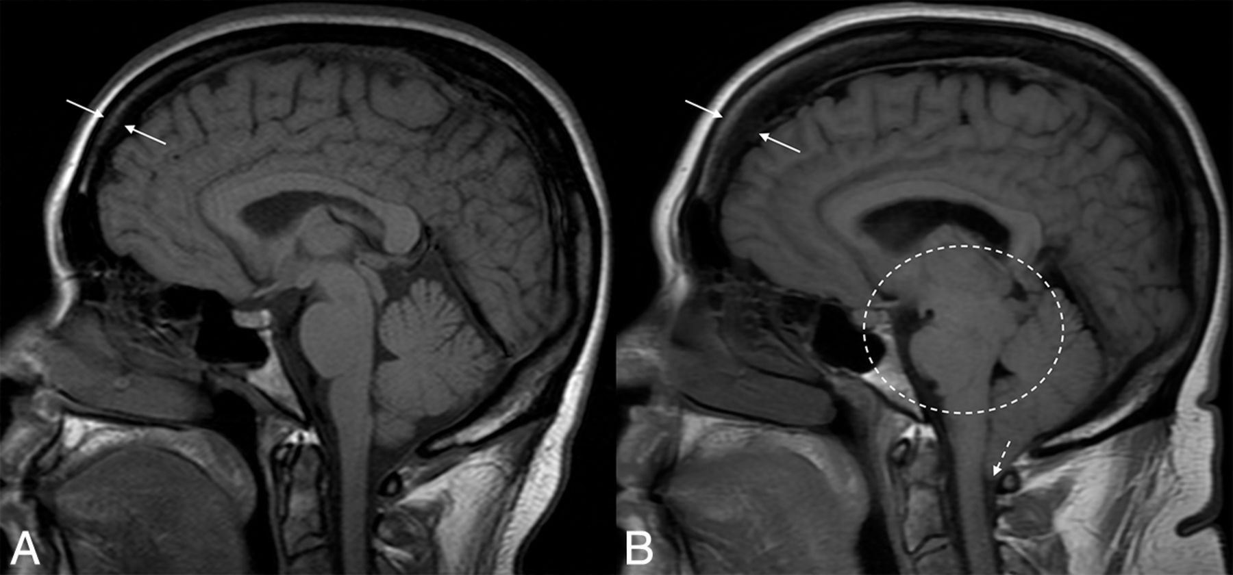
Hyperostosis related to a CSF leak. Sagittal T1-weight images before (A) and after (B) the development and diagnosis of SIH, with 16 years between examinations. Multiple classic findings of SIH are seen, including brain sag (dashed oval) and herniation of the cerebellar tonsils through the foramen magnum (dashed arrow). The patient also developed substantial frontal-predominant calvarial hyperostosis (between solid arrows), particularly along the inner table of the skull.

A similar phenomenon, termed “hyperostosis cranii ex vacuo” has been described in chronic shunting of pediatric hydrocephalus.33,34 In this condition, there is prominent growth of histologically lamellar bone, with scattered foci of woven bone.33 This growth, Moseley et al35 noted, is primarily the inner table of the calvaria. Thus, the findings are analogous to the layer cake appearance seen in adult patients with SIH. The increased inner-to-outer table measurement is important because it helps distinguish these calvarial changes from those related to expansion of the diploic space (eg, sickle cell disease or thalassemia).33

The clinical implications of these calvarial findings are currently unknown. There are, however, some potential effects that have been considered. The presence of layer cake hyperostosis in patients with SIH, for example, leads to a decrease in the volume of the calvarial vault. Conceptually, this could reduce the ability to normalize brain sag following CSF leak repair—that is, calvarial hyperostosis could impede the ability of brain to “un-sag” by acting as an unyielding intracranial barrier. Similarly, patients with SIH with layer cake skull changes might be at higher risk of rebound hypertension after leak repair because the tightened intracranial space could potentially lead to greater mass effect on the transverse sinuses. For now, these hypotheses remain conceptual in nature, though studies on these topics are ongoing.

Call For Revision

Altogether, studies have convincingly shown demonstrable changes in the calvaria in the setting of pathologic conditions of intracranial pressure. The skull expands in response to IIH and grows inward along its inner table in response to SIH. Skeptics might argue that these changes are outside the realm of the Monro-Kellie doctrine because they represent adaptive changes in response to a breakdown of pressure homeostasis. It is clear, however, that the concept of a rigid calvaria, a fundamental tenet upon which the doctrine is based, has been disproven multiple times. We should add a fourth component to the Monro-Kellie hypothesis: a nonrigid calvaria that adjusts, albeit slowly, to changes in intracranial pressure.

Footnotes

  • Disclosure forms provided by the authors are available with the full text and PDF of this article at www.ajnr.org.

Indicates open access to non-subscribers at www.ajnr.org

References

  1. 1.↵
    1. Mokri B
    . The Monro-Kellie hypothesis: applications in CSF volume depletion. Neurology 2001;56:1746–48 doi:10.1212/wnl.56.12.1746 pmid:11425944
    CrossRefPubMed
  2. 2.↵
    1. Rosenberg KI,
    2. Banik R
    . Pseudotumor cerebri syndrome associated with giant arachnoid granulation. J Neuroophthalmol 2013;33:417–19 doi:10.1097/WNO.0b013e3182a5943b pmid:24051425
    CrossRefPubMed
  3. 3.↵
    1. Babcock JC,
    2. Johnson DR,
    3. Benson JC, et al
    . Diffuse calvarial hyperostosis and spontaneous intracranial hypotension: a case-control study. AJNR Am J Neuroradiol 2022;43:978–83 doi:10.3174/ajnr.A7557 pmid:35772803
    Abstract/FREE Full Text
  4. 4.↵
    1. Wu OC,
    2. Manjila S,
    3. Malakooti N, et al
    . The remarkable medical lineage of the Monro family: contributions of Alexander primus, secundus, and tertius. J Neurosurg 2012;116:1337–46 doi:10.3171/2012.2.JNS111366 pmid:22482794
    CrossRefPubMed
  5. 5.↵
    1. Wilson MH
    . Monro-Kellie 2.0: The dynamic vascular and venous pathophysiological components of intracranial pressure. J Cereb Blood Flow Metab 2016;36:1338–50 doi:10.1177/0271678X16648711 pmid:27174995
    CrossRefPubMed
  6. 6.↵
    1. Mascarenhas S,
    2. Vilela GH,
    3. Carlotti C, et al
    . The new ICP minimally invasive method shows that the Monro-Kellie doctrine is not valid. Acta Neurochir Suppl 2012;114:117–20 doi:10.1007/978-3-7091-0956-4_21 pmid:22327675
    CrossRefPubMed
  7. 7.↵
    1. Cushing H
    . Studies in intracranial physiology & surgery: the third circulation, the hypophysis, the gliomas. Humphrey Milford, Oxford University; 1926
  8. 8.↵
    1. Greitz D,
    2. Wirestam R,
    3. Franck A, et al
    . Pulsatile brain movement and associated hydrodynamics studied by magnetic resonance phase imaging: the Monro-Kellie doctrine revisited. Neuroradiology 1992;34:370–80 doi:10.1007/BF00596493 pmid:1407513
    CrossRefPubMed
  9. 9.↵
    1. Kalisvaart AC,
    2. Wilkinson CM,
    3. Gu S, et al
    . An update to the Monro-Kellie doctrine to reflect tissue compliance after severe ischemic and hemorrhagic stroke. Sci Rep 2020;10:22013 doi:10.1038/s41598-020-78880-4 pmid:33328490
    CrossRefPubMed
  10. 10.↵
    1. Johnston I
    . The historical development of the pseudotumor concept. Neurosurg Focus 2001;11:E2 doi:10.3171/foc.2001.11.2.3 pmid:16602675
    CrossRefPubMed
  11. 11.↵
    1. Juhász J,
    2. Hensler J,
    3. Jansen O
    . MRI-findings in idiopathic intracranial hypertension (pseudotumor cerebri). Rofo 2021;193:1269–76 doi:10.1055/a-1447-0264 pmid:33979869
    CrossRefPubMed
  12. 12.↵
    1. Foley J
    . Benign forms of intracranial hypertension; toxic and otitic hydrocephalus. Brain 1955;78:1–41 doi:10.1093/brain/78.1.1 pmid:14378448
    CrossRefPubMed
  13. 13.↵
    1. Friedman DI,
    2. Liu GT,
    3. Digre KB
    . Revised diagnostic criteria for the pseudotumor cerebri syndrome in adults and children. Neurology 2013;81:1159–65 doi:10.1212/WNL.0b013e3182a55f17 pmid:23966248
    Abstract/FREE Full Text
  14. 14.↵
    1. Degnan AJ,
    2. Levy LM
    . Pseudotumor cerebri: brief review of clinical syndrome and imaging findings. AJNR Am J Neuroradiol 2011;32:1986–93 doi:10.3174/ajnr.A2404 pmid:21680652
    Abstract/FREE Full Text
  15. 15.↵
    1. Ozkal B,
    2. Ozen O
    . The effect of intracerebral structure volumes on idiopathic intracranial hypertension. Neuroreport 2021;32:16–22 doi:10.1097/WNR.0000000000001552 pmid:33196546
    CrossRefPubMed
  16. 16.↵
    1. George AE
    . Idiopathic intracranial hypertension: pathogenesis and the role of MR imaging. Radiology 1989;170:21–22 doi:10.1148/radiology.170.1.2909099 pmid:2909099
    CrossRefPubMed
  17. 17.↵
    1. Brodsky MC,
    2. Vaphiades M
    . Magnetic resonance imaging in pseudotumor cerebri. Ophthalmology 1998;105:1686–93 doi:10.1016/S0161-6420(98)99039-X pmid:9754178
    CrossRefPubMed
  18. 18.↵
    1. Morris PP,
    2. Lachman N,
    3. Black DF, et al
    . Increased curvature of the tentorium cerebelli in idiopathic intracranial hypertension. AJNR Am J Neuroradiol 2017;38:1789–93 doi:10.3174/ajnr.A5289 pmid:28663268
    Abstract/FREE Full Text
  19. 19.↵
    1. Kyung S Eun E,
    2. Botelho JV,
    3. Horton JC
    . Enlargement of the sella turcica in pseudotumor cerebri. J Neurosurg 2014;120:538–42 doi:10.3171/2013.10.JNS131265 pmid:24313606
    CrossRefPubMed
  20. 20.↵
    1. Bialer OY,
    2. Rueda MP,
    3. Bruce BB, et al
    . Meningoceles in idiopathic intracranial hypertension. AJR Am J Roentgenol 2014;202:608–13 doi:10.2214/AJR.13.10874 pmid:24555598
    CrossRefPubMed
  21. 21.↵
    1. Pérez MA,
    2. Bialer OY,
    3. Bruce BB, et al
    . Primary spontaneous cerebrospinal fluid leaks and idiopathic intracranial hypertension. J Neuroophthalmol 2013;33:330–37 doi:10.1097/WNO.0b013e318299c292 pmid:24042170
    CrossRefPubMed
  22. 22.↵
    1. Thottiyil JJ,
    2. Prasad A,
    3. Dutta DJ, et al
    . Temporal lobe encephalocele with epilepsy in a young female: an atypical presentation of idiopathic intracranial hypertension. Neurol India 2022;70:1618–21 doi:10.4103/0028-3886.355157 pmid:36076668
    CrossRefPubMed
  23. 23.↵
    1. Barke M,
    2. Castro HM,
    3. Adesina OO, et al
    . Thinning of the skull base and calvarial thickness in patients with idiopathic intracranial hypertension. J Neuroophthalmol 2022;42:192–98 doi:10.1097/WNO.0000000000001504 pmid:35195543
    CrossRefPubMed
  24. 24.↵
    1. Rabbani CC,
    2. Patel JM,
    3. Nag A, et al
    . Association of intracranial hypertension with calvarial and skull base thinning. Otol Neurotol 2019;40:e619–26 doi:10.1097/MAO.0000000000002249 pmid:31045889
    CrossRefPubMed
  25. 25.↵
    1. Schievink WI
    . Spontaneous spinal cerebrospinal fluid leaks and intracranial hypotension. JAMA 2006;295:2286–96 doi:10.1001/jama.295.19.2286 pmid:16705110
    CrossRefPubMed
  26. 26.↵
    1. Leung GK,
    2. Ho J,
    3. Pu JK
    . Pituitary enlargement in spontaneous intracranial hypotension—a diagnostic pitfall. Acta Neurochir (Wien) 2011;153:2445–46 doi:10.1007/s00701-011-1099-x pmid:21800104
    CrossRefPubMed
  27. 27.↵
    1. Bond KM,
    2. Benson JC,
    3. Cutsforth-Gregory JK, et al
    . Spontaneous intracranial hypotension: atypical radiologic appearances, imaging mimickers, and clinical look-alikes. AJNR Am J Neuroradiol 2020;41:1339–47 doi:10.3174/ajnr.A6637 pmid:32646948
    Abstract/FREE Full Text
  28. 28.↵
    1. Antony J,
    2. Hacking C,
    3. Jeffree RL
    . Pachymeningeal enhancement-a comprehensive review of literature. Neurosurg Rev 2015;38:649–59 doi:10.1007/s10143-015-0646-y pmid:26264063
    CrossRefPubMed
  29. 29.↵
    1. Dobrocky T,
    2. Grunder L,
    3. Breiding PS, et al
    . Assessing spinal cerebrospinal fluid leaks in spontaneous intracranial hypotension with a scoring system based on brain magnetic resonance imaging findings. JAMA Neurol 2019;76:580–87 doi:10.1001/jamaneurol.2018.4921 pmid:30776059
    CrossRefPubMed
  30. 30.↵
    1. Huang YM,
    2. Davidsson L
    . Sagging brain development after lumbar puncture agrees with Monro-Kellie hypothesis. J Neurol 2013;260:920–22 doi:10.1007/s00415-012-6811-0 pmid:23314405
    CrossRefPubMed
  31. 31.↵
    1. Dobrocky T,
    2. Rebsamen M,
    3. Rummel C, et al
    . Monro-Kellie hypothesis: increase of ventricular CSF volume after surgical closure of a spinal dural leak in patients with spontaneous intracranial hypotension. AJNR Am J Neuroradiol 2020;41:2055–61 doi:10.3174/ajnr.A6782 pmid:33177057
    Abstract/FREE Full Text
  32. 32.↵
    1. Johnson DR,
    2. Carr CM,
    3. Luetmer PH, et al
    . Diffuse calvarial hyperostosis in patients with spontaneous intracranial hypotension. World Neurosurg 2021;146:e848–53 doi:10.1016/j.wneu.2020.11.066 pmid:33220476
    CrossRefPubMed
  33. 33.↵
    1. Di Preta JA,
    2. Powers JM,
    3. Hicks DG
    . Hyperostosis cranii ex vacuo: a rare complication of shunting for hydrocephalus. Hum Pathol 1994;25:545–47 doi:10.1016/0046-8177(94)90130-9 pmid:8200652
    CrossRefPubMed
  34. 34.↵
    1. Lucey BP,
    2. March GP,
    3. Hutchins GM
    . Marked calvarial thickening and dural changes following chronic ventricular shunting for shaken baby syndrome. Arch Pathol Lab Med 2003;127:94–97 doi:10.5858/2003-127-94-MCTADC pmid:12521376
    CrossRefPubMed
  35. 35.↵
    1. Moseley JE,
    2. Rabinowitz JG,
    3. Dziadiw R
    . Hyperostosis cranii ex vacuo. Radiology 1966;87:1105–07 doi:10.1148/87.6.1105 pmid:5332740
    CrossRefPubMed
  • Received August 27, 2022.
  • Accepted after revision October 5, 2022.
  • © 2023 by American Journal of Neuroradiology
PreviousNext
Back to top
Advertisement
Print
Download PDF
Email Article

Thank you for your interest in spreading the word on American Journal of Neuroradiology.

NOTE: We only request your email address so that the person you are recommending the page to knows that you wanted them to see it, and that it is not junk mail. We do not capture any email address.

Enter multiple addresses on separate lines or separate them with commas.
The Monro-Kellie Doctrine: A Review and Call for Revision
(Your Name) has sent you a message from American Journal of Neuroradiology
(Your Name) thought you would like to see the American Journal of Neuroradiology web site.
CAPTCHA
This question is for testing whether or not you are a human visitor and to prevent automated spam submissions.
Cite this article
J.C. Benson, A.A. Madhavan, J.K. Cutsforth-Gregory, D.R. Johnson, C.M. Carr
The Monro-Kellie Doctrine: A Review and Call for Revision
American Journal of Neuroradiology Dec 2022, DOI: 10.3174/ajnr.A7721

Citation Manager Formats

  • BibTeX
  • Bookends
  • EasyBib
  • EndNote (tagged)
  • EndNote 8 (xml)
  • Medlars
  • Mendeley
  • Papers
  • RefWorks Tagged
  • Ref Manager
  • RIS
  • Zotero
0 Responses
Respond to this article
Share
Bookmark this article
The Monro-Kellie Doctrine: A Review and Call for Revision
J.C. Benson, A.A. Madhavan, J.K. Cutsforth-Gregory, D.R. Johnson, C.M. Carr
American Journal of Neuroradiology Dec 2022, DOI: 10.3174/ajnr.A7721
del.icio.us logo Twitter logo Facebook logo Mendeley logo
  • Tweet Widget
  • Facebook Like
  • Google Plus One
Purchase

Jump to section

  • Article
    • Abstract
    • ABBREVIATIONS:
    • BACKGROUND
    • Footnotes
    • References
  • Figures & Data
  • Info & Metrics
  • Responses
  • References
  • PDF

Related Articles

  • No related articles found.
  • PubMed
  • Google Scholar

Cited By...

  • Spinal CSF Leaks: The Neuroradiologist Transforming Care
  • Skull Base CSF Leaks: Potential Underlying Pathophysiology and Evaluation of Brain MR Imaging Findings Associated with Spontaneous Intracranial Hypotension
  • Investigating Sea-Level Brain Predictors for Acute Mountain Sickness: A Multimodal MRI Study before and after High-Altitude Exposure
  • Identifying Patients with CSF-Venous Fistula Using Brain MRI: A Deep Learning Approach
  • Likelihood of Discovering a CSF Leak Based on Intracranial MRI Findings in Patients without a Spinal Longitudinal Extradural Collection: A New Probabilistic Scoring System
  • Crossref (41)
  • Google Scholar

This article has been cited by the following articles in journals that are participating in Crossref Cited-by Linking.

  • Immune-Mediated Hypertrophic Pachymeningitis and its Mimickers: Magnetic Resonance Imaging Findings
    Thiago Bezerra Matias, Rafael Alves Cordeiro, Juliana Avila Duarte, Vinicius Menezes de Jarry, Simone Appenzeller, Luciano Villarinho, Fabiano Reis
    Academic Radiology 2023 30 11
  • Endothelial Piezo1 channel mediates mechano-feedback control of brain blood flow
    Xin Rui Lim, Mohammad M. Abd-Alhaseeb, Michael Ippolito, Masayo Koide, Amanda J. Senatore, Curtis Plante, Ashwini Hariharan, Nick Weir, Thomas A. Longden, Kathryn A. Laprade, James M. Stafford, Dorothea Ziemens, Markus Schwaninger, Jan Wenzel, Dmitry D. Postnov, Osama F. Harraz
    Nature Communications 2024 15 1
  • Regulation of brain fluid volumes and pressures: basic principles, intracranial hypertension, ventriculomegaly and hydrocephalus
    Stephen B. Hladky, Margery A. Barrand
    Fluids and Barriers of the CNS 2024 21 1
  • Skull Base CSF Leaks: Potential Underlying Pathophysiology and Evaluation of Brain MR Imaging Findings Associated with Spontaneous Intracranial Hypotension
    Ian T. Mark, Jeremy Cutsforth-Gregory, Patrick Luetmer, Ajay A. Madhavan, Michael Oien, Paul Farnsworth, Girish Bathla, Steve Messina, Michael Link, Jamie Van Gompel
    American Journal of Neuroradiology 2024 45 10
  • Decoupling Between Brain Activity and Cerebrospinal Fluid Movement in Neurological Disorders
    Xiao Liu
    Journal of Magnetic Resonance Imaging 2023
  • Likelihood of Discovering a CSF Leak Based on Intracranial MRI Findings in Patients without a Spinal Longitudinal Extradural Collection: A New Probabilistic Scoring System
    John C. Benson, Ajay A. Madhavan, Ian T. Mark, Jeremy K. Cutsforth-Gregory, Waleed Brinjikji, Jared T. Verdoorn
    American Journal of Neuroradiology 2023 44 11
  • Dural Puncture Complications
    Thomas R. Geisbush, Tomasz Matys, Tarik F. Massoud, Lotfi Hacein-Bey
    Neuroimaging Clinics of North America 2025 35 1
  • Relationship between edema and intracranial pressure following intracerebral hemorrhage in rat
    Anna C. J. Kalisvaart, Natasha A. Bahr, Frederick Colbourne
    Frontiers in Stroke 2023 2
  • The current status of noninvasive intracranial pressure monitoring: A literature review
    Dimitrios Theodoropoulos, Dimitrios A. Karabetsos, Antonios Vakis, Efrosini Papadaki, Apostolos Karantanas, Kostas Marias
    Clinical Neurology and Neurosurgery 2024 239
  • Identifying Patients with CSF-Venous Fistula Using Brain MRI: A Deep Learning Approach
    Shahriar Faghani, Mana Moassefi, Ajay A. Madhavan, Ian T. Mark, Jared T. Verdoorn, Bradley J. Erickson, John C. Benson
    American Journal of Neuroradiology 2024 45 4

More in this TOC Section

  • An Atlas of Neonatal Neurovascular Imaging Anatomy as Depicted with Microvascular Imaging: The Intracranial Arteries
  • An Atlas of Neonatal Neurovascular Imaging Anatomy as Depicted with Microvascular Imaging: The Intracranial Veins
  • Clinical Translation of Hyperpolarized 13C Metabolic Probes for Glioma Imaging
Show more Review Articles

Similar Articles

Advertisement

Indexed Content

  • Current Issue
  • Accepted Manuscripts
  • Article Preview
  • Past Issues
  • Editorials
  • Editor's Choice
  • Fellows' Journal Club
  • Letters to the Editor
  • Video Articles

Cases

  • Case Collection
  • Archive - Case of the Week
  • Archive - Case of the Month
  • Archive - Classic Case

More from AJNR

  • Trainee Corner
  • Imaging Protocols
  • MRI Safety Corner
  • Book Reviews

Multimedia

  • AJNR Podcasts
  • AJNR Scantastics

Resources

  • Turnaround Time
  • Submit a Manuscript
  • Submit a Video Article
  • Submit an eLetter to the Editor/Response
  • Manuscript Submission Guidelines
  • Statistical Tips
  • Fast Publishing of Accepted Manuscripts
  • Graphical Abstract Preparation
  • Imaging Protocol Submission
  • Evidence-Based Medicine Level Guide
  • Publishing Checklists
  • Author Policies
  • Become a Reviewer/Academy of Reviewers
  • News and Updates

About Us

  • About AJNR
  • Editorial Board
  • Editorial Board Alumni
  • Alerts
  • Permissions
  • Not an AJNR Subscriber? Join Now
  • Advertise with Us
  • Librarian Resources
  • Feedback
  • Terms and Conditions
  • AJNR Editorial Board Alumni

American Society of Neuroradiology

  • Not an ASNR Member? Join Now

© 2025 by the American Society of Neuroradiology All rights, including for text and data mining, AI training, and similar technologies, are reserved.
Print ISSN: 0195-6108 Online ISSN: 1936-959X

Powered by HighWire