
Article Figures & Data
Figures
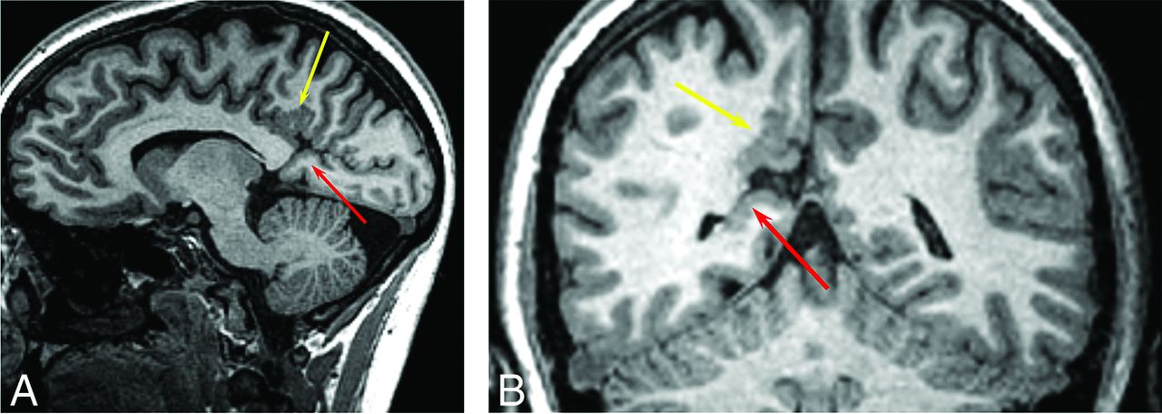
- FIG 1.
A 26-year-old woman with HHT. Sagittal 3D fast-spoiled gradient recalled imaging demonstrates polymicrogyria involving the right posterior cingulate gyrus (A, yellow arrow). There is an abnormal GM-lined cleft between the calcarine sulcus and the occipital horn of the right lateral ventricle consistent with schizencephaly (A, red arrow). Coronal T1 image shows polymicrogyria (B, yellow arrow) and schizencephaly (B, red arrow).
- FIG 2.
A 20-year-old man with HHT. 3D volume T1 images demonstrate a closed-lip schizencephaly projecting through the right inferior parietal lobe that extends to the lateral margin of the right lateral ventricle at the junction of the posterior body and atrium (A, red arrow). There is polymicrogyria involving the adjacent frontal and parietal cortex (A, yellow arrow). Coronal T1 image shows polymicrogyria (B, yellow arrow) and schizencephaly (B, red arrow).
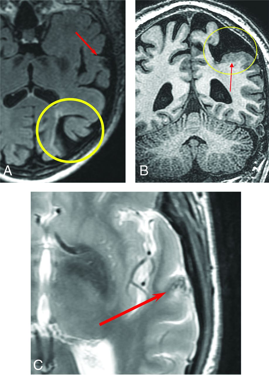
- FIG 3.
An 80-year-old man with HHT. Axial T2 FLAIR image shows porencephaly with surrounding gliosis (A, yellow circle) and an AVM in the left superior temporal gyrus (A, red arrow). Coronal volume T1 image shows porencephaly (B, yellow circle) partially lined by polymicrogyria (B, red arrow). Axial T2 FSE image shows flow voids associated with a small AVM (C, red arrow).
- FIG 4.
An 18-year-old man with HHT. Coronal 3D T1 postcontrast imaging shows right parietal polymicrogyria (A, yellow arrows), an AVM (A, yellow circle), and a small AVM (A, red arrow). Axial T1 image shows left parietal occipital polymicrogyria (B, red arrows).
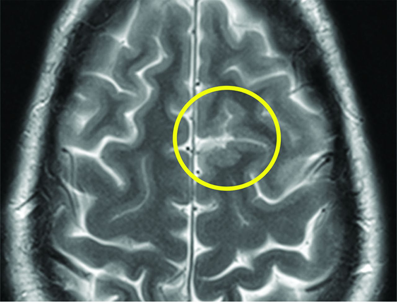
- FIG 5.
A 28-year-old man with HHT. Axial T2 FSE image shows polymicrogyria in the left superior frontal gyrus (yellow circle).


Tables
Summary of 141 patients
Demographics Summary Sex Male 36.9% Female 63.1% Age (mean) (yr) 45.3 MCD Yes 5 No 136 HHT mutation Endoglin 35.4% ALK 1a 27.7% SMAD4a 2.8% RASA1 0.7% Negative × 5 15.6% Unknown/not tested 17.7% AVMs Brain AVM 12.0% Brain AVM (possible) 2.8% Pulmonary AVM (macroscopic)b 43.3% Pulmonary AVM (microscopic)b 32.6% Spinal AVM 0.7% Brain vascular malformations Developmental venous anomaly 14.9% Capillary vascular malformation, definite 1.4% Capillary vascular malformation, possible 4.3% Curacao category Definite HHT 79.4% Possible or suspected HHT 12.1% Probable 4.3% Unlikely 4.3% ↵aALK1 includes 2 variants of unknown significance; SMAD4 includes 1 variant of unknown significance.
↵bPulmonary AVMs were defined as macroscopic if they were definitely visible on a CT scan and microscopic if contrast echocardiography showed a Grade 1 or greater delayed shunt and the CT findings were negative.


Jump to section
Related Articles
Cited By...
- No citing articles found.