Advertisement

Main menu

  • Home
  • Content
    • Current Issue
    • Accepted Manuscripts
    • Article Preview
    • Past Issue Archive
    • Video Articles
    • AJNR Case Collection
    • Case of the Week Archive
    • Case of the Month Archive
    • Classic Case Archive
  • Special Collections
    • AJNR Awards
    • Low-Field MRI
    • Alzheimer Disease
    • ASNR Foundation Special Collection
    • Photon-Counting CT
    • View All
  • Multimedia
    • AJNR Podcasts
    • AJNR SCANtastic
    • Trainee Corner
    • MRI Safety Corner
    • Imaging Protocols
  • For Authors
    • Submit a Manuscript
    • Submit a Video Article
    • Submit an eLetter to the Editor/Response
    • Manuscript Submission Guidelines
    • Statistical Tips
    • Fast Publishing of Accepted Manuscripts
    • Graphical Abstract Preparation
    • Imaging Protocol Submission
    • Author Policies
  • About Us
    • About AJNR
    • Editorial Board
    • Editorial Board Alumni
  • More
    • Become a Reviewer/Academy of Reviewers
    • Subscribers
    • Permissions
    • Alerts
    • Feedback
    • Advertisers
    • ASNR Home

User menu

  • Alerts
  • Log in

Search

  • Advanced search
American Journal of Neuroradiology

American Journal of Neuroradiology

ASHNR American Society of Functional Neuroradiology ASHNR American Society of Pediatric Neuroradiology ASSR
  • Alerts
  • Log in

Advanced Search

  • Home
  • Content
    • Current Issue
    • Accepted Manuscripts
    • Article Preview
    • Past Issue Archive
    • Video Articles
    • AJNR Case Collection
    • Case of the Week Archive
    • Case of the Month Archive
    • Classic Case Archive
  • Special Collections
    • AJNR Awards
    • Low-Field MRI
    • Alzheimer Disease
    • ASNR Foundation Special Collection
    • Photon-Counting CT
    • View All
  • Multimedia
    • AJNR Podcasts
    • AJNR SCANtastic
    • Trainee Corner
    • MRI Safety Corner
    • Imaging Protocols
  • For Authors
    • Submit a Manuscript
    • Submit a Video Article
    • Submit an eLetter to the Editor/Response
    • Manuscript Submission Guidelines
    • Statistical Tips
    • Fast Publishing of Accepted Manuscripts
    • Graphical Abstract Preparation
    • Imaging Protocol Submission
    • Author Policies
  • About Us
    • About AJNR
    • Editorial Board
    • Editorial Board Alumni
  • More
    • Become a Reviewer/Academy of Reviewers
    • Subscribers
    • Permissions
    • Alerts
    • Feedback
    • Advertisers
    • ASNR Home
  • Follow AJNR on Twitter
  • Visit AJNR on Facebook
  • Follow AJNR on Instagram
  • Join AJNR on LinkedIn
  • RSS Feeds

AJNR Awards, New Junior Editors, and more. Read the latest AJNR updates

Research ArticleAdult Brain
Open Access

SWI by 7T MR Imaging for the Microscopic Imaging Diagnosis of Astrocytic and Oligodendroglial Tumors

M. Natsumeda, H. Matsuzawa, M. Watanabe, K. Motohashi, R. Gabdulkhaev, Y. Tsukamoto, Y. Kanemaru, J. Watanabe, R. Ogura, M. Okada, S. Kurabe, K. Okamoto, A. Kakita, H. Igarashi and Y. Fujii
American Journal of Neuroradiology October 2022, DOI: https://doi.org/10.3174/ajnr.A7666
M. Natsumeda
aFrom the Department of Neurosurgery (M.N., K.M., Y.T., Y.K., J.W., R.O., M.O., S.K., Y.F.)
  • Find this author on Google Scholar
  • Find this author on PubMed
  • Search for this author on this site
  • ORCID record for M. Natsumeda
H. Matsuzawa
bCenter for Integrated Human Brain Science (H.M., M.W., H.I.)
  • Find this author on Google Scholar
  • Find this author on PubMed
  • Search for this author on this site
  • ORCID record for H. Matsuzawa
M. Watanabe
bCenter for Integrated Human Brain Science (H.M., M.W., H.I.)
  • Find this author on Google Scholar
  • Find this author on PubMed
  • Search for this author on this site
  • ORCID record for M. Watanabe
K. Motohashi
aFrom the Department of Neurosurgery (M.N., K.M., Y.T., Y.K., J.W., R.O., M.O., S.K., Y.F.)
  • Find this author on Google Scholar
  • Find this author on PubMed
  • Search for this author on this site
  • ORCID record for K. Motohashi
R. Gabdulkhaev
cDepartment of Pathology (R.G., A.K.)
  • Find this author on Google Scholar
  • Find this author on PubMed
  • Search for this author on this site
  • ORCID record for R. Gabdulkhaev
Y. Tsukamoto
aFrom the Department of Neurosurgery (M.N., K.M., Y.T., Y.K., J.W., R.O., M.O., S.K., Y.F.)
  • Find this author on Google Scholar
  • Find this author on PubMed
  • Search for this author on this site
  • ORCID record for Y. Tsukamoto
Y. Kanemaru
aFrom the Department of Neurosurgery (M.N., K.M., Y.T., Y.K., J.W., R.O., M.O., S.K., Y.F.)
  • Find this author on Google Scholar
  • Find this author on PubMed
  • Search for this author on this site
  • ORCID record for Y. Kanemaru
J. Watanabe
aFrom the Department of Neurosurgery (M.N., K.M., Y.T., Y.K., J.W., R.O., M.O., S.K., Y.F.)
  • Find this author on Google Scholar
  • Find this author on PubMed
  • Search for this author on this site
  • ORCID record for J. Watanabe
R. Ogura
aFrom the Department of Neurosurgery (M.N., K.M., Y.T., Y.K., J.W., R.O., M.O., S.K., Y.F.)
  • Find this author on Google Scholar
  • Find this author on PubMed
  • Search for this author on this site
  • ORCID record for R. Ogura
M. Okada
aFrom the Department of Neurosurgery (M.N., K.M., Y.T., Y.K., J.W., R.O., M.O., S.K., Y.F.)
  • Find this author on Google Scholar
  • Find this author on PubMed
  • Search for this author on this site
  • ORCID record for M. Okada
S. Kurabe
aFrom the Department of Neurosurgery (M.N., K.M., Y.T., Y.K., J.W., R.O., M.O., S.K., Y.F.)
  • Find this author on Google Scholar
  • Find this author on PubMed
  • Search for this author on this site
  • ORCID record for S. Kurabe
K. Okamoto
dDepartment of Translational Research (K.O.), Brain Research Institute, Niigata University, Niigata, Japan
  • Find this author on Google Scholar
  • Find this author on PubMed
  • Search for this author on this site
  • ORCID record for K. Okamoto
A. Kakita
cDepartment of Pathology (R.G., A.K.)
  • Find this author on Google Scholar
  • Find this author on PubMed
  • Search for this author on this site
  • ORCID record for A. Kakita
H. Igarashi
bCenter for Integrated Human Brain Science (H.M., M.W., H.I.)
  • Find this author on Google Scholar
  • Find this author on PubMed
  • Search for this author on this site
  • ORCID record for H. Igarashi
Y. Fujii
aFrom the Department of Neurosurgery (M.N., K.M., Y.T., Y.K., J.W., R.O., M.O., S.K., Y.F.)
  • Find this author on Google Scholar
  • Find this author on PubMed
  • Search for this author on this site
  • ORCID record for Y. Fujii
  • Article
  • Figures & Data
  • Supplemental
  • Info & Metrics
  • Responses
  • References
  • PDF
Loading

Article Figures & Data

Figures

  • FIG 1.
    • Download figure
    • Open in new tab
    • Download powerpoint
    FIG 1.

    Comparison of 7T SWI and 3T MR imaging. Displacement of the corticomedullary junction, seen as a hypointense band (triangles), and elongation of medullary veins running perpendicular to the cortical surface (brackets) can be observed on 7T SWI (spatial resolution, 0.156 × 0.156 × 3 mm; total scan time, 3 minutes and 48 seconds; number of slices, 5) (A), which cannot be appreciated on 3T SWI (0.653 × 0.653 × 2 mm; 4 minutes and 12 seconds; 64 slices) (B), 3T T1-weighted images (C), or 3T FLAIR images (D) obtained on a 3T clinical scanner. Apparent cortical thickening was observed in this diffuse astrocytoma, giving the impression of an oligodendroglioma. However, on close inspection, tapering of the cortex is observed at the edges of the lesion (A, arrows).
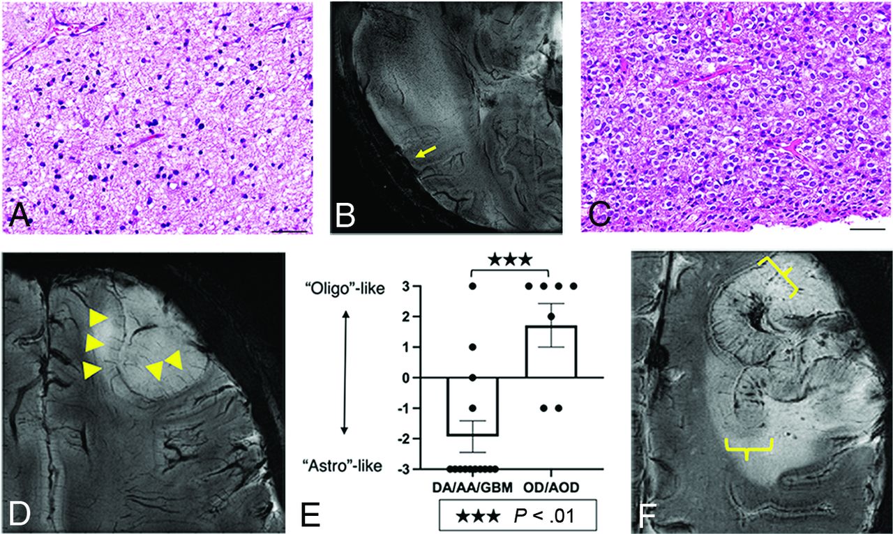
  • FIG 2.
    • Download figure
    • Open in new tab
    • Download powerpoint
    FIG 2.

    7T SWI characteristics of astrocytic and oligodendroglial lesions. Astrocytic lesions (A) were predominantly located in the WM and displayed infiltrative growth and focal obscuring of the corticomedullary junction (arrow) without displacing medullary vessels and other normal structures on 7T SWI (B). An oligodendroglioma (WHO grade II) with classic perinuclear halos and chicken wire-like vessels (C) shows thickening of the cortex (triangles), elongation but not thickening of the medullary vessels of the cortex, and expansive growth, displacing the medullary vessels on 7T-SWI (D). Scoring yielded a significant difference between astrocytomas and oligodendrogliomas (mean −1.93 versus +1.71, P < .01) (E). In a case of anaplastic oligodendroglioma, IDH-mutant and 1p/19q-codeleted, diffuse thickening of the cortex without tapering, thickening of medullary vessels, and microbleeds were observed (F). Scale bars = 50 µm. DA/AA/GBM indicates diffuse astrocytoma/anaplastic astrocytoma/glioblastoma; OD/AOD, oligodendroglioma/anaplastic oligodendroglioma.

  • FIG 3.
    • Download figure
    • Open in new tab
    • Download powerpoint
    FIG 3.

    Malignancy scoring of gliomas. A diffuse astrocytoma, IDH mutant (WHO grade II) with a malignancy score of 0 (A), and a glioblastoma, IDH wild-type (WHO grade IV) with a malignancy score of 3, with evidence of thickening of medullary vessels (bracket), microbleeds (arrows) and necrosis (surrounded by triangles) (B). Malignancy scoring was significantly elevated in WHO grade III tumors compared with grade II (mean, 1.38 versus 0.20, P < .01), and grade IV tumors compared with grade III (mean, 2.79 versus 1.38, P < .01) and grade II (P < .01) (C).

  • FIG 4.
    • Download figure
    • Open in new tab
    • Download powerpoint
    FIG 4.

    A diffuse astrocytoma, IDH-mutant, displaying thickening of the cortex with elongation of medullary vessels (arrows) and expansive growth on 7T MR imaging, mimicking an oligodendroglioma (A). Morphologically, the tumor displayed predominantly oligodendroglioma-like pathology (B). Fluorescence in situ hybridization (FISH) revealed 1p-intact (C), 19q-loss (D). ATRX staining was lost in tumor cells (E), and P53 was immunopositive (>10% positive) (F), suggesting astrocytic lineage. Scale bars = 100 µm. Circles indicate individual tumor cells.

PreviousNext
Back to top
Advertisement
Print
Download PDF
Email Article

Thank you for your interest in spreading the word on American Journal of Neuroradiology.

NOTE: We only request your email address so that the person you are recommending the page to knows that you wanted them to see it, and that it is not junk mail. We do not capture any email address.

Enter multiple addresses on separate lines or separate them with commas.
SWI by 7T MR Imaging for the Microscopic Imaging Diagnosis of Astrocytic and Oligodendroglial Tumors
(Your Name) has sent you a message from American Journal of Neuroradiology
(Your Name) thought you would like to see the American Journal of Neuroradiology web site.
CAPTCHA
This question is for testing whether or not you are a human visitor and to prevent automated spam submissions.
Cite this article
M. Natsumeda, H. Matsuzawa, M. Watanabe, K. Motohashi, R. Gabdulkhaev, Y. Tsukamoto, Y. Kanemaru, J. Watanabe, R. Ogura, M. Okada, S. Kurabe, K. Okamoto, A. Kakita, H. Igarashi, Y. Fujii
SWI by 7T MR Imaging for the Microscopic Imaging Diagnosis of Astrocytic and Oligodendroglial Tumors
American Journal of Neuroradiology Oct 2022, DOI: 10.3174/ajnr.A7666

Citation Manager Formats

  • BibTeX
  • Bookends
  • EasyBib
  • EndNote (tagged)
  • EndNote 8 (xml)
  • Medlars
  • Mendeley
  • Papers
  • RefWorks Tagged
  • Ref Manager
  • RIS
  • Zotero
0 Responses
Respond to this article
Share
Bookmark this article
SWI by 7T MR Imaging for the Microscopic Imaging Diagnosis of Astrocytic and Oligodendroglial Tumors
M. Natsumeda, H. Matsuzawa, M. Watanabe, K. Motohashi, R. Gabdulkhaev, Y. Tsukamoto, Y. Kanemaru, J. Watanabe, R. Ogura, M. Okada, S. Kurabe, K. Okamoto, A. Kakita, H. Igarashi, Y. Fujii
American Journal of Neuroradiology Oct 2022, DOI: 10.3174/ajnr.A7666
del.icio.us logo Twitter logo Facebook logo Mendeley logo
  • Tweet Widget
  • Facebook Like
  • Google Plus One
Purchase

Jump to section

  • Article
    • Abstract
    • ABBREVIATIONS:
    • MATERIALS AND METHODS
    • RESULTS
    • DISCUSSION
    • CONCLUSIONS
    • Acknowledgments
    • Footnotes
    • References
  • Figures & Data
  • Supplemental
  • Info & Metrics
  • Responses
  • References
  • PDF

Related Articles

  • PubMed
  • Google Scholar

Cited By...

  • No citing articles found.
  • Crossref
  • Google Scholar

This article has not yet been cited by articles in journals that are participating in Crossref Cited-by Linking.

More in this TOC Section

  • Diagnostic Neuroradiology of Monoclonal Antibodies
  • Clinical Outcomes After Chiari I Decompression
  • Segmentation of Brain Metastases with BLAST
Show more Adult Brain

Similar Articles

Advertisement

Indexed Content

  • Current Issue
  • Accepted Manuscripts
  • Article Preview
  • Past Issues
  • Editorials
  • Editor's Choice
  • Fellows' Journal Club
  • Letters to the Editor
  • Video Articles

Cases

  • Case Collection
  • Archive - Case of the Week
  • Archive - Case of the Month
  • Archive - Classic Case

More from AJNR

  • Trainee Corner
  • Imaging Protocols
  • MRI Safety Corner
  • Book Reviews

Multimedia

  • AJNR Podcasts
  • AJNR Scantastics

Resources

  • Turnaround Time
  • Submit a Manuscript
  • Submit a Video Article
  • Submit an eLetter to the Editor/Response
  • Manuscript Submission Guidelines
  • Statistical Tips
  • Fast Publishing of Accepted Manuscripts
  • Graphical Abstract Preparation
  • Imaging Protocol Submission
  • Evidence-Based Medicine Level Guide
  • Publishing Checklists
  • Author Policies
  • Become a Reviewer/Academy of Reviewers
  • News and Updates

About Us

  • About AJNR
  • Editorial Board
  • Editorial Board Alumni
  • Alerts
  • Permissions
  • Not an AJNR Subscriber? Join Now
  • Advertise with Us
  • Librarian Resources
  • Feedback
  • Terms and Conditions
  • AJNR Editorial Board Alumni

American Society of Neuroradiology

  • Not an ASNR Member? Join Now

© 2025 by the American Society of Neuroradiology All rights, including for text and data mining, AI training, and similar technologies, are reserved.
Print ISSN: 0195-6108 Online ISSN: 1936-959X

Powered by HighWire