Advertisement

Main menu

  • Home
  • Content
    • Current Issue
    • Accepted Manuscripts
    • Article Preview
    • Past Issue Archive
    • Video Articles
    • AJNR Case Collection
    • Case of the Week Archive
    • Case of the Month Archive
    • Classic Case Archive
  • Special Collections
    • AJNR Awards
    • Low-Field MRI
    • Alzheimer Disease
    • ASNR Foundation Special Collection
    • Photon-Counting CT
    • View All
  • Multimedia
    • AJNR Podcasts
    • AJNR SCANtastic
    • Trainee Corner
    • MRI Safety Corner
    • Imaging Protocols
  • For Authors
    • Submit a Manuscript
    • Submit a Video Article
    • Submit an eLetter to the Editor/Response
    • Manuscript Submission Guidelines
    • Statistical Tips
    • Fast Publishing of Accepted Manuscripts
    • Graphical Abstract Preparation
    • Imaging Protocol Submission
    • Author Policies
  • About Us
    • About AJNR
    • Editorial Board
    • Editorial Board Alumni
  • More
    • Become a Reviewer/Academy of Reviewers
    • Subscribers
    • Permissions
    • Alerts
    • Feedback
    • Advertisers
    • ASNR Home

User menu

  • Alerts
  • Log in

Search

  • Advanced search
American Journal of Neuroradiology

American Journal of Neuroradiology

ASHNR American Society of Functional Neuroradiology ASHNR American Society of Pediatric Neuroradiology ASSR
  • Alerts
  • Log in

Advanced Search

  • Home
  • Content
    • Current Issue
    • Accepted Manuscripts
    • Article Preview
    • Past Issue Archive
    • Video Articles
    • AJNR Case Collection
    • Case of the Week Archive
    • Case of the Month Archive
    • Classic Case Archive
  • Special Collections
    • AJNR Awards
    • Low-Field MRI
    • Alzheimer Disease
    • ASNR Foundation Special Collection
    • Photon-Counting CT
    • View All
  • Multimedia
    • AJNR Podcasts
    • AJNR SCANtastic
    • Trainee Corner
    • MRI Safety Corner
    • Imaging Protocols
  • For Authors
    • Submit a Manuscript
    • Submit a Video Article
    • Submit an eLetter to the Editor/Response
    • Manuscript Submission Guidelines
    • Statistical Tips
    • Fast Publishing of Accepted Manuscripts
    • Graphical Abstract Preparation
    • Imaging Protocol Submission
    • Author Policies
  • About Us
    • About AJNR
    • Editorial Board
    • Editorial Board Alumni
  • More
    • Become a Reviewer/Academy of Reviewers
    • Subscribers
    • Permissions
    • Alerts
    • Feedback
    • Advertisers
    • ASNR Home
  • Follow AJNR on Twitter
  • Visit AJNR on Facebook
  • Follow AJNR on Instagram
  • Join AJNR on LinkedIn
  • RSS Feeds

AJNR Awards, New Junior Editors, and more. Read the latest AJNR updates

Research ArticleExtracranial Vascular
Open Access

High-Resolution MRI for Evaluation of the Possibility of Successful Recanalization in Symptomatic Chronic ICA Occlusion: A Retrospective Study

M. Tang, X. Yan, J. Gao, L. Li, X. Zhe, Xin Zhang, F. Jiang, J. Hu, N. Ma, K. Ai and Xiaoling Zhang
American Journal of Neuroradiology July 2022, DOI: https://doi.org/10.3174/ajnr.A7576
M. Tang
aFrom the Departments of MRI (M.T., X.Y., J.G., L.L., X. Zhe., X. Zhang., N.M., X. Zhang)
  • Find this author on Google Scholar
  • Find this author on PubMed
  • Search for this author on this site
  • ORCID record for M. Tang
X. Yan
aFrom the Departments of MRI (M.T., X.Y., J.G., L.L., X. Zhe., X. Zhang., N.M., X. Zhang)
  • Find this author on Google Scholar
  • Find this author on PubMed
  • Search for this author on this site
  • ORCID record for X. Yan
J. Gao
aFrom the Departments of MRI (M.T., X.Y., J.G., L.L., X. Zhe., X. Zhang., N.M., X. Zhang)
  • Find this author on Google Scholar
  • Find this author on PubMed
  • Search for this author on this site
  • ORCID record for J. Gao
L. Li
aFrom the Departments of MRI (M.T., X.Y., J.G., L.L., X. Zhe., X. Zhang., N.M., X. Zhang)
  • Find this author on Google Scholar
  • Find this author on PubMed
  • Search for this author on this site
  • ORCID record for L. Li
X. Zhe
aFrom the Departments of MRI (M.T., X.Y., J.G., L.L., X. Zhe., X. Zhang., N.M., X. Zhang)
  • Find this author on Google Scholar
  • Find this author on PubMed
  • Search for this author on this site
  • ORCID record for X. Zhe
Xin Zhang
aFrom the Departments of MRI (M.T., X.Y., J.G., L.L., X. Zhe., X. Zhang., N.M., X. Zhang)
  • Find this author on Google Scholar
  • Find this author on PubMed
  • Search for this author on this site
  • ORCID record for Xin Zhang
F. Jiang
bNeurology (F.J., J.H.), Shaanxi Provincial People’s Hospital, Beilin District, Xi'an City, Shaanxi Province, China
  • Find this author on Google Scholar
  • Find this author on PubMed
  • Search for this author on this site
  • ORCID record for F. Jiang
J. Hu
bNeurology (F.J., J.H.), Shaanxi Provincial People’s Hospital, Beilin District, Xi'an City, Shaanxi Province, China
  • Find this author on Google Scholar
  • Find this author on PubMed
  • Search for this author on this site
  • ORCID record for J. Hu
N. Ma
aFrom the Departments of MRI (M.T., X.Y., J.G., L.L., X. Zhe., X. Zhang., N.M., X. Zhang)
  • Find this author on Google Scholar
  • Find this author on PubMed
  • Search for this author on this site
  • ORCID record for N. Ma
K. Ai
cDepartment of Clinical Science (K.A.), Philips Healthcare, Xìan, China
  • Find this author on Google Scholar
  • Find this author on PubMed
  • Search for this author on this site
  • ORCID record for K. Ai
Xiaoling Zhang
aFrom the Departments of MRI (M.T., X.Y., J.G., L.L., X. Zhe., X. Zhang., N.M., X. Zhang)
  • Find this author on Google Scholar
  • Find this author on PubMed
  • Search for this author on this site
  • ORCID record for Xiaoling Zhang
  • Article
  • Figures & Data
  • Info & Metrics
  • Responses
  • References
  • PDF
Loading

Article Figures & Data

Figures

  • Tables
  • FIG 1.
    • Download figure
    • Open in new tab
    • Download powerpoint
    FIG 1.

    A–C, T2WI, T1WI, and CE-T1WI show the measurement of the proximal segment; the loose matrix (purple) shows hyperintensity on T2WI, iso- or hypointensity on T1WI, and enhancement on CE-T1WI. Calcification (blue) appears as hypointensity on all sequences; hemorrhage (orange) appears as hyperintensity on T1WI, iso- or hypointensity on T2WI, and no enhancement on CE-T1WI. The lipid-rich necrotic core (yellow) shows iso- or hyperintensity on T1WI, iso- or hyperintensity on T2WI, and enhancement on CE-T1WI. D, The lumen and wall area were measured in the distal occlusion.
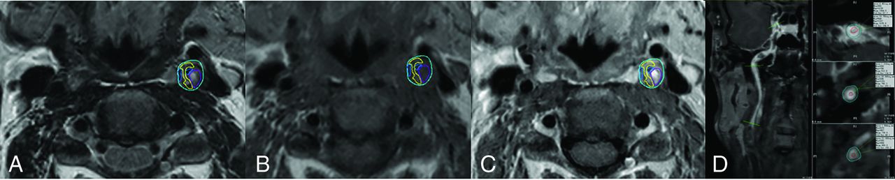
  • FIG 2.
    • Download figure
    • Open in new tab
    • Download powerpoint
    FIG 2.

    Successful recanalization cases. A, Curved planar reformation (CPR) on T1WI-volume isotropic turbo spin-echo acquisition (VISTA) shows moderate hypointensity in the proximal occlusion and iso- or hypointensity in the lumen of the distal occlusion. B, CPR on T1WI-VISTA-CE shows inhomogeneous enhancement in the origin of the left ICA and hypointensity in the distal occlusion. C and D, CE-MRA and preoperative DSA show a tapered stump (triangle) and reversal of flow above the clinoid segment of left ICA (arrow). E and F, Postoperative DSA shows successful recanalization. G–I, Plaque composition is shown in the proximal occlusion on T1WI and T2WI and enhancement on T1WI (blue, calcification; orange, hemorrhage; yellow, lipid-rich/necrotic core; purple, loose matrix).
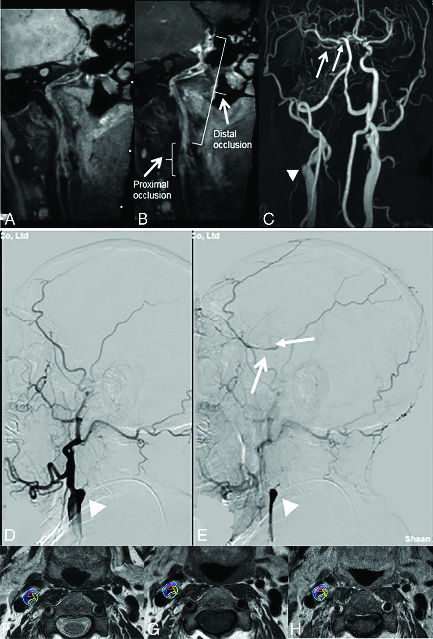
  • FIG 3.
    • Download figure
    • Open in new tab
    • Download powerpoint
    FIG 3.

    Unsuccessful recanalization cases. A, Curved planar reformation (CPR) on T1WI-volume isotropic turbo spin-echo acquisition (VISTA) shows mixed signal and wall collapse in the lumen distal to the occlusion. B, CPR on T1WI-VISTA-CE shows inhomogeneous enhancement in the proximal occlusion and distal segment of the right ICA (arrowhead). C–E, CE-MRA and DSA show a blunt stump (triangle), secondary collateral circulation (arrowhead), and reversal flow above the communicating segment of right ICA (arrows). F–H, Plaque composition is shown in the proximal occlusion (blue, calcification; orange, hemorrhage; yellow, lipid-rich/necrotic core; purple, loose matrix).

Tables

  • Figures
    • View popup
    Table 1:

    Characteristics of patients with CICAO

    Successful (n = 76)Unsuccessful (n = 38)P
    Age (mean) (yr)53.9 (SD, 15.9)58.5 (SD, 11.0).115
    Male (No.) (%)49 (64.5)23 (60.5).680
    Smoking (No.) (%)44 (57.9)26 (68.4).276
    Hypertension (No.) (%)29 (38.1)14 (36.8).891
    Diabetes mellitus (No.) (%)22 (28.9)5 (13.2).062
    Hyperlipidemia (No.) (%)39 (51.3)15 (39.5).233
    History of stroke (No.) (%)12 (15.8)8 (21.1).500
    Stroke (No.) (%)55 (72.4)32 (84.2).161
    • View popup
    Table 2:

    HR-MR imaging characteristics of lesions in symptomatic patients with CICAO

    Total (n = 114)Successful (n = 76)Unsuccessful (n = 38)P
    Right (No.) (%)67 (58.8)42 (55.2)25 (65.8).282
    Tapered stump (No.) (%)64 (56.1)48 (63.2)16 (42.1).033
    Level of reversed distal ICA flow<.001
     Communicating segment (No.) (%)8 (7.0)2 (2.6)6 (15.8)
     Ophthalmic segment (No.) (%)12 (10.5)2 (2.6)10 (26.3)
     Clinoid segment (No.) (%)23 (20.2)14 (18.4)9 (23.7)
     Cavernous segment (No.) (%)32 (28.1)24 (31.6)8 (21.1)
     Petrous segment or below (No.) (%)39 (34.2)34 (44.7)5 (13.2)
    Collateral circulation
     Primary (No.) (%)64 (56.1)39 (51.3)25 (65.8).142
     Secondary (No.) (%)25 (21.9)12 (15.7)13 (34.2).025
    Proximal occlusion
     Lesion length (mean) (mm)21.9 (SD, 4.2)20.7 (SD, 5.4)23.9 (SD, 4.8)<.005
     Lesion volume (mean) (mm3)1084.8 (SD, 472.5)1007.3 (SD, 453.4)1159.1 (SD, 486.1).110
     Lipid-rich necrotic core (mean) (mm3)143.3 (SD, 76.6)167.2 (SD, 74.2)139.8 (SD, 54.5)<.050
     Hemorrhage (mean) (mm354.5 (SD, 43.1)58.4 (SD, 46.7)53.6 (SD, 40.4).510
     Calcification (mean) (mm3)112.9 (SD, 96.4)41.3 (SD, 39.5)141.9 (SD, 107.8)<.001
     Dense fibrous tissue (mean) (mm3)708.2 (SD, 391.1)689.6 (SD, 372.3)738.8 (SD, 437.6).611
     Loose matrix (mean) (mm3)250.3 (SD, 130.5)219.7 (SD, 103.8)257.9 (SD, 141.6).120
    Distal occlusion
     Lumen iso -to hypointensity (No.) (%)60 (52.6)45 (59.2)15 (39.5).047
     Lumen area (mean) (mm2)6.28 (SD, 3.5)7.4 (SD, 3.4)3.9 (SD, 2.3)<.001
     Wall area (mean) (mm2)32.7 (SD, 6.9)32.0 (SD, 6.8)33.4 (SD, 8.7).340
    • View popup
    Table 3:

    Factors associated with recanalization success in univariable and multivariable analysis

    CharacteristicUnivariable AnalysisMultivariable Analysis
    OR (95% CI)POR (95% CI)P
    Right0.64 (0.51–1.33).282
    Tapered stump2.36 (1.02–3.11).0352.23 (0.87–3.36).091
    Level of reversed distal ICA flow
     Communicating segment0.14 (0.07–0.21).0010.13 (0.07–0.24).010
     Ophthalmic segment0.08 (0.03–0.44)<.0010.14 (0.08–0.48).001
     Clinoid segment0.73 (0.63–1.21).2140.62 (0.36–1.22).160
     Cavernous segment1.73 (0.96–3.89).071.66 (0.87–3.54).550
     Petrous segment or below5.34 (1.41–11.37)<.0014.07 (1.65–8.38).001
    Collateral circulation
     Primary0.55 (0.35–1.59).143
     Secondary0.36 (0.14–0.71).0271.32 (0.98–1.47).210
    Proximal occlusion
     Lesion length0.51 (0.13–0.74).0050.41 (0.36–0.55).009
     Lesion volume2.11 (0.71–4.49).110
     Lipid-rich necrotic core3.26 (1.45–6.36).0242.36 (0.94–5.33).060
     Hemorrhage1.35 (0.65–2.38).510
     Calcification0.71 (0.44–0.90)<.0010.56 (0.37–0.68).002
     Dense fibrous tissue3.31 (0.93–10.36).615
     Loose matrix1.66 (0.87–3.88).120
    Distal occlusion
     Lumen iso- to hypointensity2.23 (1.06–5.29).0471.87 (0.91–4.66).091
     Lumen area1.32 (1.02–2.17)<.0011.13 (1.04–1.61).002
     Wall area1.76 (0.71–3.38).230
PreviousNext
Back to top
Advertisement
Print
Download PDF
Email Article

Thank you for your interest in spreading the word on American Journal of Neuroradiology.

NOTE: We only request your email address so that the person you are recommending the page to knows that you wanted them to see it, and that it is not junk mail. We do not capture any email address.

Enter multiple addresses on separate lines or separate them with commas.
High-Resolution MRI for Evaluation of the Possibility of Successful Recanalization in Symptomatic Chronic ICA Occlusion: A Retrospective Study
(Your Name) has sent you a message from American Journal of Neuroradiology
(Your Name) thought you would like to see the American Journal of Neuroradiology web site.
CAPTCHA
This question is for testing whether or not you are a human visitor and to prevent automated spam submissions.
Cite this article
M. Tang, X. Yan, J. Gao, L. Li, X. Zhe, Xin Zhang, F. Jiang, J. Hu, N. Ma, K. Ai, Xiaoling Zhang
High-Resolution MRI for Evaluation of the Possibility of Successful Recanalization in Symptomatic Chronic ICA Occlusion: A Retrospective Study
American Journal of Neuroradiology Jul 2022, DOI: 10.3174/ajnr.A7576

Citation Manager Formats

  • BibTeX
  • Bookends
  • EasyBib
  • EndNote (tagged)
  • EndNote 8 (xml)
  • Medlars
  • Mendeley
  • Papers
  • RefWorks Tagged
  • Ref Manager
  • RIS
  • Zotero
0 Responses
Respond to this article
Share
Bookmark this article
High-Resolution MRI for Evaluation of the Possibility of Successful Recanalization in Symptomatic Chronic ICA Occlusion: A Retrospective Study
M. Tang, X. Yan, J. Gao, L. Li, X. Zhe, Xin Zhang, F. Jiang, J. Hu, N. Ma, K. Ai, Xiaoling Zhang
American Journal of Neuroradiology Jul 2022, DOI: 10.3174/ajnr.A7576
del.icio.us logo Twitter logo Facebook logo Mendeley logo
  • Tweet Widget
  • Facebook Like
  • Google Plus One
Purchase

Jump to section

  • Article
    • Abstract
    • ABBREVIATIONS:
    • MATERIALS AND METHODS
    • RESULTS
    • DISCUSSION
    • CONCLUSIONS
    • Footnotes
    • References
  • Figures & Data
  • Info & Metrics
  • Responses
  • References
  • PDF

Related Articles

  • No related articles found.
  • PubMed
  • Google Scholar

Cited By...

  • Vessel wall MRI evaluation for the safety of endovascular recanalization of non-acute intracranial anterior circulation artery occlusions
  • MR-VWI concentric ring sign: a potential imaging feature of internal carotid artery pseudo occlusion and predictive value for successful recanalization
  • Endovascular Recanalization of Symptomatic Chronic ICA Occlusion: Procedural Outcomes and Radiologic Predictors
  • Crossref (6)
  • Google Scholar

This article has been cited by the following articles in journals that are participating in Crossref Cited-by Linking.

  • Endovascular Recanalization of Symptomatic Chronic ICA Occlusion: Procedural Outcomes and Radiologic Predictors
    C. Zhou, Y.-Z. Cao, Z.-Y. Jia, L.-B. Zhao, S.-S. Lu, H.-B. Shi, S. Liu
    American Journal of Neuroradiology 2023 44 3
  • Endovascular recanalization for symptomatic chronic internal carotid artery occlusion: proposal of a modified angiographic classification and clinical outcomes
    Chun Zhou, Yue-Zhou Cao, Sheng Liu, Zhen-Yu Jia, Lin-Bo Zhao, Hai-Bin Shi, Yang Zhao
    Clinical Neurology and Neurosurgery 2023 233
  • Dynamic real‐time 3D roadmap: A navigational tool for endovascular recanalization in chronic intracranial arterial occlusion
    Wen Zhao, Ling Chen, Honggen Guo, Qi Jia, Xiaojian Lu, Feng Zhang, Zhengwen Chen, Peicheng Li
    Brain‐X 2024 2 4
  • Endovascular Surgery Revascularization of Chronic Cervical Carotid Occlusions: Systematic Review and Meta‐Analysis
    Santiago Ortega‐Gutierrez, Milagros Galecio‐Castillo, Cynthia B. Zevallos, Aaron Rodriguez‐Calienes, Juan Vivanco‐Suarez, Julie Weng, Edgar A. Samaniego, Mudassir Farooqui, Colin Derdeyn
    Stroke: Vascular and Interventional Neurology 2024 4 1
  • MR-VWI concentric ring sign: a potential imaging feature of internal carotid artery pseudo occlusion and predictive value for successful recanalization
    Wenxuan Lu, Wenhui Zhang, Dongxu Zhao, Yilin Zhang, Huili Gao, Tianxiao Li, Zhaoshuo Li
    Journal of NeuroInterventional Surgery 2025
  • Vessel wall MRI evaluation for the safety of endovascular recanalization of non-acute intracranial anterior circulation artery occlusions
    Yao Meng, Yun Yang, Miyengi Cosmas Magigi, Weili Li, Yun Song, Wei Zhao, Meimei Zheng, Lili Sun, Hao Yin, Wei Wang, Xiao He, Jun Zhang, Ju Han
    Journal of NeuroInterventional Surgery 2025 17 e2

More in this TOC Section

  • Proximal Vertebral Artery Variants and Embryology
  • High-Risk Plaque Features in Carotid MRI
  • Nonstenotic Carotid Plaques and Stroke Review
Show more Extracranial Vascular

Similar Articles

Advertisement

Indexed Content

  • Current Issue
  • Accepted Manuscripts
  • Article Preview
  • Past Issues
  • Editorials
  • Editor's Choice
  • Fellows' Journal Club
  • Letters to the Editor
  • Video Articles

Cases

  • Case Collection
  • Archive - Case of the Week
  • Archive - Case of the Month
  • Archive - Classic Case

More from AJNR

  • Trainee Corner
  • Imaging Protocols
  • MRI Safety Corner
  • Book Reviews

Multimedia

  • AJNR Podcasts
  • AJNR Scantastics

Resources

  • Turnaround Time
  • Submit a Manuscript
  • Submit a Video Article
  • Submit an eLetter to the Editor/Response
  • Manuscript Submission Guidelines
  • Statistical Tips
  • Fast Publishing of Accepted Manuscripts
  • Graphical Abstract Preparation
  • Imaging Protocol Submission
  • Evidence-Based Medicine Level Guide
  • Publishing Checklists
  • Author Policies
  • Become a Reviewer/Academy of Reviewers
  • News and Updates

About Us

  • About AJNR
  • Editorial Board
  • Editorial Board Alumni
  • Alerts
  • Permissions
  • Not an AJNR Subscriber? Join Now
  • Advertise with Us
  • Librarian Resources
  • Feedback
  • Terms and Conditions
  • AJNR Editorial Board Alumni

American Society of Neuroradiology

  • Not an ASNR Member? Join Now

© 2025 by the American Society of Neuroradiology All rights, including for text and data mining, AI training, and similar technologies, are reserved.
Print ISSN: 0195-6108 Online ISSN: 1936-959X

Powered by HighWire