
Article Figures & Data
Figures
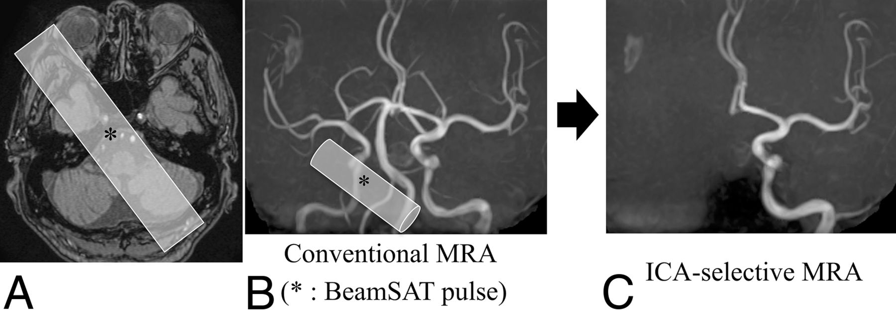
- FIG 1.
A, The BeamSAT pulse is positioned to cover the unilateral petrous portion of the ICA and bilateral vertebral arteries in an axial TOF source image obtained with conventional MRA (A). By adding the BeamSAT pulse to the unilateral ICA and bilateral vertebral arteries on 3D TOF-MRA (B), we performed ICA-selective MRA (C). The asterisk indicates BeamSAT.
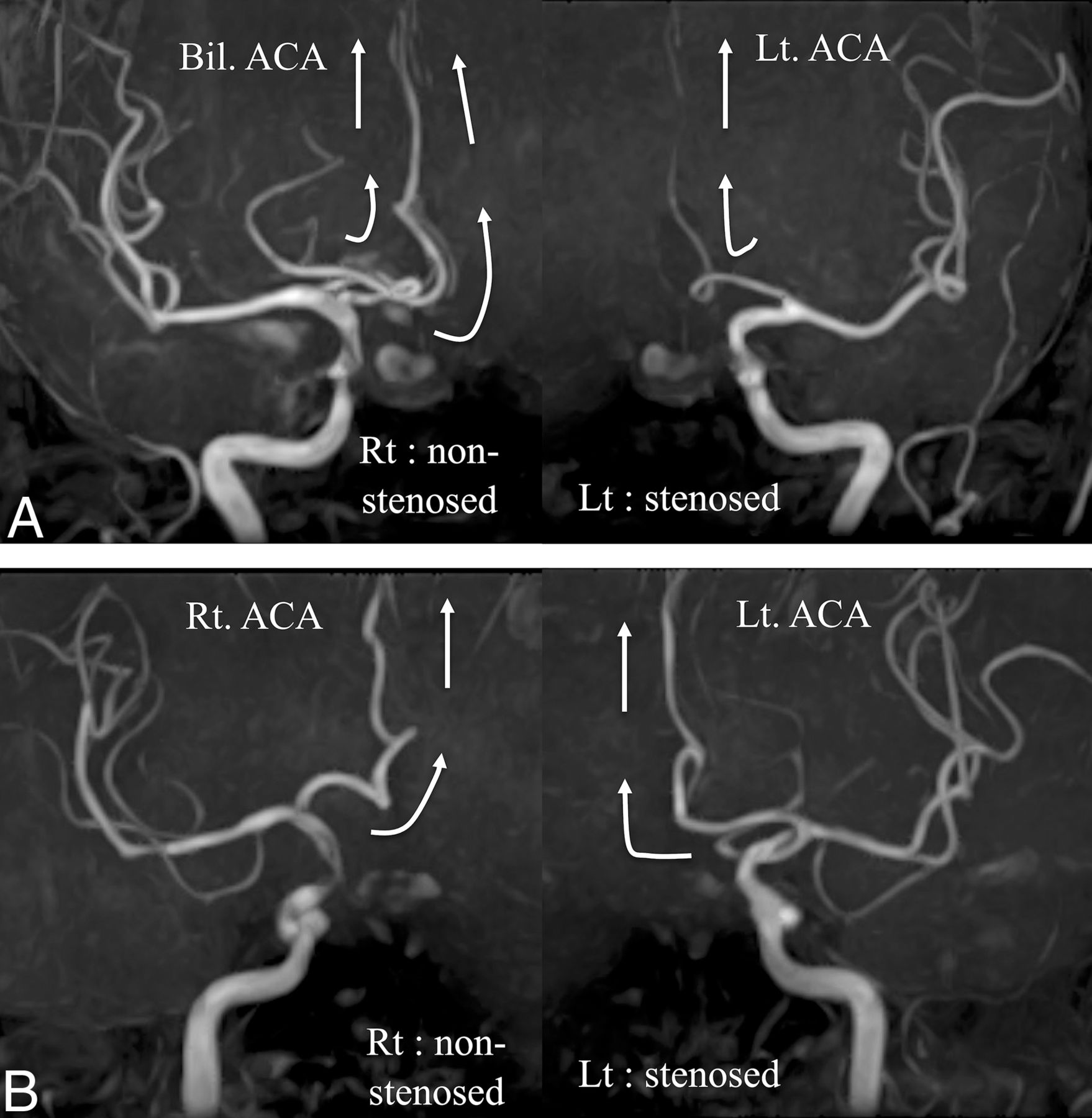
- FIG 2.
A, A case with an AcomA. The bilateral anterior cerebral arteries are perfused from the ICA of the nonstenotic side. Only the ipsilateral anterior cerebral artery is perfused by the ICA of the stenotic side. B, A case without an AcomA. Each ICA perfuses only the ipsilateral anterior cerebral artery. White arrows show the direction of blood flow. Bil. indicates bilateral; Rt., right; Lt., left.
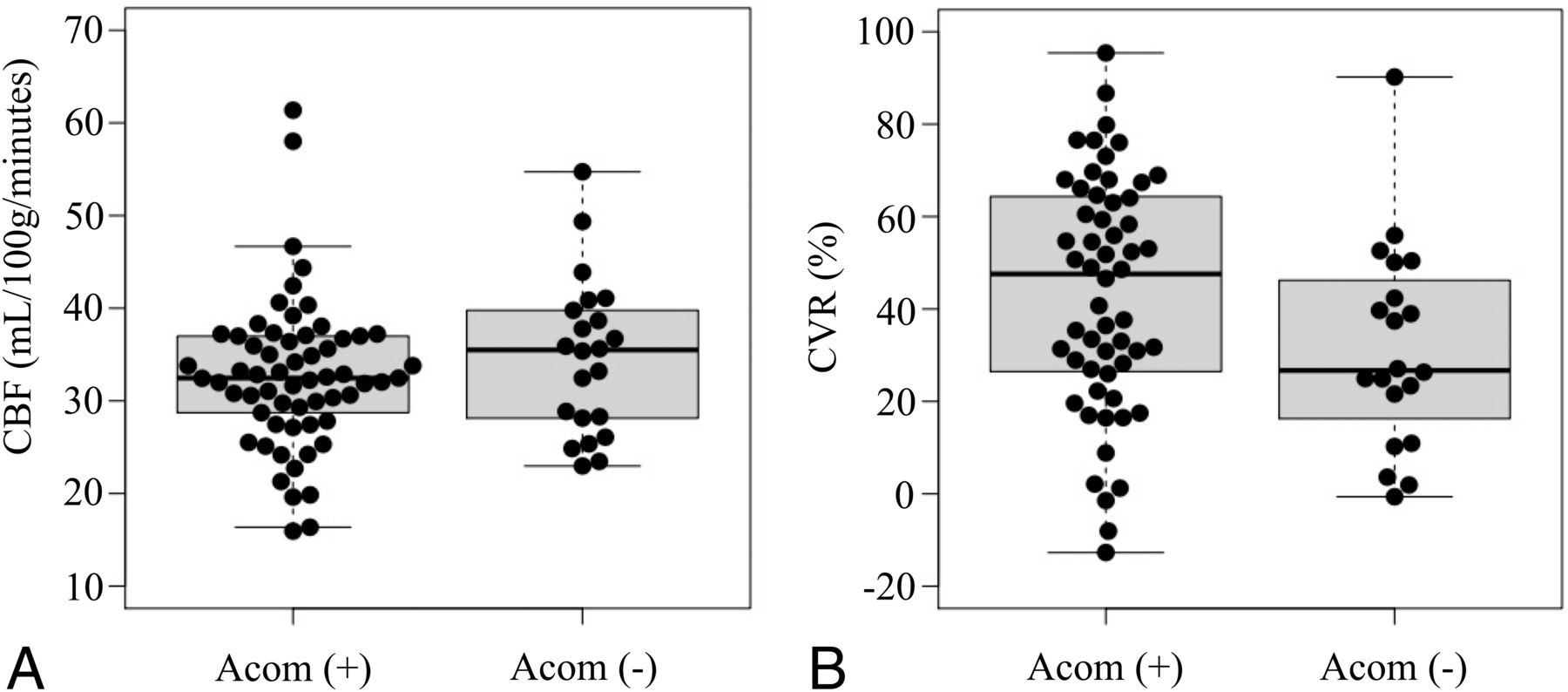
- FIG 3.
A, Box-and-whisker plots of preoperative CBF on the stenotic side in the AcomA (+) group and the AcomA (−) group. Preoperative CBF is not significantly different between the 2 groups. B, Box-and-whisker plots of preoperative CVR on the stenotic side in the AcomA (+) group and the AcomA (−) group. The CVR to the acetazolamide challenge is not significantly different between the 2 groups. The thick horizontal lines divide the boxes at the median values. The bottom and top of the boxes indicate the first and third quartiles. The whiskers extend to the most extreme data points, which are no more than 1.5 times the interquartile range from the box.
- FIG 4.
A, Box-and-whisker plots of preoperative CBF on the stenotic side in the AcomA (+)/PSV ≥200 cm/s group and the AcomA (−) / PSV ≥200 cm/s group. Preoperative CBF is not significantly different between the 2 groups. B, Box-and-whisker plots of preoperative CVR on the stenotic side in the AcomA (+) / PSV ≥200 cm/s group and the AcomA (−) / PSV ≥200 cm/s group. The CVR to the acetazolamide challenge is significantly lower in the AcomA (−) / PSV ≥200 cm/s group than in the AcomA (+) / PSV ≥200 cm/s group. The thick horizontal lines divide the boxes at the median values. The bottom and top of the boxes indicate the first and third quartiles. The whiskers extend to the most extreme data points, which are no more than 1.5 times the interquartile range from the box. The asterisk indicates P < . 05.
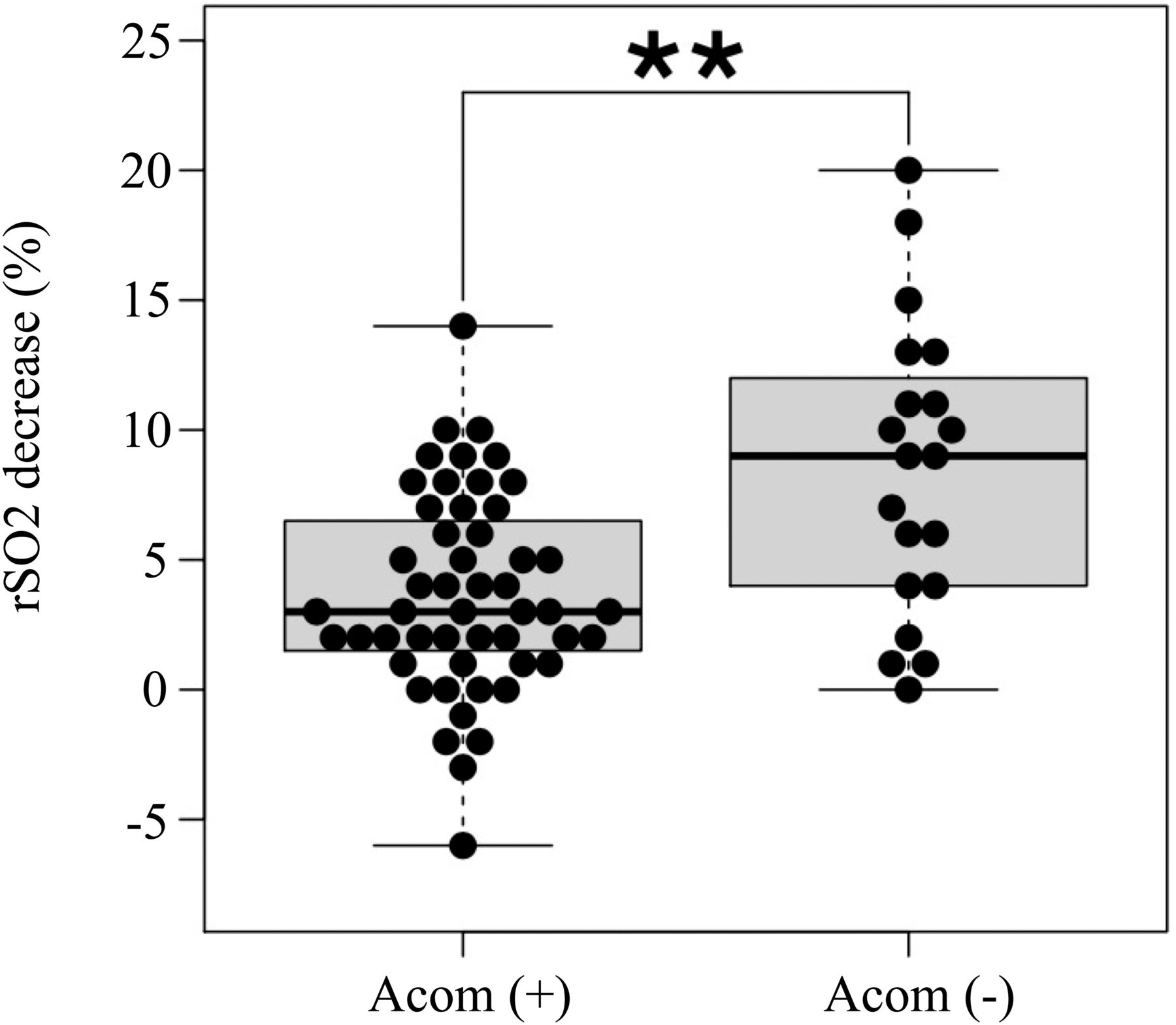
- FIG 5.
Box-and-whisker plots of the decrease in rSO2 after temporary ICA occlusion on the stenotic side during CEA or CAS. The decrease in rSO2 is significantly greater in the AcomA (−) group than in the AcomA (+) group. The thick horizontal lines divide the boxes at the median values. The bottom and top of the boxes indicate the first and third quartiles. The whiskers extend to the most extreme data points, which are no more than 1.5 times the interquartile range from the box. Double asterisks indicate P < . 01.


Tables
AcomA (+) (n = 61) AcomA (–) (n = 22) P Value Age (yr) 75.1 (SD, 7.4) 76.9 (SD, 5.9) .24 Male 53 (87%) 19 (86%) 1 Rt. ICS 30 (49%) 8 (36%) .33 CAS 26 (43%) 6 (27%) .31 Symptomatic 24 (39%) 12 (55%) .32 Degree of stenosis (%) 74.7 (SD, 12.7) 76.0 (SD, 10.4) .77 PSV (cm/s) 281.1 (SD, 120.5) 250.8 (SD, 138.5) .37 Hypertension 45 (74%) 19 (86%) .37 Hyperlipidemia 33 (54%) 16 (73%) .14 Diabetes mellitus 21 (34%) 7 (32%) 1 Ischemic heart disease 11 (18%) 2 (9%) .50 Smoking 31 (51%) 13 (59%) .62 COPD 6 (10%) 2 (9%) 1 Postoperative DWI high 12 (20%) 11 (50%) .011 Note:—Rt indicates right; ICS, ICA stenosis; COPD, chronic obstructive pulmonary disease.
↵a Values are presented as mean (SD) or number (%).
- Table 2:
Univariate and multivariate logistic regression analyses of factors for new ischemic lesions on MR imaginga
Predictor Univariate Analysis Multivariate Analysis DWI-Positive (n = 23) DWI-Negative (n = 57) P Value OR (95% CI) P Value Presence of AcomA 12 (52%) 46 (81%) .014 0.07 (0.012–0.45) .005 Age (mean) 78.0 (SD, 5.7) 74.4 (SD, 7.3) .023 1.14 (1.01–1.29) .039 CAS 15 (65%) 17 (30%) .005 12.99 (2.01–80.86) .006 Preoperative CVR (mean) (%) 43.4 (SD, 25.9) 37.8 (SD, 24.1) .40 1.02 (0.99–1.05) .20 Ulcerated plaque 7 (30%) 15 (26%) .78 3.07 (0.60–15.76) .18 ↵a ORs of age and CVR are presented as estimated odds of outcome for a 1 year increase in age and a 1% increase in percentage.
- Table 3:
Univariate and multivariate logistic regression analyses of factors for new ischemic lesions on MR imaging in patients with a PSV of ≥200 cm/sa
Predictor Univariate Analysis Multivariate Analysis DWI-Positive (n = 13) DWI-Negative (n = 39) P Value OR (95% CI) P Value Presence of AcomA 6 (46%) 34 (87%) .005 0.08 (0.011–0.64) .017 Age (mean) 78.7 (SD, 4.3) 75.0 (SD, 7.4) .032 1.13 (0.96–1.32) .13 CAS 7 (54%) 14 (36%) .33 4.23 (0.62–28.89) .14 Preoperative CVR (mean) (%) 35.7 (SD, 18.7) 36.7 (SD, 24.5) .88 1.02 (0.98–1.06) .34 Ulcerated plaque 5 (38%) 8 (21%) .27 4.71(0.77–28.83) .094 ↵a ORs of age and CVR are presented as estimated odds of outcome for a 1 year increase in age and a 1% increase in percentage.


