Advertisement

Main menu

  • Home
  • Content
    • Current Issue
    • Accepted Manuscripts
    • Article Preview
    • Past Issue Archive
    • Video Articles
    • AJNR Case Collection
    • Case of the Week Archive
    • Case of the Month Archive
    • Classic Case Archive
  • Special Collections
    • AJNR Awards
    • Low-Field MRI
    • Alzheimer Disease
    • ASNR Foundation Special Collection
    • Photon-Counting CT
    • View All
  • Multimedia
    • AJNR Podcasts
    • AJNR SCANtastic
    • Trainee Corner
    • MRI Safety Corner
    • Imaging Protocols
  • For Authors
    • Submit a Manuscript
    • Submit a Video Article
    • Submit an eLetter to the Editor/Response
    • Manuscript Submission Guidelines
    • Statistical Tips
    • Fast Publishing of Accepted Manuscripts
    • Graphical Abstract Preparation
    • Imaging Protocol Submission
    • Author Policies
  • About Us
    • About AJNR
    • Editorial Board
    • Editorial Board Alumni
  • More
    • Become a Reviewer/Academy of Reviewers
    • Subscribers
    • Permissions
    • Alerts
    • Feedback
    • Advertisers
    • ASNR Home

User menu

  • Alerts
  • Log in

Search

  • Advanced search
American Journal of Neuroradiology

American Journal of Neuroradiology

ASHNR American Society of Functional Neuroradiology ASHNR American Society of Pediatric Neuroradiology ASSR
  • Alerts
  • Log in

Advanced Search

  • Home
  • Content
    • Current Issue
    • Accepted Manuscripts
    • Article Preview
    • Past Issue Archive
    • Video Articles
    • AJNR Case Collection
    • Case of the Week Archive
    • Case of the Month Archive
    • Classic Case Archive
  • Special Collections
    • AJNR Awards
    • Low-Field MRI
    • Alzheimer Disease
    • ASNR Foundation Special Collection
    • Photon-Counting CT
    • View All
  • Multimedia
    • AJNR Podcasts
    • AJNR SCANtastic
    • Trainee Corner
    • MRI Safety Corner
    • Imaging Protocols
  • For Authors
    • Submit a Manuscript
    • Submit a Video Article
    • Submit an eLetter to the Editor/Response
    • Manuscript Submission Guidelines
    • Statistical Tips
    • Fast Publishing of Accepted Manuscripts
    • Graphical Abstract Preparation
    • Imaging Protocol Submission
    • Author Policies
  • About Us
    • About AJNR
    • Editorial Board
    • Editorial Board Alumni
  • More
    • Become a Reviewer/Academy of Reviewers
    • Subscribers
    • Permissions
    • Alerts
    • Feedback
    • Advertisers
    • ASNR Home
  • Follow AJNR on Twitter
  • Visit AJNR on Facebook
  • Follow AJNR on Instagram
  • Join AJNR on LinkedIn
  • RSS Feeds

AJNR Awards, New Junior Editors, and more. Read the latest AJNR updates

Research ArticleAdult Brain
Open Access

Diagnostic Accuracy of High-Resolution 3D T2-SPACE in Detecting Cerebral Venous Sinus Thrombosis

A. Hakim, C. Kurmann, K. Pospieszny, T.R. Meinel, M.A. Shahin, M.R. Heldner, R. Umarova, S. Jung, M. Arnold and M. El-Koussy
American Journal of Neuroradiology May 2022, DOI: https://doi.org/10.3174/ajnr.A7530
A. Hakim
aFrom the University Institute of Diagnostic and Interventional Neuroradiology (A.H., C.K., K.P., M.E.)
  • Find this author on Google Scholar
  • Find this author on PubMed
  • Search for this author on this site
  • ORCID record for A. Hakim
C. Kurmann
aFrom the University Institute of Diagnostic and Interventional Neuroradiology (A.H., C.K., K.P., M.E.)
  • Find this author on Google Scholar
  • Find this author on PubMed
  • Search for this author on this site
  • ORCID record for C. Kurmann
K. Pospieszny
aFrom the University Institute of Diagnostic and Interventional Neuroradiology (A.H., C.K., K.P., M.E.)
  • Find this author on Google Scholar
  • Find this author on PubMed
  • Search for this author on this site
  • ORCID record for K. Pospieszny
T.R. Meinel
bDepartment of Neurology (T.R.M., M.R.H., R.U., S.J., M.A.), Bern University Hospital, Inselspital, University of Bern, Bern, Switzerland
  • Find this author on Google Scholar
  • Find this author on PubMed
  • Search for this author on this site
  • ORCID record for T.R. Meinel
M.A. Shahin
cDepartment of Radiodiagnosis (M.A.S.), Faculty of Medicine, Cairo University Hospitals, Cairo, Egypt
  • Find this author on Google Scholar
  • Find this author on PubMed
  • Search for this author on this site
  • ORCID record for M.A. Shahin
M.R. Heldner
bDepartment of Neurology (T.R.M., M.R.H., R.U., S.J., M.A.), Bern University Hospital, Inselspital, University of Bern, Bern, Switzerland
  • Find this author on Google Scholar
  • Find this author on PubMed
  • Search for this author on this site
  • ORCID record for M.R. Heldner
R. Umarova
bDepartment of Neurology (T.R.M., M.R.H., R.U., S.J., M.A.), Bern University Hospital, Inselspital, University of Bern, Bern, Switzerland
  • Find this author on Google Scholar
  • Find this author on PubMed
  • Search for this author on this site
  • ORCID record for R. Umarova
S. Jung
bDepartment of Neurology (T.R.M., M.R.H., R.U., S.J., M.A.), Bern University Hospital, Inselspital, University of Bern, Bern, Switzerland
  • Find this author on Google Scholar
  • Find this author on PubMed
  • Search for this author on this site
  • ORCID record for S. Jung
M. Arnold
bDepartment of Neurology (T.R.M., M.R.H., R.U., S.J., M.A.), Bern University Hospital, Inselspital, University of Bern, Bern, Switzerland
  • Find this author on Google Scholar
  • Find this author on PubMed
  • Search for this author on this site
  • ORCID record for M. Arnold
M. El-Koussy
aFrom the University Institute of Diagnostic and Interventional Neuroradiology (A.H., C.K., K.P., M.E.)
  • Find this author on Google Scholar
  • Find this author on PubMed
  • Search for this author on this site
  • ORCID record for M. El-Koussy
  • Article
  • Figures & Data
  • Supplemental
  • Info & Metrics
  • Responses
  • References
  • PDF
Loading

Article Figures & Data

Figures

  • Tables
  • FIG 1.
    • Download figure
    • Open in new tab
    • Download powerpoint
    FIG 1.

    T2-SPACE obtained from different patients. The normal segments (green arrows) show a homogeneous physiologic flow void. In the thrombosed segments (red arrows, A), superior sagittal sinus and torcular; left sigmoid sinus and jugular vein (B); and the left vein of Labbé (C), the flow void is absent and the signal is mostly inhomogeneous. According to the stage of the thrombus, expansion of the sinus may be seen (superior sagittal sinus in A).
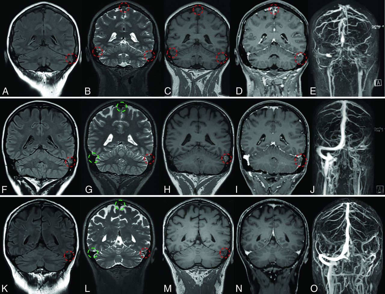
  • FIG 2.
    • Download figure
    • Open in new tab
    • Download powerpoint
    FIG 2.

    Coronal MR imaging from 3 different patients showing 3 phases of CVST. From left to right, FLAIR (A, F, K), T2-SPACE (B, G, L), T1-SPACE (C, H, M), CE T1-MPRAGE (D, I, N), and MIP of CE MRA (E, J, O). Thrombosed and patent veins are marked with red and green circles, respectively. The latter exhibit a physiologic flow void, clearly visible on SPACE images. Upper row, A–E, Acute CVST in a 45-year-old woman with headache and vomiting. MR imaging, performed on the day of symptom onset shows extensive thrombosis (red circles) of the transverse and sigmoid sinuses, jugular vein, torcular Herophili, and superior sagittal sinus. The flow void is absent on the T1 and T2 sequences. There are filling defects on CE-MPRAGE. The extension of the thrombosis is seen on the CE-MRA. Middle row, F–J, Subacute CVST in a 30-year-old woman who presented initially with headache. A follow-up MR imaging 13 days after the initial presentation shows thrombosis with an absent flow void on SPACE of the left transverse/sigmoid sinus and jugular vein. Lower row, K–O, Chronic CVST in a 63-year-old male patient who initially presented with headache. A follow-up MR imaging after 200 days shows a thrombus in the left transverse sinus. The flow void is absent on T2 and T1, but CVST is not detectable on CE-MPRAGE.
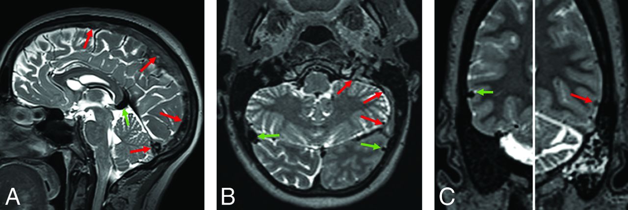
  • FIG 3.
    • Download figure
    • Open in new tab
    • Download powerpoint
    FIG 3.

    Examples from 4 different patients showing a thrombus (red arrows) on the T2-SPACE in the superior sagittal sinus (A), the vein of Labbé (C), the sigmoid sinus (E), and the cortical vein (G). CVST was missed on the CE T1-MPRAGE (B, D, F, and H).

Tables

  • Figures
    • View popup
    Table 1:

    Characteristics of patients included in the analysis

    ControlCVST
    No. of patients4135
    Male/female ratio14:2714:21
    Age (median) (interquartile range) (yr)38.5 (27–56)43.5 (34–61)
    No. of MR imaging examinations5163
    1.5T/3T34:1737:26
    • View popup
    Table 2:

    Results of T2-SPACE and CE-MPRAGE based on the first available MR imaging examinationsa

    T2-SPACECE-MPRAGE
    Sensitivity1 (0.9–1)0.91 (0.8–1)
    Specificity1 (0.9–1)0.98 (0.9–1)
    Accuracy1 (0.9–1)0.95 (0.9–1)
    Positive predictive value1 (0.9–1)0.97 (0.8–1)
    Negative predictive value1 (0.9–1)0.93 (0.8–1)
    • ↵a Data in parentheses are 95% confidence intervals.

PreviousNext
Back to top
Advertisement
Print
Download PDF
Email Article

Thank you for your interest in spreading the word on American Journal of Neuroradiology.

NOTE: We only request your email address so that the person you are recommending the page to knows that you wanted them to see it, and that it is not junk mail. We do not capture any email address.

Enter multiple addresses on separate lines or separate them with commas.
Diagnostic Accuracy of High-Resolution 3D T2-SPACE in Detecting Cerebral Venous Sinus Thrombosis
(Your Name) has sent you a message from American Journal of Neuroradiology
(Your Name) thought you would like to see the American Journal of Neuroradiology web site.
CAPTCHA
This question is for testing whether or not you are a human visitor and to prevent automated spam submissions.
Cite this article
A. Hakim, C. Kurmann, K. Pospieszny, T.R. Meinel, M.A. Shahin, M.R. Heldner, R. Umarova, S. Jung, M. Arnold, M. El-Koussy
Diagnostic Accuracy of High-Resolution 3D T2-SPACE in Detecting Cerebral Venous Sinus Thrombosis
American Journal of Neuroradiology May 2022, DOI: 10.3174/ajnr.A7530

Citation Manager Formats

  • BibTeX
  • Bookends
  • EasyBib
  • EndNote (tagged)
  • EndNote 8 (xml)
  • Medlars
  • Mendeley
  • Papers
  • RefWorks Tagged
  • Ref Manager
  • RIS
  • Zotero
0 Responses
Respond to this article
Share
Bookmark this article
Diagnostic Accuracy of High-Resolution 3D T2-SPACE in Detecting Cerebral Venous Sinus Thrombosis
A. Hakim, C. Kurmann, K. Pospieszny, T.R. Meinel, M.A. Shahin, M.R. Heldner, R. Umarova, S. Jung, M. Arnold, M. El-Koussy
American Journal of Neuroradiology May 2022, DOI: 10.3174/ajnr.A7530
del.icio.us logo Twitter logo Facebook logo Mendeley logo
  • Tweet Widget
  • Facebook Like
  • Google Plus One
Purchase

Jump to section

  • Article
    • Abstract
    • ABBREVIATIONS:
    • MATERIALS AND METHODS
    • RESULTS
    • DISCUSSION
    • CONCLUSIONS
    • Footnotes
    • References
  • Figures & Data
  • Supplemental
  • Info & Metrics
  • Responses
  • References
  • PDF

Related Articles

  • PubMed
  • Google Scholar

Cited By...

  • No citing articles found.
  • Crossref
  • Google Scholar

This article has not yet been cited by articles in journals that are participating in Crossref Cited-by Linking.

More in this TOC Section

  • Diagnostic Neuroradiology of Monoclonal Antibodies
  • Clinical Outcomes After Chiari I Decompression
  • Segmentation of Brain Metastases with BLAST
Show more Adult Brain

Similar Articles

Advertisement

Indexed Content

  • Current Issue
  • Accepted Manuscripts
  • Article Preview
  • Past Issues
  • Editorials
  • Editor's Choice
  • Fellows' Journal Club
  • Letters to the Editor
  • Video Articles

Cases

  • Case Collection
  • Archive - Case of the Week
  • Archive - Case of the Month
  • Archive - Classic Case

More from AJNR

  • Trainee Corner
  • Imaging Protocols
  • MRI Safety Corner
  • Book Reviews

Multimedia

  • AJNR Podcasts
  • AJNR Scantastics

Resources

  • Turnaround Time
  • Submit a Manuscript
  • Submit a Video Article
  • Submit an eLetter to the Editor/Response
  • Manuscript Submission Guidelines
  • Statistical Tips
  • Fast Publishing of Accepted Manuscripts
  • Graphical Abstract Preparation
  • Imaging Protocol Submission
  • Evidence-Based Medicine Level Guide
  • Publishing Checklists
  • Author Policies
  • Become a Reviewer/Academy of Reviewers
  • News and Updates

About Us

  • About AJNR
  • Editorial Board
  • Editorial Board Alumni
  • Alerts
  • Permissions
  • Not an AJNR Subscriber? Join Now
  • Advertise with Us
  • Librarian Resources
  • Feedback
  • Terms and Conditions
  • AJNR Editorial Board Alumni

American Society of Neuroradiology

  • Not an ASNR Member? Join Now

© 2025 by the American Society of Neuroradiology All rights, including for text and data mining, AI training, and similar technologies, are reserved.
Print ISSN: 0195-6108 Online ISSN: 1936-959X

Powered by HighWire