
Article Figures & Data
Figures
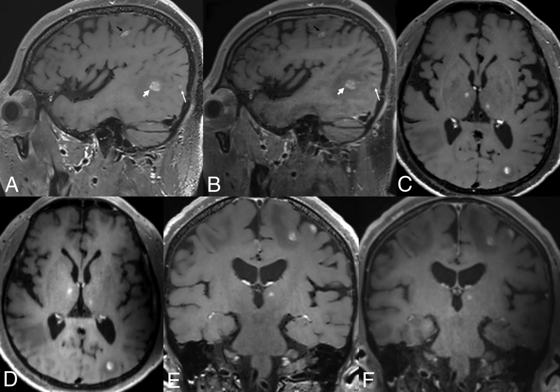
- FIG 1.
Brain MR imaging of a 62-year-old male patient with lung cancer. Postcontrast standard SPACE images (A, C, and E) show multiple variable-sized, enhancing lesions in both cerebral hemispheres. Multiple enhancing lesions are also well-visualized on postcontrast wave-T1-SPACE (B, D, and F), though more noise was present than in standard SPACE.
- FIG 2.
Brain MR imaging of a 68-year-old male patient with lung cancer. Postcontrast standard SPACE (A, C, and E) shows multiple tiny enhancing lesions in both cerebral hemispheres. Although these tiny enhancing lesions are also visible on postcontrast wave-T1-SPACE (B, D, and F), they are somewhat less pronounced (arrows).
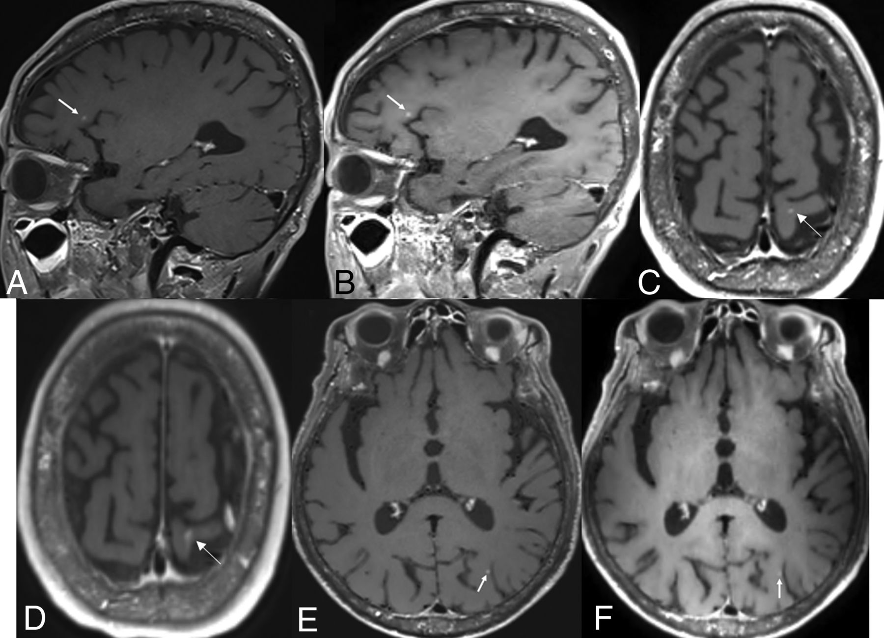
- FIG 3.
Brain MR imaging of a 54-year-old male patient with lung cancer. Postcontrast standard SPACE (A, C, and E) shows a tiny enhancing lesion (arrows) in the right frontal lobe, but it is almost invisible on postcontrast wave-T1-SPACE (B, D, and F).
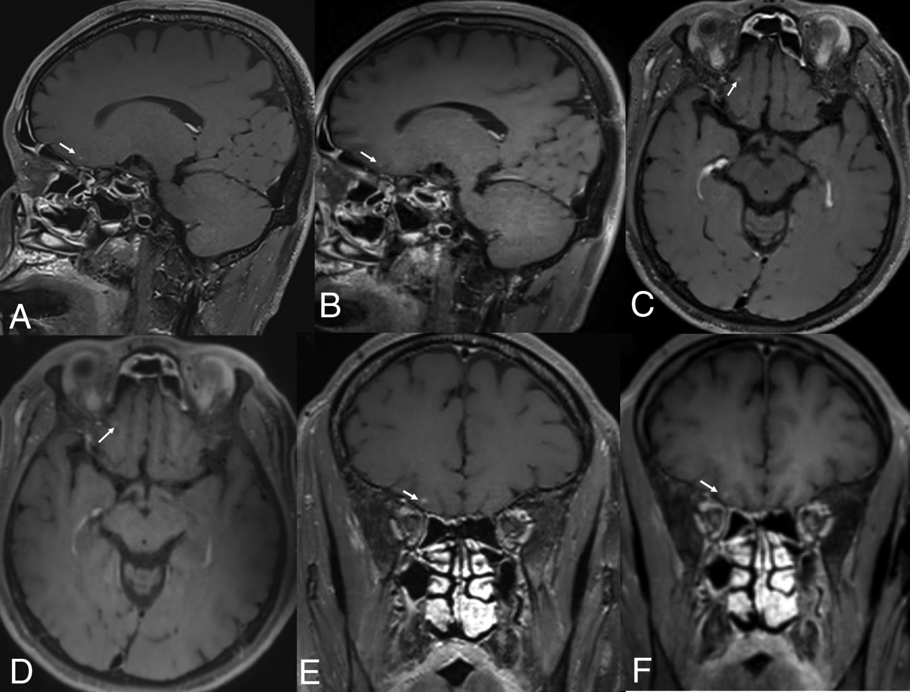
- FIG 4.
Brain MR imaging of a 77-year-old male patient with colorectal cancer. Postcontrast standard (A, C, and E) and wave-T1-SPACE (B, D, and F) show multiple large-sized, enhancing masses in both cerebral hemispheres, and there was no significant difference in the visualization of enhancing lesions, though more noise was observed in postcontrast wave-T1-SPACE.


Tables
Wave-T1-SPACE Standard SPACE FOV (mm) 230 × 230 230 × 230 Matrix size 256 × 256 256 × 256 Section thickness (mm) 0.9 0.9 TR/TE (ms) 700/12 700/28 Flip angle T1 variable T1 variable Echo-train length 60 60 Acceleration factor 4 (phase: 2; section: 2) 2 (phase: 2) No. of slices 200 192 Scan time 2 min 2 seconds 3 min 55 seconds - Table 2:
Comparison of lesion detectability according to the types of MR images and enhancing lesion size
Wave-T1-SPACE Standard SPACE Pa No. of enhancing lesions (<5 mm) (mean) Observer 1 1.61 (SD, 0.29) 2.84 (SD, 0.47) <.001 Observer 2 1.41 (SD, 0.19) 2.68 (SD, 0.43) <.001 Interobserver agreementb 0.72 (0.59–0.84) 0.81 (0.74–0.88) <.001 No. of enhancing lesions (>5 mm) (mean) Observer 1 1.34 (SD, 0.28) 1.32 (SD, 0.27) .66 Observer 2 1.29 (SD, 0.27) 1.29 (SD, 0.27) .26 Interobserver agreementb 0.95 (0.91–1.00) 1.00 (0.99–1.00) <.001 - Table 3:
CNRlesion/parenchyma, CNRwhite matter/gray matter, and CRlesion/parenchyma and image quality of postcontrast wave-T1-SPACE and standard SPACE
Wave-T1-SPACE Standard SPACE P Valuea CNR (n = 21) (mean) Lesion/parenchyma 23.03 (SD, 3.63) 53.74 (SD, 8.15) <.001 White matter/gray matter 2.86 (SD, 0.47) −0.99 (SD, 0.40) <.001 CR, lesion/parenchyma (n = 21) 63.42 (SD, 8.68) 99.75 (SD, 10.88) <.001 Overall image quality (mean) 4.27 (SD, 0.49) 4.98 (SD, 0.13) <.001 Grade V 16 (28.6)b 55 (98.2)b Grade IV 39 (69.6)b 1 (0.18)b Grade III 1 (1.78)b 0 (0)b Grade II 0 (0)b 0 (0)b Grade I 0 (0)b 0 (0)b


