Advertisement

Main menu

  • Home
  • Content
    • Current Issue
    • Accepted Manuscripts
    • Article Preview
    • Past Issue Archive
    • Video Articles
    • AJNR Case Collection
    • Case of the Week Archive
    • Case of the Month Archive
    • Classic Case Archive
  • Special Collections
    • AJNR Awards
    • Low-Field MRI
    • Alzheimer Disease
    • ASNR Foundation Special Collection
    • Photon-Counting CT
    • View All
  • Multimedia
    • AJNR Podcasts
    • AJNR SCANtastic
    • Trainee Corner
    • MRI Safety Corner
    • Imaging Protocols
  • For Authors
    • Submit a Manuscript
    • Submit a Video Article
    • Submit an eLetter to the Editor/Response
    • Manuscript Submission Guidelines
    • Statistical Tips
    • Fast Publishing of Accepted Manuscripts
    • Graphical Abstract Preparation
    • Imaging Protocol Submission
    • Author Policies
  • About Us
    • About AJNR
    • Editorial Board
    • Editorial Board Alumni
  • More
    • Become a Reviewer/Academy of Reviewers
    • Subscribers
    • Permissions
    • Alerts
    • Feedback
    • Advertisers
    • ASNR Home

User menu

  • Alerts
  • Log in

Search

  • Advanced search
American Journal of Neuroradiology

American Journal of Neuroradiology

ASHNR American Society of Functional Neuroradiology ASHNR American Society of Pediatric Neuroradiology ASSR
  • Alerts
  • Log in

Advanced Search

  • Home
  • Content
    • Current Issue
    • Accepted Manuscripts
    • Article Preview
    • Past Issue Archive
    • Video Articles
    • AJNR Case Collection
    • Case of the Week Archive
    • Case of the Month Archive
    • Classic Case Archive
  • Special Collections
    • AJNR Awards
    • Low-Field MRI
    • Alzheimer Disease
    • ASNR Foundation Special Collection
    • Photon-Counting CT
    • View All
  • Multimedia
    • AJNR Podcasts
    • AJNR SCANtastic
    • Trainee Corner
    • MRI Safety Corner
    • Imaging Protocols
  • For Authors
    • Submit a Manuscript
    • Submit a Video Article
    • Submit an eLetter to the Editor/Response
    • Manuscript Submission Guidelines
    • Statistical Tips
    • Fast Publishing of Accepted Manuscripts
    • Graphical Abstract Preparation
    • Imaging Protocol Submission
    • Author Policies
  • About Us
    • About AJNR
    • Editorial Board
    • Editorial Board Alumni
  • More
    • Become a Reviewer/Academy of Reviewers
    • Subscribers
    • Permissions
    • Alerts
    • Feedback
    • Advertisers
    • ASNR Home
  • Follow AJNR on Twitter
  • Visit AJNR on Facebook
  • Follow AJNR on Instagram
  • Join AJNR on LinkedIn
  • RSS Feeds

AJNR Awards, New Junior Editors, and more. Read the latest AJNR updates

Research ArticleHead and Neck Imaging

Phosphaturic Mesenchymal Tumor

J.C. Benson, J.A. Trejo-Lopez, A.M. Nassiri, K. Eschbacher, M.J. Link, C.L. Driscoll, R.D. Tiegs, J. Sfeir and D.R. DeLone
American Journal of Neuroradiology May 2022, DOI: https://doi.org/10.3174/ajnr.A7513
J.C. Benson
aFrom the Departments of Radiology (J.C.B., D.R.D.)
  • Find this author on Google Scholar
  • Find this author on PubMed
  • Search for this author on this site
  • ORCID record for J.C. Benson
J.A. Trejo-Lopez
bLaboratory Medicine and Pathology (J.A.T.-L., K.E.)
  • Find this author on Google Scholar
  • Find this author on PubMed
  • Search for this author on this site
  • ORCID record for J.A. Trejo-Lopez
A.M. Nassiri
cOtorhinolaryngology (A.M.N., C.L.D.)
  • Find this author on Google Scholar
  • Find this author on PubMed
  • Search for this author on this site
  • ORCID record for A.M. Nassiri
K. Eschbacher
bLaboratory Medicine and Pathology (J.A.T.-L., K.E.)
  • Find this author on Google Scholar
  • Find this author on PubMed
  • Search for this author on this site
  • ORCID record for K. Eschbacher
M.J. Link
dNeurosurgery (M.J.L.)
  • Find this author on Google Scholar
  • Find this author on PubMed
  • Search for this author on this site
  • ORCID record for M.J. Link
C.L. Driscoll
cOtorhinolaryngology (A.M.N., C.L.D.)
  • Find this author on Google Scholar
  • Find this author on PubMed
  • Search for this author on this site
  • ORCID record for C.L. Driscoll
R.D. Tiegs
eEndocrinology (R.D.T., J.S.), Mayo Clinic, Rochester, Minnesota
  • Find this author on Google Scholar
  • Find this author on PubMed
  • Search for this author on this site
  • ORCID record for R.D. Tiegs
J. Sfeir
eEndocrinology (R.D.T., J.S.), Mayo Clinic, Rochester, Minnesota
  • Find this author on Google Scholar
  • Find this author on PubMed
  • Search for this author on this site
  • ORCID record for J. Sfeir
D.R. DeLone
aFrom the Departments of Radiology (J.C.B., D.R.D.)
  • Find this author on Google Scholar
  • Find this author on PubMed
  • Search for this author on this site
  • ORCID record for D.R. DeLone
  • Article
  • Figures & Data
  • Info & Metrics
  • Responses
  • References
  • PDF
Loading

Article Figures & Data

Figures

  • Tables
  • FIG 1.
    • Download figure
    • Open in new tab
    • Download powerpoint
    FIG 1.

    68Ga DOTATATE PET/CT demonstrates marked radiotracer uptake within the petrous aspect of the left temporal bone.

  • FIG 2.
    • Download figure
    • Open in new tab
    • Download powerpoint
    FIG 2.

    From superior to inferior, axial (A–C) MR images show an avidly enhancing mass (solid arrows) extending from the middle ear along the floor of the middle cranial fossa, with substantial intraosseous involvement. A small portion of the mass (dashed straight arrow) closely approximates the geniculate ganglion of the facial nerve (solid curved arrow) (circles in B and C denote the external auditory canal). The mastoid air cells are completely opacified (asterisks). Coronal image (D) shows the mass extending intracranially, with associated dural thickening (dashed curved arrow).

  • FIG 3.
    • Download figure
    • Open in new tab
    • Download powerpoint
    FIG 3.

    Noncontrast temporal bone CT performed 2 days after MR imaging again shows the mass (solid straight arrows) extending from the mesotympanum (A) into the anterior hypotympanum (B), which is widened. The mass is locally destructive (C), causing erosion of the skull base with involvement of the foramen spinosum (dashed straight arrow) and left temporomandibular joint (curved arrow). The margins of the mass are indistinguishable from fluid density within the middle ear and mastoid air cells (asterisks), which remained opacified.
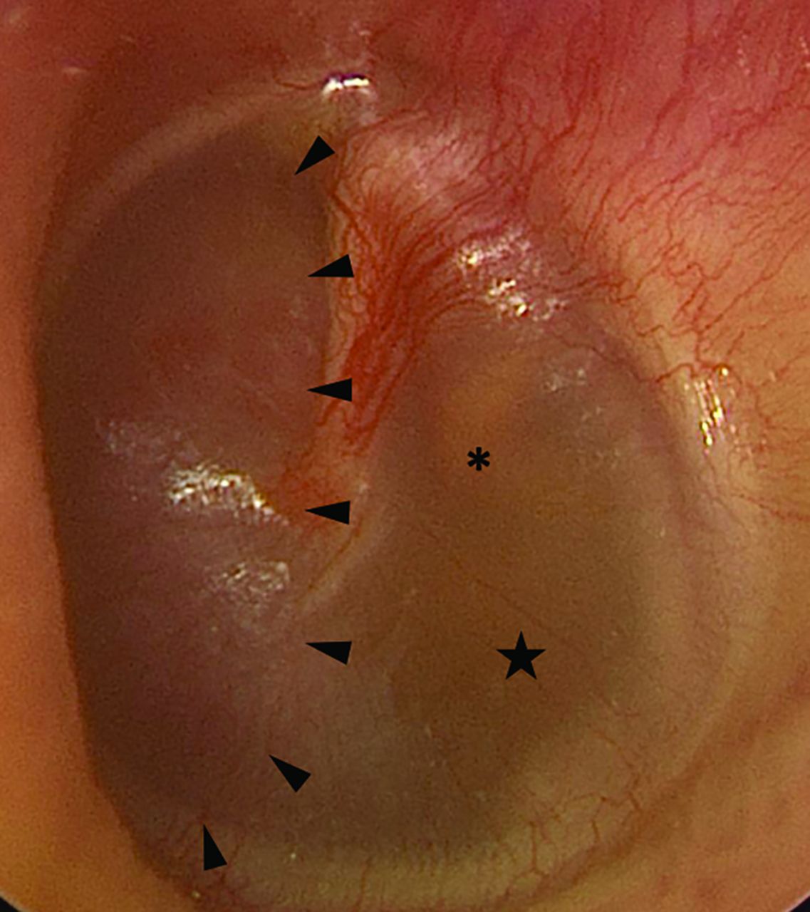
  • FIG 4.
    • Download figure
    • Open in new tab
    • Download powerpoint
    FIG 4.

    Preoperative otoscopy of the left tympanic membrane reveals a reddish mass (arrowheads) within the anterior mesotympanum. An amber effusion (star) is seen posterior to the mass, surrounding the incudostapedial joint (asterisk).
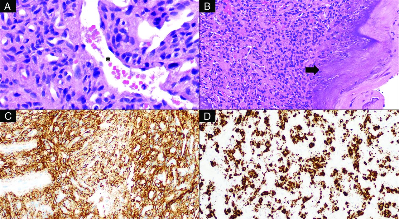
  • FIG 5.
    • Download figure
    • Open in new tab
    • Download powerpoint
    FIG 5.

    Histologic examination of this tumor reveals bland, spindled-to-stellate neoplastic cells (A, H&E, original magnification ×400) situated in a hyalinized matrix with a well-developed capillary network including ectatic, staghorn vessels (asterisk) as well as characteristic deposition of coarse calcification (B, H&E, original magnification ×200; arrow). The tumor is reactive for SSTR2A (C, immunohistochemistry, original magnification ×200) and shows evidence of FGF23 mRNA expression (D, chromogenic in situ hybridization, original magnification ×200).

Tables

  • Figures
  • Patient laboratory testing on presentation

    LevelsResultsReference Range
    Total serum calcium9.08.8–10.2 mg/dL
    Albumin4.43.5–5.0 g/dL
    Phosphorus1.52.5–4.5 mg/dL
    Magnesium2.11.7–2.3 mg/dL
    Creatinine0.970.74–1.35 mg/dL
    Alkaline phosphatase10235–104 U/L
    Parathyroid hormone6415–65 pg/mL
    25 (hydroxyvitamin) Vitamin D8330–50 ng/mL
    1, 25 (hydroxyvitamin)2 Vitamin D2918–64 pg/mL
    Intact FGF2382≤59 pg/mL
    Tubular reabsorption of phosphate62%>85%
PreviousNext
Back to top
Advertisement
Print
Download PDF
Email Article

Thank you for your interest in spreading the word on American Journal of Neuroradiology.

NOTE: We only request your email address so that the person you are recommending the page to knows that you wanted them to see it, and that it is not junk mail. We do not capture any email address.

Enter multiple addresses on separate lines or separate them with commas.
Phosphaturic Mesenchymal Tumor
(Your Name) has sent you a message from American Journal of Neuroradiology
(Your Name) thought you would like to see the American Journal of Neuroradiology web site.
CAPTCHA
This question is for testing whether or not you are a human visitor and to prevent automated spam submissions.
Cite this article
J.C. Benson, J.A. Trejo-Lopez, A.M. Nassiri, K. Eschbacher, M.J. Link, C.L. Driscoll, R.D. Tiegs, J. Sfeir, D.R. DeLone
Phosphaturic Mesenchymal Tumor
American Journal of Neuroradiology May 2022, DOI: 10.3174/ajnr.A7513

Citation Manager Formats

  • BibTeX
  • Bookends
  • EasyBib
  • EndNote (tagged)
  • EndNote 8 (xml)
  • Medlars
  • Mendeley
  • Papers
  • RefWorks Tagged
  • Ref Manager
  • RIS
  • Zotero
0 Responses
Respond to this article
Share
Bookmark this article
Phosphaturic Mesenchymal Tumor
J.C. Benson, J.A. Trejo-Lopez, A.M. Nassiri, K. Eschbacher, M.J. Link, C.L. Driscoll, R.D. Tiegs, J. Sfeir, D.R. DeLone
American Journal of Neuroradiology May 2022, DOI: 10.3174/ajnr.A7513
del.icio.us logo Twitter logo Facebook logo Mendeley logo
  • Tweet Widget
  • Facebook Like
  • Google Plus One
Purchase

Jump to section

  • Article
    • Abstract
    • ABBREVIATIONS:
    • DISCUSSION
    • Footnotes
    • References
  • Figures & Data
  • Info & Metrics
  • Responses
  • References
  • PDF

Related Articles

  • No related articles found.
  • PubMed
  • Google Scholar

Cited By...

  • No citing articles found.
  • Crossref (17)
  • Google Scholar

This article has been cited by the following articles in journals that are participating in Crossref Cited-by Linking.

  • Phosphaturic Mesenchymal Tumors: Rethinking the Clinical Diagnosis and Surgical Treatment
    Yupeng Liu, Hongbo He, Can Zhang, Hao Zeng, Xiaopeng Tong, Qing Liu
    Journal of Clinical Medicine 2022 12 1
  • Tumor‐induced Osteomalacia: A Case Report and Etiological Analysis with Literature Review
    Zhenhao Zhang, Jiaxin Li, Zhicai Zhang, Zengwu Shao
    Orthopaedic Surgery 2023 15 12
  • Intracranial Phosphaturic Mesenchymal Tumor Detected by 68Ga-DOTATATE PET/CT
    Qinwen Shi, Junjun Cheng, Yue Zhang, Minggang Su
    Clinical Nuclear Medicine 2024 49 5
  • Lung Nodule as Culprit Lesion Causing Recurrent Tumor-Induced Osteomalacia Revealed by 68Ga-DOTATATE PET/CT
    Yuwei Zhang, Peipei Wang, Hongli Jing
    Clinical Nuclear Medicine 2023 48 9
  • Phosphaturic mesenchymal tumor: management and outcomes of ten patients treated at a single institution
    Marcos R. Gonzalez, Neel Patel, Joseph J. Connolly, Yin P. Hung, Connie Y. Chang, Santiago A. Lozano-Calderon
    Skeletal Radiology 2024 53 8
  • Tumor-induced Osteomalacia Secondary to Phosphaturic Mesenchymal Tumor
    Amit Agarwal, Girish Bathla, Vivek Gupta
    Radiology: Imaging Cancer 2023 5 2
  • A Rare Case of Skull Base Phosphaturic Mesenchymal Tumor
    Mirza Athar Ali, Muntimadugu Babaiah, Prabhakar Mariappan, Saadvik Raghuram, Krishna Reddy Thaduri, Muthulingam Shunmugavel, Soundarya YSB, Oviya Manohar, Surya Simhareddy Molakala
    Applied Radiation Oncology 2024 13 3
  • Advanced tumour-induced osteomalacia secondary to sinonasal phosphaturic mesenchymal tumour
    Monica Van Wijk, Leon Janse van Rensburg, Bianca D. Berndorfler, Johan F. Opperman, Johan Grobbelaar, Amir H. Afrogheh, Sarah Versveld, Razaan Davis
    South African Journal of Radiology 2024 28 1
  • CT and MRI features of phosphaturic mesenchymal tumor
    S. A. Buryakina, S. A. Gronskaia, N. V. Tarbaeva, Zh. E. Belaya, N. G. Mokrysheva
    Medical Visualization 2025 29 2
  • Case Report: Phosphaturic mesenchymal tumor presenting solely as knee pain without hypophosphatemia
    Qianqian Cao, Cheng Cheng, Zhipeng Dai
    Frontiers in Oncology 2025 15

More in this TOC Section

  • ASL Sensitivity for Head and Neck Paraganglioma
  • Post SRS Peritumoral Hyperintense Signal of VSs
  • Contrast Enhanced Pituitary CISS/FIESTA
Show more Head and Neck Imaging

Similar Articles

Advertisement

Indexed Content

  • Current Issue
  • Accepted Manuscripts
  • Article Preview
  • Past Issues
  • Editorials
  • Editor's Choice
  • Fellows' Journal Club
  • Letters to the Editor
  • Video Articles

Cases

  • Case Collection
  • Archive - Case of the Week
  • Archive - Case of the Month
  • Archive - Classic Case

More from AJNR

  • Trainee Corner
  • Imaging Protocols
  • MRI Safety Corner
  • Book Reviews

Multimedia

  • AJNR Podcasts
  • AJNR Scantastics

Resources

  • Turnaround Time
  • Submit a Manuscript
  • Submit a Video Article
  • Submit an eLetter to the Editor/Response
  • Manuscript Submission Guidelines
  • Statistical Tips
  • Fast Publishing of Accepted Manuscripts
  • Graphical Abstract Preparation
  • Imaging Protocol Submission
  • Evidence-Based Medicine Level Guide
  • Publishing Checklists
  • Author Policies
  • Become a Reviewer/Academy of Reviewers
  • News and Updates

About Us

  • About AJNR
  • Editorial Board
  • Editorial Board Alumni
  • Alerts
  • Permissions
  • Not an AJNR Subscriber? Join Now
  • Advertise with Us
  • Librarian Resources
  • Feedback
  • Terms and Conditions
  • AJNR Editorial Board Alumni

American Society of Neuroradiology

  • Not an ASNR Member? Join Now

© 2025 by the American Society of Neuroradiology All rights, including for text and data mining, AI training, and similar technologies, are reserved.
Print ISSN: 0195-6108 Online ISSN: 1936-959X

Powered by HighWire