Advertisement

Main menu

  • Home
  • Content
    • Current Issue
    • Accepted Manuscripts
    • Article Preview
    • Past Issue Archive
    • Video Articles
    • AJNR Case Collection
    • Case of the Week Archive
    • Case of the Month Archive
    • Classic Case Archive
  • Special Collections
    • AJNR Awards
    • Low-Field MRI
    • Alzheimer Disease
    • ASNR Foundation Special Collection
    • Photon-Counting CT
    • View All
  • Multimedia
    • AJNR Podcasts
    • AJNR SCANtastic
    • Trainee Corner
    • MRI Safety Corner
    • Imaging Protocols
  • For Authors
    • Submit a Manuscript
    • Submit a Video Article
    • Submit an eLetter to the Editor/Response
    • Manuscript Submission Guidelines
    • Statistical Tips
    • Fast Publishing of Accepted Manuscripts
    • Graphical Abstract Preparation
    • Imaging Protocol Submission
    • Author Policies
  • About Us
    • About AJNR
    • Editorial Board
    • Editorial Board Alumni
  • More
    • Become a Reviewer/Academy of Reviewers
    • Subscribers
    • Permissions
    • Alerts
    • Feedback
    • Advertisers
    • ASNR Home

User menu

  • Alerts
  • Log in

Search

  • Advanced search
American Journal of Neuroradiology

American Journal of Neuroradiology

ASHNR American Society of Functional Neuroradiology ASHNR American Society of Pediatric Neuroradiology ASSR
  • Alerts
  • Log in

Advanced Search

  • Home
  • Content
    • Current Issue
    • Accepted Manuscripts
    • Article Preview
    • Past Issue Archive
    • Video Articles
    • AJNR Case Collection
    • Case of the Week Archive
    • Case of the Month Archive
    • Classic Case Archive
  • Special Collections
    • AJNR Awards
    • Low-Field MRI
    • Alzheimer Disease
    • ASNR Foundation Special Collection
    • Photon-Counting CT
    • View All
  • Multimedia
    • AJNR Podcasts
    • AJNR SCANtastic
    • Trainee Corner
    • MRI Safety Corner
    • Imaging Protocols
  • For Authors
    • Submit a Manuscript
    • Submit a Video Article
    • Submit an eLetter to the Editor/Response
    • Manuscript Submission Guidelines
    • Statistical Tips
    • Fast Publishing of Accepted Manuscripts
    • Graphical Abstract Preparation
    • Imaging Protocol Submission
    • Author Policies
  • About Us
    • About AJNR
    • Editorial Board
    • Editorial Board Alumni
  • More
    • Become a Reviewer/Academy of Reviewers
    • Subscribers
    • Permissions
    • Alerts
    • Feedback
    • Advertisers
    • ASNR Home
  • Follow AJNR on Twitter
  • Visit AJNR on Facebook
  • Follow AJNR on Instagram
  • Join AJNR on LinkedIn
  • RSS Feeds

AJNR Awards, New Junior Editors, and more. Read the latest AJNR updates

Research ArticleHead and Neck Imaging

Phosphaturic Mesenchymal Tumor

J.C. Benson, J.A. Trejo-Lopez, A.M. Nassiri, K. Eschbacher, M.J. Link, C.L. Driscoll, R.D. Tiegs, J. Sfeir and D.R. DeLone
American Journal of Neuroradiology May 2022, DOI: https://doi.org/10.3174/ajnr.A7513
J.C. Benson
aFrom the Departments of Radiology (J.C.B., D.R.D.)
  • Find this author on Google Scholar
  • Find this author on PubMed
  • Search for this author on this site
  • ORCID record for J.C. Benson
J.A. Trejo-Lopez
bLaboratory Medicine and Pathology (J.A.T.-L., K.E.)
  • Find this author on Google Scholar
  • Find this author on PubMed
  • Search for this author on this site
  • ORCID record for J.A. Trejo-Lopez
A.M. Nassiri
cOtorhinolaryngology (A.M.N., C.L.D.)
  • Find this author on Google Scholar
  • Find this author on PubMed
  • Search for this author on this site
  • ORCID record for A.M. Nassiri
K. Eschbacher
bLaboratory Medicine and Pathology (J.A.T.-L., K.E.)
  • Find this author on Google Scholar
  • Find this author on PubMed
  • Search for this author on this site
  • ORCID record for K. Eschbacher
M.J. Link
dNeurosurgery (M.J.L.)
  • Find this author on Google Scholar
  • Find this author on PubMed
  • Search for this author on this site
  • ORCID record for M.J. Link
C.L. Driscoll
cOtorhinolaryngology (A.M.N., C.L.D.)
  • Find this author on Google Scholar
  • Find this author on PubMed
  • Search for this author on this site
  • ORCID record for C.L. Driscoll
R.D. Tiegs
eEndocrinology (R.D.T., J.S.), Mayo Clinic, Rochester, Minnesota
  • Find this author on Google Scholar
  • Find this author on PubMed
  • Search for this author on this site
  • ORCID record for R.D. Tiegs
J. Sfeir
eEndocrinology (R.D.T., J.S.), Mayo Clinic, Rochester, Minnesota
  • Find this author on Google Scholar
  • Find this author on PubMed
  • Search for this author on this site
  • ORCID record for J. Sfeir
D.R. DeLone
aFrom the Departments of Radiology (J.C.B., D.R.D.)
  • Find this author on Google Scholar
  • Find this author on PubMed
  • Search for this author on this site
  • ORCID record for D.R. DeLone
  • Article
  • Figures & Data
  • Info & Metrics
  • Responses
  • References
  • PDF
Loading

Abstract

Phosphaturic mesenchymal tumors (PMTs) are neoplasms associated with tumor-induced osteomalacia. Patients typically present with pathologic fractures in the setting of chronic hypophosphatemic hyperphosphaturic osteomalacia, as well as gradual muscle weakness, bone pain, and difficulty walking. Because of their rarity and nonspecific symptomatology, phosphaturic mesenchymal tumors often go undiagnosed for years. Even when discovered on imaging, the tumors can be diagnostically challenging for radiologists. Phosphaturic mesenchymal tumors often tend to be small and can be located nearly anywhere in the body, and, therefore, can mimic many other tumors. This case highlights the imaging and pathologic markers of a phosphaturic mesenchymal tumor, often found in a patient with tumor-induced osteomalacia.

ABBREVIATIONS:

FGF23
fibroblast growth factor 23
PMT
phosphaturic mesenchymal tumor
TIO
tumor-induced osteomalacia

The patient is a 57-year-old man who presented to our institution for evaluation with a 4-year history of recurrent insufficiency fractures involving his femurs and ribs, associated with considerable disability. His initial symptom was right-hip pain that failed to resolve with conservative treatment. An MR imaging at that time showed a left subtrochanteric insufficiency fracture. Two years later, after developing pain in both thighs, the patient was diagnosed with insufficiency fractures involving the subtrochanteric regions of both proximal femurs. At the time of presentation, a review of his laboratory records revealed chronic hypophosphatemia, with serum phosphorus levels ranging between 1.7 and 2.1 mg/dL during the preceding 4 years. His serum calcium and parathyroid hormone levels have been consistently within the normal range.

Given his history and laboratory abnormalities, the diagnosis of tumor-induced osteomalacia (TIO) was entertained. Confirmatory testing (Table) demonstrated hypophosphatemia due to renal phosphate wasting, as well as elevated fibroblast growth factor 23 (FGF23).

View this table:
  • View inline
  • View popup

Patient laboratory testing on presentation

The patient did not have skeletal deformities or a family history of bone disorders, and his bone density readings were in the osteopenia range. He then underwent a PET gallium (68Ga) DOTATATE study to examine for a mesenchymal tumor, which is the usual source of the excess FGF23 production.

Imaging

The 68Ga DOTATATE examination demonstrated a focal lesion in the petrous aspect of the left temporal bone (Fig 1), considered highly suspicious for the patient’s culprit lesion. Dedicated imaging of the temporal bone was therefore performed.

FIG 1.
  • Download figure
  • Open in new tab
  • Download powerpoint
FIG 1.

68Ga DOTATATE PET/CT demonstrates marked radiotracer uptake within the petrous aspect of the left temporal bone.

On MR imaging, an avidly enhancing lesion was seen along the cochlear promontory in the middle ear, extending into the anterior hypotympanum along the course of the Eustachian tube (Fig 2). Anteriorly, the mass involved the skull base, encroaching on the foramen spinosum. Posteriorly, the mass closely approximated the geniculate ganglion of the facial nerve. When read in conjunction with the 68Ga DOTATATE examination, this was initially thought to represent a glomus tympanicum tumor with unusual anterior growth. A glomus faciale tumor growing from the geniculate ganglion, phosphaturic mesenchymal tumor (PMT), primary neuroendocrine carcinoma, or metastasis were also considered.

FIG 2.
  • Download figure
  • Open in new tab
  • Download powerpoint
FIG 2.

From superior to inferior, axial (A–C) MR images show an avidly enhancing mass (solid arrows) extending from the middle ear along the floor of the middle cranial fossa, with substantial intraosseous involvement. A small portion of the mass (dashed straight arrow) closely approximates the geniculate ganglion of the facial nerve (solid curved arrow) (circles in B and C denote the external auditory canal). The mastoid air cells are completely opacified (asterisks). Coronal image (D) shows the mass extending intracranially, with associated dural thickening (dashed curved arrow).

On a subsequent CT, the tumoral margins were not well-demarcated because the density of the mass matched that of the fluid within the adjacent middle ear and mastoid air cells. However, the CT scans did reveal the mass to be permeative with destruction of the adjacent skull base (Fig 3). In retrospect, the patient did note a long-standing history of left-sided hearing loss and ear fullness without drainage.

FIG 3.
  • Download figure
  • Open in new tab
  • Download powerpoint
FIG 3.

Noncontrast temporal bone CT performed 2 days after MR imaging again shows the mass (solid straight arrows) extending from the mesotympanum (A) into the anterior hypotympanum (B), which is widened. The mass is locally destructive (C), causing erosion of the skull base with involvement of the foramen spinosum (dashed straight arrow) and left temporomandibular joint (curved arrow). The margins of the mass are indistinguishable from fluid density within the middle ear and mastoid air cells (asterisks), which remained opacified.

Operative Report

On preoperative otoscopy, the mass was easily visible within the middle ear and less intensely red than a typical paraganglioma (which is typically deep red) (Fig 4). Considering the imaging findings and clinical history, the likely diagnosis was PMT, and surgical treatment was recommended. Tumoral resection was accomplished via a left middle cranial fossa craniotomy. Intraoperatively, the tumor was noted to have eroded through the middle fossa floor just posterior and lateral to foramen spinosum. No involvement of the subtemporal dura was noted intraoperatively, though this may have been due to the dural thickening noted on imaging being reactive in nature rather than tumoral extension. It appeared to originate at the anterior aspect of the Eustachian tube and extended along the Eustachian tube inferiorly into the middle ear. The tumor was quite vascular and required piecemeal excision with frequent use of hemostatic agents and bipolar cautery during resection. Angled instruments and otologic endoscopes were used to release the inferior extent of the tumor from the middle ear. Ultimately, gross total resection was achieved. The patient had an uneventful postoperative course without signs of CSF leak or cranial nerve deficits.

FIG 4.
  • Download figure
  • Open in new tab
  • Download powerpoint
FIG 4.

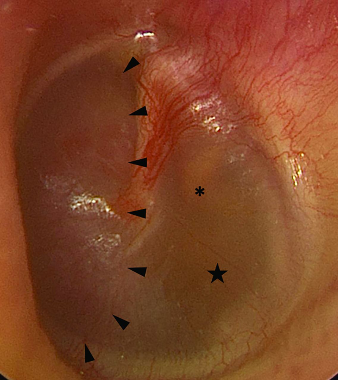
Preoperative otoscopy of the left tympanic membrane reveals a reddish mass (arrowheads) within the anterior mesotympanum. An amber effusion (star) is seen posterior to the mass, surrounding the incudostapedial joint (asterisk).

Pathology

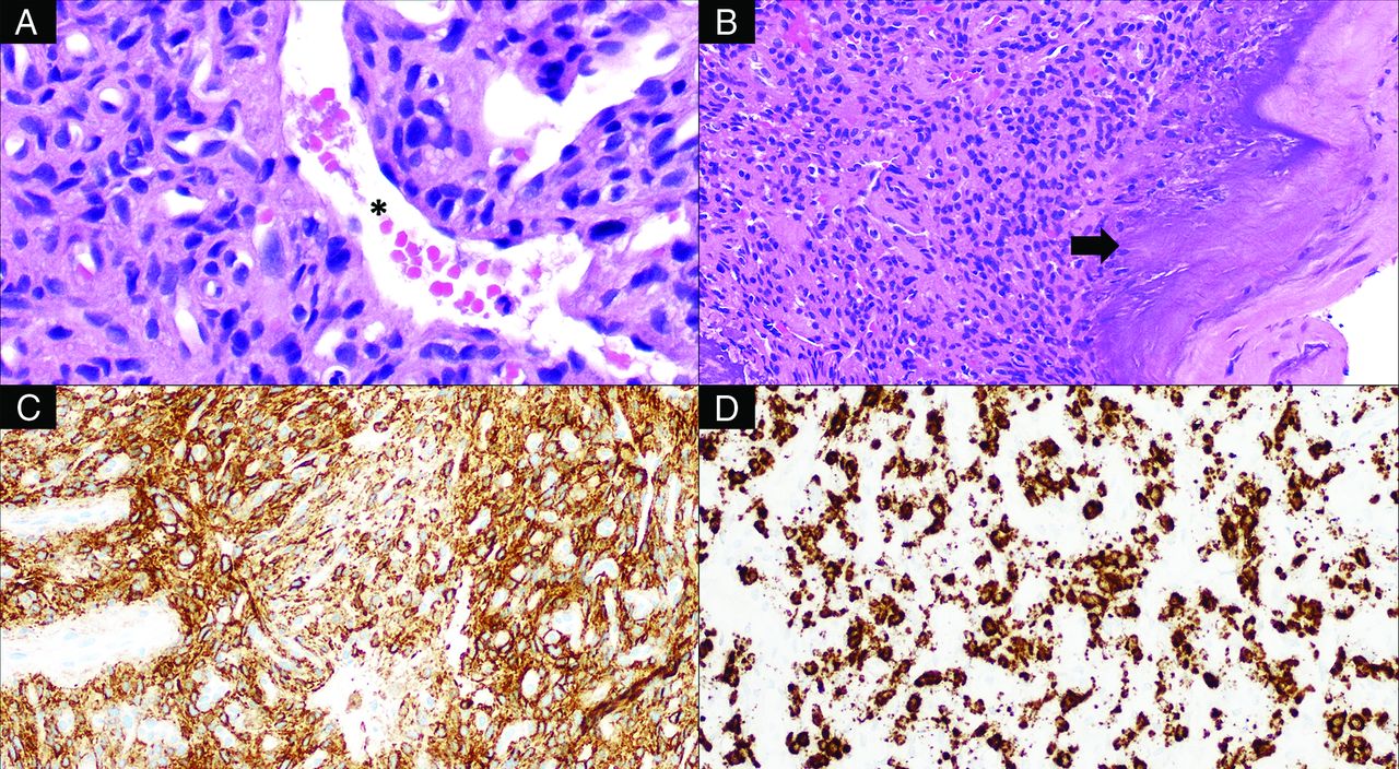
Histologic examination of the tumor revealed a highly vascular neoplasm composed of predominantly spindled tumor cells with bland, ovoid nuclei with interspersed staghorn-like vessels (Fig 5). There were scattered coarse, flocculent calcifications. Neoplastic cells showed overall monotonous nuclei, with low proliferative activity and no tumor necrosis. Immunohistochemical studies revealed tumor cells to be diffusely positive for SSTR2A, while negative for chromogranin and synaptophysin. In addition, there was widespread expression by the tumor cells of FGF23 mRNA by chromogenic in situ hybridization. Overall, these findings supported the final diagnosis of a morphologically benign PMT.

FIG 5.
  • Download figure
  • Open in new tab
  • Download powerpoint
FIG 5.

Histologic examination of this tumor reveals bland, spindled-to-stellate neoplastic cells (A, H&E, original magnification ×400) situated in a hyalinized matrix with a well-developed capillary network including ectatic, staghorn vessels (asterisk) as well as characteristic deposition of coarse calcification (B, H&E, original magnification ×200; arrow). The tumor is reactive for SSTR2A (C, immunohistochemistry, original magnification ×200) and shows evidence of FGF23 mRNA expression (D, chromogenic in situ hybridization, original magnification ×200).

Histologically, PMTs are highly vascular and composed of bland, spindled-to-stellate cells in a distinctive myxoid or myxochondroid matrix with flocculent calcification.1⇓-3 FN1-FGFR1 or FN1-FGF1 fusions are common in PMTs (up to approximately 50%), which may activate the FGFR1 pathway leading to increased expression of the FGF23 gene and protein.1⇓-3 FGF23 expression can be detected by reverse-transcription polymerase chain reaction, chromogenic in situ hybridization, or immunohistochemistry.1,2

The histologic differential diagnosis of this tumor includes solitary fibrous tumor, mesenchymal chondrosarcoma, and paraganglioma. Solitary fibrous tumors can have staghorn vessels and have a similar, bland, spindle cell appearance. However, solitary fibrous tumors lack the characteristic matrix and calcifications of PMTs and would not express SSTR2A or FGF23 mRNA.2,3 Mesenchymal chondrosarcomas have malignant, small, round cells; lack the characteristic calcified matrix of a PMT; and would not express FGF23 mRNA. A highly vascular paraganglioma may, at times, be morphologically reminiscent of PMT. However, the immunophenotype of PMT (positive for SSTR2A and FGF23 mRNA, negative for chromogranin and synaptophysin) excluded this possibility. Furthermore, although paragangliomas and PMTs can both exhibit high vascularity, paragangliomas lack the characteristic chondromyxoid matrix and calcifications of PMT. Unlike PMTs, paragangliomas have a characteristic nested (zellballen) architecture composed of neuroendocrine cells and are positive for neuroendocrine markers (eg, synaptophysin and chromogranin). The tumor presented here did not have the classic morphology of paraganglioma and was negative for synaptophysin and chromogranin.

DISCUSSION

PMTs are exceedingly rare tumors that arise primarily from bones and connective tissue. They are typically seen in middle-aged adults without an apparent sex predilection. Pediatric cases have been infrequently reported.4 The median age at the time of diagnosis is 44–48 years of age.5,6 Tumor locality is unpredictable; PMTs can be found in any osseous or soft-tissue location and uncommonly involve the skin.7 More than half of the tumors are found within the extremities, with the femur being the most common site.8 Within the head and neck, the sinonasal region is most common, followed by the mandible and maxilla. Although histologically benign, rare cases of malignant transformation and metastases have been reported.8⇓-10

Clinically, PMTs are associated with tumor-induced osteomalacia (TIO), also known as oncogenic osteomalacia, a paraneoplastic syndrome characterized by widespread reduction of osteoblastic activity. PMTs secrete FGF23, a peptide hormone-like regulator of phosphate levels, which decreases re-absorption of phosphate in the proximal tubules of the kidneys, causing it to be wasted within the urine. The downstream suppression of osteoblasts and mobilization of calcium and phosphate from the bones lead to widespread hypophosphatemic osteomalacia.11 Patients present with bone pain, insufficiency fractures, and gradual-onset muscular weakness.12

A rare subtype of PMT has recently been identified as being nonphosphaturic.13 The descriptions of these tumors are heterogeneous. Some might produce negligible amounts of FGF23 or are thought to exist in patients with a compensatory mechanism in place.3 Some are thought to produce a deformed and/or inactive form of the FGF23 protein.14 Other tumors may simply have been identified in patients before symptomatic osteomalacia had developed.15

Unfortunately, the rarity of PMTs makes them notoriously under-recognized by clinicians. Many patients are symptomatic for years or decades before the tumor is diagnosed.16,17 The diagnosis typically is based on a combination of clinical history, laboratory values, and imaging. Specifically, elevated serum FGF23 levels in a patient with hypophosphatemia and normal renal function is highly suggestive of a PMT. However, TIO has a known association with multiple other tumors, typically of mesenchymal origin, including solitary fibrous tumors, giant cell tumors, chondrosarcomas, and osteosarcomas.18,19 Most, but not all, of these tumors result in TIO through the secretion of FGF23; some secrete various other factors such as frizzled-related protein-4, FGF7, and matrix extracellular phosphoglycoprotein.20

Once the diagnosis of TIO has been established, various nuclear medicine studies can be used to search for the culprit lesion, including 68Ga DOTATATE, technetium Tc99m-octreotide, and FDG PET/CT scans.21 68Ga DOTATATE is now the favored technique, having been shown to have the greatest sensitivity for tumoral detection.15, 22⇓-24 This superiority of 68Ga DOTATATE over other modalities is thought to be due to the higher affinity of its radiotracer to somatostatin receptors 2 and 5, which are expressed by PMTs.23 Nevertheless, the sensitivity of these examinations is suboptimal. A series by El-Maouche et al23 found that 68Ga DOTATATE examinations found a PMT in just more than half of patients with hypophosphatemic TIO, while indium-111 (111In) pentetreotide (OctreoScan) SPECT/CT and [18F] FDG identified <50%.

PMTs are not encapsulated and tend to be locally infiltrative.12 On CT, intraosseous PMTs are osteolytic with a narrow zone of transition and internal matrix; soft-tissue masses are hypodense and enhance.25 On MR imaging, tumors are isointense to soft tissues on T1-weighted imaging and demonstrate enhancement.26 Intralesional T2 signal is variable. Most commonly, tumors are T2-hyperintense, though Broski et al22 noted that PMTs characteristically have small foci of intralesional T2 hypointensity. Some may be partially cystic. Larger tumors may have intralesional flow voids.27 However, most PMTs are small and slow-growing, and tumor localization is highly variable. Thus, they may present a diagnostic conundrum for radiologists, particularly when the diagnosis of TIO is unknown, or, as in this case, if the PMT mimics the appearance of a more common entity.26

The only known definitive treatment for TIO is surgical resection of the tumor. Fortunately, removal of PMTs results in complete resolution of the biochemical and physical sequelae of the paraneoplastic syndrome.7 Because PMTs are often locally invasive, a wide excision is needed to successfully obtain tumor-free margins.28 Although local recurrence may occur, surgical resection is considered curative in approximately 90% of cases.27,29 Cryoablation has been successfully used to treat residual tumor after resection.30 For patients with unresectable tumors, a fully human monoclonal antibody against FGF23, burosumab, has been shown to provide biochemical and symptomatic improvement and was recently FDA-approved.31

Difficult cases are excellent learning opportunities, and this case is no exception. Despite the difficulty of the resection, the surgeons indicated that the available preoperative imaging was excellent, and they stated that additional imaging would not have changed the surgical approach. In retrospect, the differential considerations given in the initial radiology report could have been more precise. The case was challenging due to its location in an anatomic region that can support the growth of paragangliomas. Nevertheless, the clinical history of TIO should have served as a more convincing indicator that the tumor identified on imaging represented a PMT. Incorrectly favoring a paraganglioma over a PMT could have delayed or even prevented surgery because the purpose of the examination was to search for a lesion responsible for the patient’s TIO. Fortunately, the patient ultimately underwent the best treatment course. He is expected to make a full recovery and will undergo routine surveillance imaging to monitor for recurrence.

Case Summary

  • PMTs are extremely rare entities that are associated with TIO.

  • The tumors are small and can be found nearly anywhere in the body, making them a common mimic of more common malignancies.

  • Surgical resection of the tumor results in resolution of a patient’s osteomalacia.

  • Histologically, PMTs have an unusual, hyalinized matrix that undergoes calcification with frequent expression of FGF23 mRNA.

Footnotes

  • Disclosure forms provided by the authors are available with the full text and PDF of this article at www.ajnr.org.

References

  1. 1.↵
    1. Lee J,
    2. Folpe AL
    . Phosphaturic mesenchymal tumor. In: Editorial Board. WHO Classification of Tumours: Soft Tissue and Bone Tumours. International Agency for Research on Cancer; 2020
  2. 2.↵
    1. Bahrami A,
    2. Weiss SW,
    3. Montgomery E, et al
    . RT-PCR analysis for FGF23 using paraffin sections in the diagnosis of phosphaturic mesenchymal tumors with and without known tumor-induced osteomalacia. Am J Surg Pathol 2009;33:1348–54 doi:10.1097/PAS.0b013e3181aa2311 pmid:19609206
    CrossRefPubMed
  3. 3.↵
    1. Folpe AL,
    2. Fanburg-Smith JC,
    3. Billings SD, et al
    . Most osteomalacia-associated mesenchymal tumors are a single histopathologic entity: an analysis of 32 cases and a comprehensive review of the literature. Am J Surg Pathol 2004;28:1–30 doi:10.1097/00000478-200401000-00001 pmid:14707860
    CrossRefPubMed
  4. 4.↵
    1. Kumar S,
    2. Shah R,
    3. Patil V, et al
    . Tumor-induced rickets-osteomalacia: an enigma. J Pediatr Endocrinol Metab 2020 July 20. [Epub ahead of print] doi:10.1515/jpem-2020-0079 pmid:32681779
    CrossRefPubMed
  5. 5.↵
    1. Honda R,
    2. Kawabata Y,
    3. Ito S, et al
    . Phosphaturic mesenchymal tumor, mixed connective tissue type, non-phosphaturic variant: report of a case and review of 32 cases from the Japanese published work. J Dermatol 2014;41:845–49 doi:10.1111/1346-8138.12602 pmid:25182295
    CrossRefPubMed
  6. 6.↵
    1. Wu H,
    2. Bui MM,
    3. Zhou L, et al
    . Phosphaturic mesenchymal tumor with an admixture of epithelial and mesenchymal elements in the jaws: clinicopathological and immunohistochemical analysis of 22 cases with literature review. Mod Pathol 2019;32:189–204 doi:10.1038/s41379-018-0100-0 pmid:30206408
    CrossRefPubMed
  7. 7.↵
    1. Hodgson SF,
    2. Clarke BL,
    3. Tebben PJ, et al
    . Oncogenic osteomalacia: localization of underlying peripheral mesenchymal tumors with use of Tc 99m sestamibi scintigraphy. Endocr Pract 2006;12:35–42 doi:10.4158/EP.12.1.35 pmid:16524861
    CrossRefPubMed
  8. 8.↵
    1. Oyama N,
    2. Kojima-Ishii K,
    3. Toda N, et al
    . Malignant transformation of phosphaturic mesenchymal tumor: a case report and literature review. Clin Pediatr Endocrinol 2020;29:69–75 doi:10.1297/cpe.29.69 pmid:32313375
    CrossRefPubMed
  9. 9.↵
    1. Morimoto T,
    2. Takenaka S,
    3. Hashimoto N, et al
    . Malignant phosphaturic mesenchymal tumor of the pelvis: a report of two cases. Oncol Lett 2014;8:67–71 doi:10.3892/ol.2014.2081 pmid:24959220
    CrossRefPubMed
  10. 10.↵
    1. Qiu S,
    2. Cao LL,
    3. Qiu Y, et al
    . Malignant phosphaturic mesenchymal tumor with pulmonary metastasis: a case report. Medicine (Baltimore) 2017;96:e6750 doi:10.1097/MD.0000000000006750 pmid:28445300
    CrossRefPubMed
  11. 11.↵
    1. Ghorbani-Aghbolaghi A,
    2. Darrow MA,
    3. Wang T
    . Phosphaturic mesenchymal tumor (PMT): exceptionally rare disease, yet crucial not to miss. Autops Case Rep 2017;7:32–37 doi:10.4322/acr.2017.031 pmid:28445300
    CrossRefPubMed
  12. 12.↵
    1. Pelo S,
    2. Gasparini G,
    3. Garagiola U, et al
    . Phosphaturic mesenchymal tumor, an unusual localization in head and neck. J Surg Case Rep 2018;2018:rjy091 doi:10.1093/jscr/rjy091 pmid:29942463
    CrossRefPubMed
  13. 13.↵
    1. Shiba E,
    2. Matsuyama A,
    3. Shibuya R, et al
    . Immunohistochemical and molecular detection of the expression of FGF23 in phosphaturic mesenchymal tumors including the non-phosphaturic variant. Diagn Pathol 2016;11:26 doi:10.1186/s13000-016-0477-3 pmid:26956379
    CrossRefPubMed
  14. 14.↵
    1. Winters R,
    2. Bihlmeyer S,
    3. McCahill L, et al
    . Phosphaturic mesenchymal tumour-mixed connective tissue variant without oncogenic osteomalacia. J Clin Pathol 2009;62:760–61 doi:10.1136/jcp.2008.055996 pmid:19638551
    Abstract/FREE Full Text
  15. 15.↵
    1. Folpe AL
    . Phosphaturic mesenchymal tumors: a review and update. Semin Diagn Pathol 2019;36:260–68 doi:10.1053/j.semdp.2019.07.002 pmid:31301876
    CrossRefPubMed
  16. 16.↵
    1. Adnan Z,
    2. Nikomarov D,
    3. Weiler-Sagie M, et al
    . N. Phosphaturic mesenchymal tumors among elderly patients: a case report and review of literature. Endocrinol Diabetes Metab Case Rep 2019;2019:18–139 doi:10.1530/EDM-18-01396] pmid:31051470
    CrossRefPubMed
  17. 17.↵
    1. Drezner MK
    . Tumor-induced osteomalacia. Rev Endocr Metab Disord 2001;2:175–86 doi:10.1023/A:1010006811394 pmid:11705323
    CrossRefPubMed
  18. 18.↵
    1. Cho SI,
    2. Do NY,
    3. Yu SW, et al
    . Nasal hemangiopericytoma causing oncogenic osteomalacia. Clin Exp Otorhinolaryngol 2012;5:173–76 doi:10.3342/ceo.2012.5.3.173 pmid:22977716
    CrossRefPubMed
  19. 19.↵
    1. Edmister KA,
    2. Sundaram M
    . Oncogenic osteomalacia. Semin Musculoskelet Radiol 2002;6:191–96 doi:10.1055/s-2002-36716 pmid:12541196
    CrossRefPubMed
  20. 20.↵
    1. Jagtap VS,
    2. Sarathi V,
    3. Lila AR, et al
    . Tumor-induced osteomalacia: a single center experience. Endocr Pract 2011;17:177–84 doi:10.4158/EP10151.OR pmid:20713341
    CrossRefPubMed
  21. 21.↵
    1. Liu S,
    2. Zhou X,
    3. Song A, et al
    . Successful treatment of tumor-induced osteomalacia causing by phosphaturic mesenchymal tumor of the foot. Medicine (Baltimore) 2019;98:e16296 doi:10.1097/MD.0000000000016296 pmid:31277164
    CrossRefPubMed
  22. 22.↵
    1. Broski SM,
    2. Folpe AL,
    3. Wenger DE
    . Imaging features of phosphaturic mesenchymal tumors. Skeletal Radiol 2019;48:119–27 doi:10.1007/s00256-018-3014-5 pmid:29987349
    CrossRefPubMed
  23. 23.↵
    1. El-Maouche D,
    2. Sadowski SM,
    3. Papadakis GZ, et al
    . 68Ga-DOTATATE for tumor localization in tumor-induced osteomalacia. J Clin Endocrinol Metab 2016;101:3575–81 doi:10.1210/jc.2016-2052 pmid:27533306
    CrossRefPubMed
  24. 24.↵
    1. Ha S,
    2. Park S,
    3. Kim H, et al
    . Successful localization using 68Ga-DOTATOC PET/CT of a phosphaturic mesenchymal tumor causing osteomalacia in a patient with concurrent follicular lymphoma. Nucl Med Mol Imaging 2018;52:462–67 doi:10.1007/s13139-018-0546-5 pmid:30538778
    CrossRefPubMed
  25. 25.↵
    1. Kawthalkar AS,
    2. Janu AK,
    3. Deshpande MS, et al
    . Phosphaturic mesenchymal tumors from head to toe: imaging findings and role of the radiologist in diagnosing tumor-induced osteomalacia. Indian J Orthop 2020;54:215–23 doi:10.1007/s43465-019-00005-5 pmid:32257040
    CrossRefPubMed
  26. 26.↵
    1. Walsh EM,
    2. Jacob J,
    3. Lucas DR, et al
    . Phosphaturic mesenchymal tumor of the cerebellopontine angle. JAMA Otolaryngol Head Neck Surg 2019;145:287–88 doi:10.1001/jamaoto.2018.4045 pmid:30703204
    CrossRefPubMed
  27. 27.↵
    1. Richardson AL,
    2. Richardson OK
    . Phosphaturic mesenchymal tumor: case report. Radiol Case Rep 2019;14:1518–24 doi:10.1016/j.radcr.2019.09.027 pmid:31681451
    CrossRefPubMed
  28. 28.↵
    1. Kane SV,
    2. Kakkar A,
    3. Oza N, et al
    . Phosphaturic mesenchymal tumor of the nasal cavity and paranasal sinuses: a clinical curiosity presenting a diagnostic challenge. Auris Nasus Larynx 2018;45:377–83 doi:10.1016/j.anl.2017.05.006 pmid:28579442
    CrossRefPubMed
  29. 29.↵
    1. Radaideh AR,
    2. Jaradat D,
    3. Abu-Kalaf MM, et al
    . Resolution of severe oncogenic hypophosphatemic osteomalacia after resection of a deeply located soft-tissue tumour. Curr Oncol 2009;16:87–90 doi:10.3747/co.v16i5.412 pmid:19862367
    CrossRefPubMed
  30. 30.↵
    1. Horng JC,
    2. Van Eperen E,
    3. Tutton S, et al
    . Persistent phosphaturic mesenchymal tumor causing tumor-induced osteomalacia treated with image-guided ablation. Osteoporos Int J Int 2021;32:1895–98 doi:10.1007/s00198-020-05795-1 pmid:33655402
    CrossRefPubMed
  31. 31.↵
    1. Brandi ML,
    2. Clunie GP,
    3. Houillier P, et al
    . Challenges in the management of tumor-induced osteomalacia (TIO). Bone 2021;152:116064 doi:10.1016/j.bone.2021.116064 pmid:34147708
    CrossRefPubMed
  • Received January 3, 2022.
  • Accepted after revision March 17, 2022.
  • © 2022 by American Journal of Neuroradiology
PreviousNext
Back to top
Advertisement
Print
Download PDF
Email Article

Thank you for your interest in spreading the word on American Journal of Neuroradiology.

NOTE: We only request your email address so that the person you are recommending the page to knows that you wanted them to see it, and that it is not junk mail. We do not capture any email address.

Enter multiple addresses on separate lines or separate them with commas.
Phosphaturic Mesenchymal Tumor
(Your Name) has sent you a message from American Journal of Neuroradiology
(Your Name) thought you would like to see the American Journal of Neuroradiology web site.
CAPTCHA
This question is for testing whether or not you are a human visitor and to prevent automated spam submissions.
Cite this article
J.C. Benson, J.A. Trejo-Lopez, A.M. Nassiri, K. Eschbacher, M.J. Link, C.L. Driscoll, R.D. Tiegs, J. Sfeir, D.R. DeLone
Phosphaturic Mesenchymal Tumor
American Journal of Neuroradiology May 2022, DOI: 10.3174/ajnr.A7513

Citation Manager Formats

  • BibTeX
  • Bookends
  • EasyBib
  • EndNote (tagged)
  • EndNote 8 (xml)
  • Medlars
  • Mendeley
  • Papers
  • RefWorks Tagged
  • Ref Manager
  • RIS
  • Zotero
0 Responses
Respond to this article
Share
Bookmark this article
Phosphaturic Mesenchymal Tumor
J.C. Benson, J.A. Trejo-Lopez, A.M. Nassiri, K. Eschbacher, M.J. Link, C.L. Driscoll, R.D. Tiegs, J. Sfeir, D.R. DeLone
American Journal of Neuroradiology May 2022, DOI: 10.3174/ajnr.A7513
del.icio.us logo Twitter logo Facebook logo Mendeley logo
  • Tweet Widget
  • Facebook Like
  • Google Plus One
Purchase

Jump to section

  • Article
    • Abstract
    • ABBREVIATIONS:
    • DISCUSSION
    • Footnotes
    • References
  • Figures & Data
  • Info & Metrics
  • Responses
  • References
  • PDF

Related Articles

  • No related articles found.
  • PubMed
  • Google Scholar

Cited By...

  • No citing articles found.
  • Crossref (16)
  • Google Scholar

This article has been cited by the following articles in journals that are participating in Crossref Cited-by Linking.

  • Phosphaturic Mesenchymal Tumors: Rethinking the Clinical Diagnosis and Surgical Treatment
    Yupeng Liu, Hongbo He, Can Zhang, Hao Zeng, Xiaopeng Tong, Qing Liu
    Journal of Clinical Medicine 2022 12 1
  • Tumor‐induced Osteomalacia: A Case Report and Etiological Analysis with Literature Review
    Zhenhao Zhang, Jiaxin Li, Zhicai Zhang, Zengwu Shao
    Orthopaedic Surgery 2023 15 12
  • Intracranial Phosphaturic Mesenchymal Tumor Detected by 68Ga-DOTATATE PET/CT
    Qinwen Shi, Junjun Cheng, Yue Zhang, Minggang Su
    Clinical Nuclear Medicine 2024 49 5
  • Lung Nodule as Culprit Lesion Causing Recurrent Tumor-Induced Osteomalacia Revealed by 68Ga-DOTATATE PET/CT
    Yuwei Zhang, Peipei Wang, Hongli Jing
    Clinical Nuclear Medicine 2023 48 9
  • Phosphaturic mesenchymal tumor: management and outcomes of ten patients treated at a single institution
    Marcos R. Gonzalez, Neel Patel, Joseph J. Connolly, Yin P. Hung, Connie Y. Chang, Santiago A. Lozano-Calderon
    Skeletal Radiology 2024 53 8
  • Tumor-induced Osteomalacia Secondary to Phosphaturic Mesenchymal Tumor
    Amit Agarwal, Girish Bathla, Vivek Gupta
    Radiology: Imaging Cancer 2023 5 2
  • A Rare Case of Skull Base Phosphaturic Mesenchymal Tumor
    Mirza Athar Ali, Muntimadugu Babaiah, Prabhakar Mariappan, Saadvik Raghuram, Krishna Reddy Thaduri, Muthulingam Shunmugavel, Soundarya YSB, Oviya Manohar, Surya Simhareddy Molakala
    Applied Radiation Oncology 2024 13 3
  • Advanced tumour-induced osteomalacia secondary to sinonasal phosphaturic mesenchymal tumour
    Monica Van Wijk, Leon Janse van Rensburg, Bianca D. Berndorfler, Johan F. Opperman, Johan Grobbelaar, Amir H. Afrogheh, Sarah Versveld, Razaan Davis
    South African Journal of Radiology 2024 28 1
  • CT and MRI features of phosphaturic mesenchymal tumor
    S. A. Buryakina, S. A. Gronskaia, N. V. Tarbaeva, Zh. E. Belaya, N. G. Mokrysheva
    Medical Visualization 2025 29 2
  • Case Report: Phosphaturic mesenchymal tumor presenting solely as knee pain without hypophosphatemia
    Qianqian Cao, Cheng Cheng, Zhipeng Dai
    Frontiers in Oncology 2025 15

More in this TOC Section

  • FDG Uptake and Thyroid Cancer Post-BRAF Therapy
  • Normal Facial Nerve Enhancement on VIBE Sequence
  • Comprehensive Review of Inner Ear Anatomy on Photon-Counting CT
Show more Head and Neck Imaging

Similar Articles

Advertisement

Indexed Content

  • Current Issue
  • Accepted Manuscripts
  • Article Preview
  • Past Issues
  • Editorials
  • Editor's Choice
  • Fellows' Journal Club
  • Letters to the Editor
  • Video Articles

Cases

  • Case Collection
  • Archive - Case of the Week
  • Archive - Case of the Month
  • Archive - Classic Case

More from AJNR

  • Trainee Corner
  • Imaging Protocols
  • MRI Safety Corner

Multimedia

  • AJNR Podcasts
  • AJNR Scantastics

Resources

  • Turnaround Time
  • Submit a Manuscript
  • Submit a Video Article
  • Submit an eLetter to the Editor/Response
  • Manuscript Submission Guidelines
  • Statistical Tips
  • Fast Publishing of Accepted Manuscripts
  • Graphical Abstract Preparation
  • Imaging Protocol Submission
  • Evidence-Based Medicine Level Guide
  • Publishing Checklists
  • Author Policies
  • Become a Reviewer/Academy of Reviewers
  • News and Updates

About Us

  • About AJNR
  • Editorial Board
  • Editorial Board Alumni
  • Alerts
  • Permissions
  • Not an AJNR Subscriber? Join Now
  • Advertise with Us
  • Librarian Resources
  • Feedback
  • Terms and Conditions
  • AJNR Editorial Board Alumni

American Society of Neuroradiology

  • Not an ASNR Member? Join Now

© 2025 by the American Society of Neuroradiology All rights, including for text and data mining, AI training, and similar technologies, are reserved.
Print ISSN: 0195-6108 Online ISSN: 1936-959X

Powered by HighWire