
Article Figures & Data
Figures
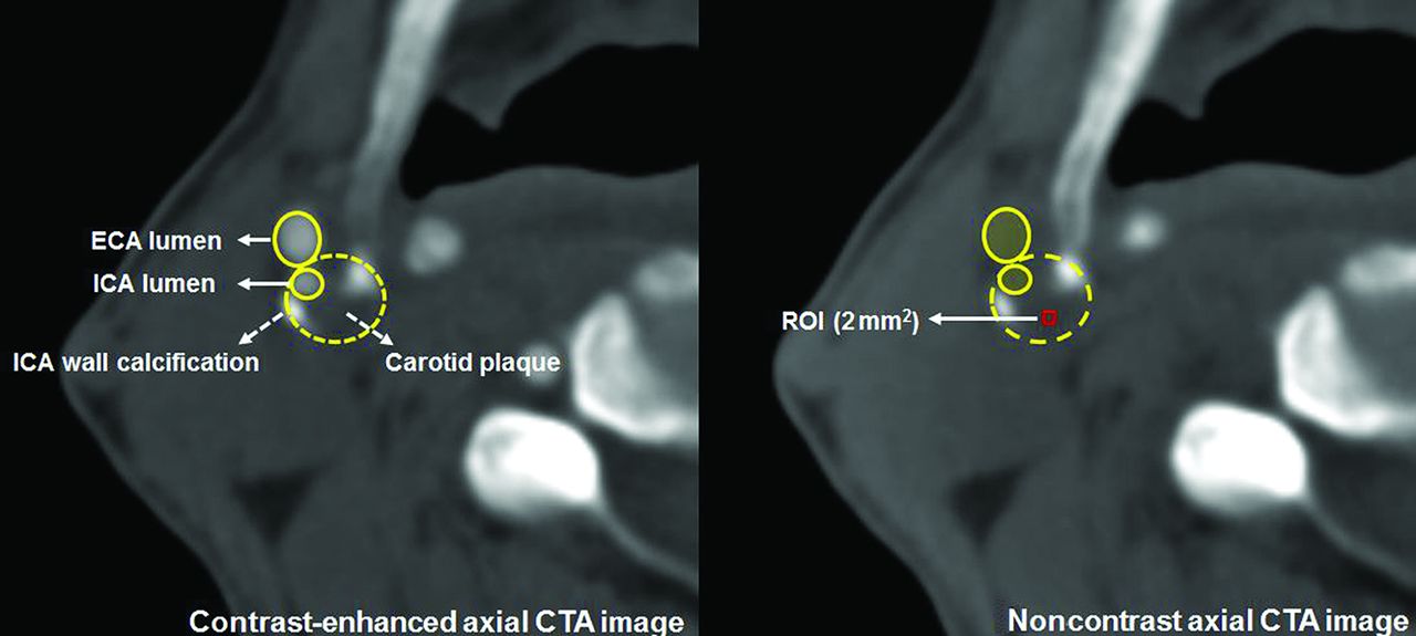
- FIG 1.
Representative figure of plaque Hounsfield unit value measurement. The plaque Hounsfield unit values were measured from noncontrast axial CTA images. An ROI of 2 mm2 was preselected on the visually least attenuated area of the plaque at the most stenotic level of contrast-enhanced axial images. Measurements were performed 5 times from the noncontrast axial images using prepositioned ROIs on the plaque. The lowest Hounsfield unit value in each plaque was recorded. ECA indicates external carotid artery.
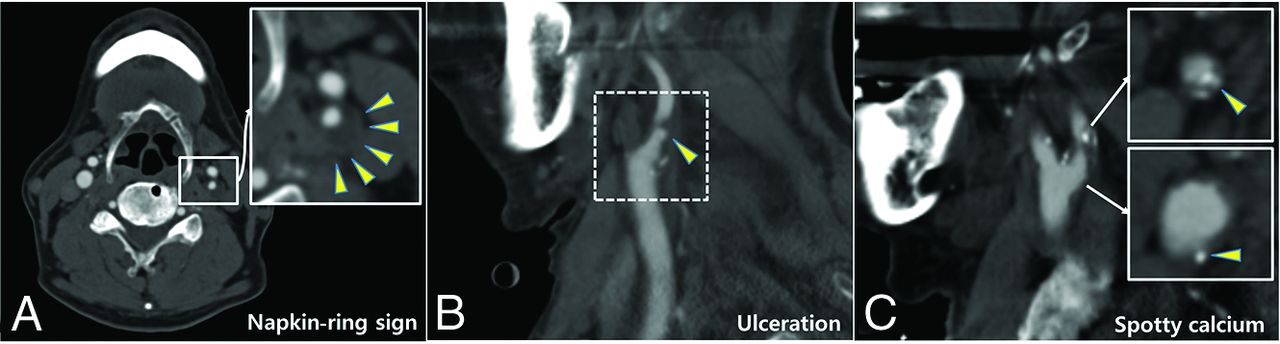
- FIG 2.
Representative figures of high-risk features of carotid plaque (arrowheads) on CTA. Napkin-ring sign (A), carotid plaque ulceration (B), and spotty calcium (C).
- FIG 3.
Kaplan-Meier survival analyses of the cumulative event-free rates based on the dichotomization of Hounsfield units at the median value of the cohort (36 HU). Cumulative event-free rates of MACE (A), stroke (B), ACS (C), and CV mortality (D), according to the carotid plaque density. CV indicates cardiovascular.


Tables
- Table 1:
Baseline and clinical characteristics of the study sample, stratified by the occurrence of MACEsa
Subjects Total (n = 176) Non-MACE Group (n = 142) MACE Group (n = 34) P Value Age (yr) 69.3 (SD, 7.6) 69.3 (SD, 7.7) 69.2 (SD, 6.8) .49 Median (IQR) (yr) 70 (64–75) 69 (64–75) 71 (66–75) Male sex 156 (88.6) 126 (88.7) 30 (88.2) .998 Body mass index (kg/m2) 24.5 (SD, 3.1) 24.5 (SD, 3.0) 24.4 (SD, 3.4) .96 Risk factors Diabetes mellitus 76 (43.2) 66 (46.5) 10 (29.4) .096 Hypertension 126 (71.6) 101 (71.1) 25 (73.5) .80 Dyslipidemia 96 (54.6) 77 (54.2) 19 (55.9) .60 Current smoking 43 (24.4) 32 (22.5) 11 (32.4) .42 Past smoking 68 (38.6) 56 (39.4) 12 (35.3) .91 Atrial fibrillation 9 (5.1) 5 (3.5) 4 (11.8) .001 Medical history History of strokeb 56 (31.8) 44 (31.0) 12 (35.3) .66 History of ACSb 30 (17.1) 21 (14.8) 9 (26.5) .30 Antiplatelet 154 (87.5) 124 (87.3) 30 (88.2) .72 Statins 142 (80.7) 115 (81.0) 27 (79.4) .47 Plaques Total (n = 194) Non-MACE Group (n = 156) MACE Group (n = 38) P Value Stenosis (%) 68.6 (SD, 10.8) 67.9 (SD, 10.0) 71.3 (SD, 13.4) .49 Median (IQR) 68 (60–75) 68 (60–75) 71 (62–80) Mixed plaque 129 (66.5) 102 (65.4) 27 (71.1) .18 Plaque ulceration 15 (7.7) 13 (8.3) 2 (5.3) .53 Hounsfield unit 36.9 (SD, 15.4) 39.2 (SD, 15.2) 27.6 (SD, 12.7) <.001 Median (IQR) 36 (28–45) 40 (30–47) 28 (19–36) Spotty calcium 40 (20.6) 23 (14.7) 17 (44.7) <.001 Napkin-ring sign 56 (28.9) 40 (25.6) 16 (42.1) .09 Calcium score 443.5 (SD, 443.8) 416.3 (SD, 428.0) 551.2 (SD, 493.0) .06 Median (IQR) 306 (101–639) 294 (74–564) 367 (165–1018) Follow-up (mo) (median) (IQR) 41 (29–60) 41 (30–59) 47 (22–71) .76 ↵a Any stroke, ACS, or cardiovascular mortality. Continuous data are presented as means (SD) or median (IQR); categoric data are given as (No.) (%).
Model 1b Model 2c HR (95% CI) P Value HR (95% CI) P Value Diabetes mellitus 0.60 (0.28–1.24) .17 NA NA Atrial fibrillation 1.07 (0.33–3.48) .91 NA NA Hounsfield unit 0.96 (0.94–0.98) <.001 0.96 (0.94–0.98) <.001 Spotty calcium 4.00 (2.00–8.00) <.001 3.97 (2.08–7.60) <.001 Napkin-ring sign 0.75 (0.36–1.55) .45 NA NA Calcium score 1.00 (1.00–1.001) .25 NA NA - Table 4:
Cox proportional hazards model for the association of the plaque Hounsfield unit values with the occurrence of the MACE composite outcome and the individual MACE components (n = 194)a
MACE (–) MACE (+) HR (95% CI) P Value MACEb (n = 38) 39.2 (SD, 15.2) 27.6 (SD, 12.7) 0.96 (0.95–0.98) <.001 Any stroke (n = 19) 37.9 (SD, 15.4) 27.8 (SD, 12.0) 0.97 (0.94–0.99) .01 Ipsilateral stroke (n = 14) 37.6 (SD, 15.4) 27.4 (SD, 12.4) 0.96 (0.93–0.99) .02 ACS (n = 16) 37.8 (SD, 15.2) 26.6 (SD, 14.0) 0.96 (0.93–0.99) .01 Cardiovascular mortality (n = 12) 37.7 (SD, 15.4) 24.9 (SD, 8.8) 0.95 (0.92–0.99) .01 All-cause mortality (n = 32) 37.7 (SD, 15.6) 32.8 (SD, 13.9) 0.98 (0.96–1.01) .14 - Table 5:
Cox proportional hazards model for the association of the presence of spotty calcium on CTA with the occurrence of the MACE composite outcome and the individual MACE components (n = 194)a
MACE (–) MACE (+) HR (95% CI) P Value MACEb (n = 38) 23 (14.7) 17 (44.7) 4.08 (2.13–7.83) <.001 Any stroke (n = 19) 32 (18.3) 8 (42.1) 3.25 (1.30–8.11) .01 Ipsilateral stroke (n = 14) 35 (19.4) 5 (35.7) 2.61 (0.87–7.82) .09 ACS (n = 16) 31 (17.4) 9 (56.3) 6.39 (2.34–17.45) <.001 Cardiovascular mortality (n = 12) 35 (19.2) 5 (41.7) 3.01 (0.95–9.48) .06 All-cause mortality (n = 32) 28 (17.3) 12 (37.5) 2.57 (1.25–5.26) .01


Jump to section
Related Articles
Cited By...
- No citing articles found.