
Article Figures & Data
Figures
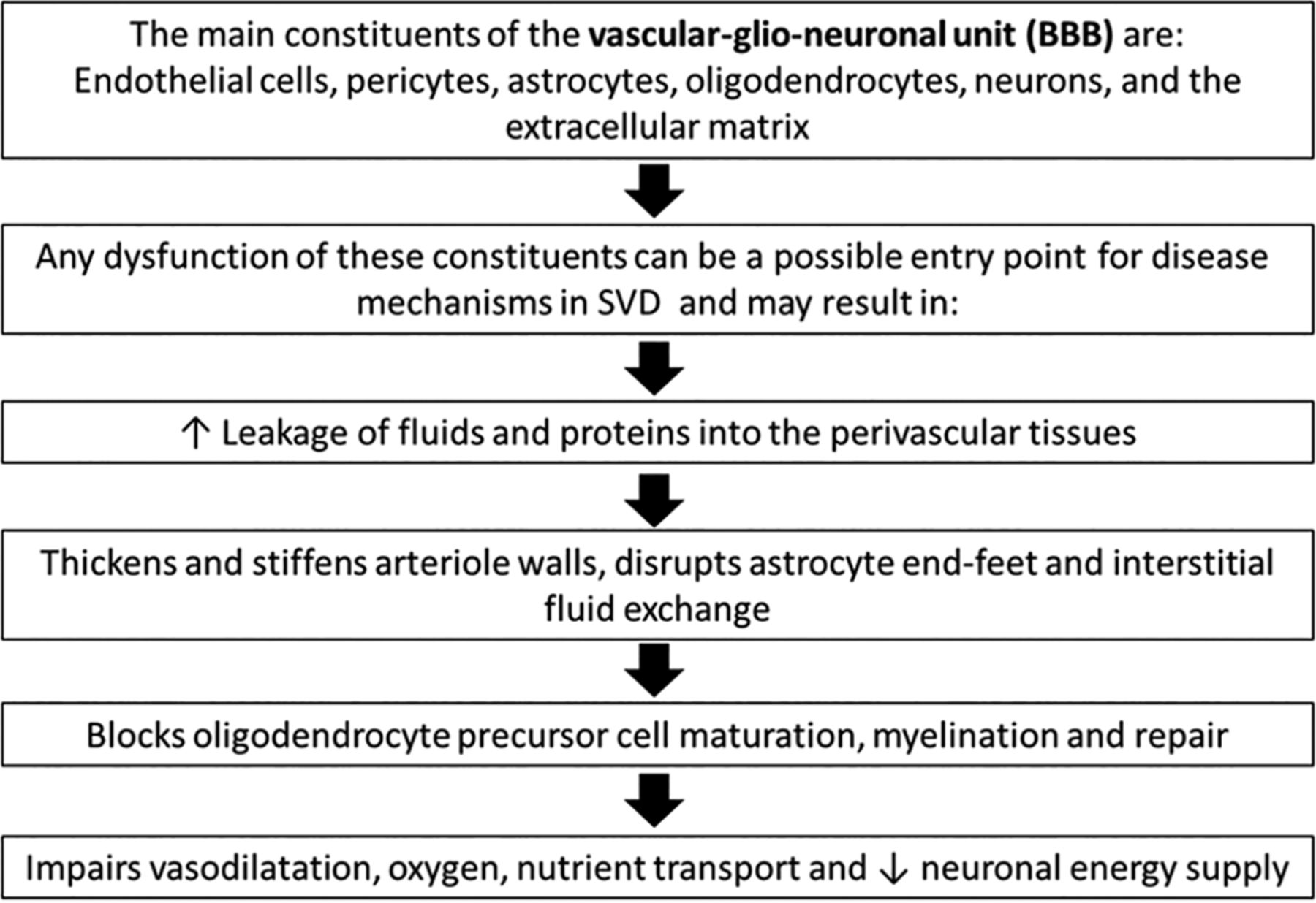
- FIG 1.
Possible entry points in the pathophysiology of SVD. The order of these events has not been determined.
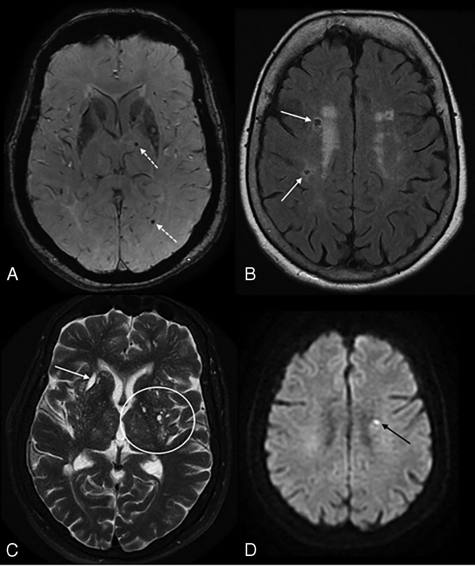
- FIG 2.
A, Recent subcortical infarct in the right lentiform nucleus in a 59-year-old woman with a history of hypertension. From left to right, axial DWI, ADC map, FLAIR, and coronal T2WI show restricted diffusion (solid white arrow, A). On coronal T2WI, the elongated morphology of the acute infarct in the craniocaudal axis (dashed white arrow, A) is related to the territory of a perforating artery. B, A lacune of presumed vascular origin in a 67-year-old man with a history of dementia. From left to right, axial DWI, T2WI, FLAIR, and T1WI show a remote lacunar infarct in the left frontal corona radiata, which demonstrates T2 hyperintensity with a peripheral rim of gliosis, best seen on FLAIR image (solid white arrow, B). C, Prominent perivascular spaces in a 63-year-old man with a history of dementia. A prominent perivascular space is noted in the left insular region with a centrally traversing vessel (solid white arrow, C), without peripheral gliosis on the FLAIR image (dashed arrow, C). Additionally, there are >20 dilated perivascular spaces in the bilateral basal ganglia on axial T2WI (circles, C). D, Cerebral microbleeds with cSS in an 87-year-old man with a history of CAA who presented with a worsening mental status. Axial SWI demonstrate multiple foci of susceptibility artifacts predominantly involving the basal ganglia and thalami, consistent with microbleeds. Additional areas of scattered cortical subarachnoid hemorrhagic staining (arrows, D) indicate cortical superficial siderosis.
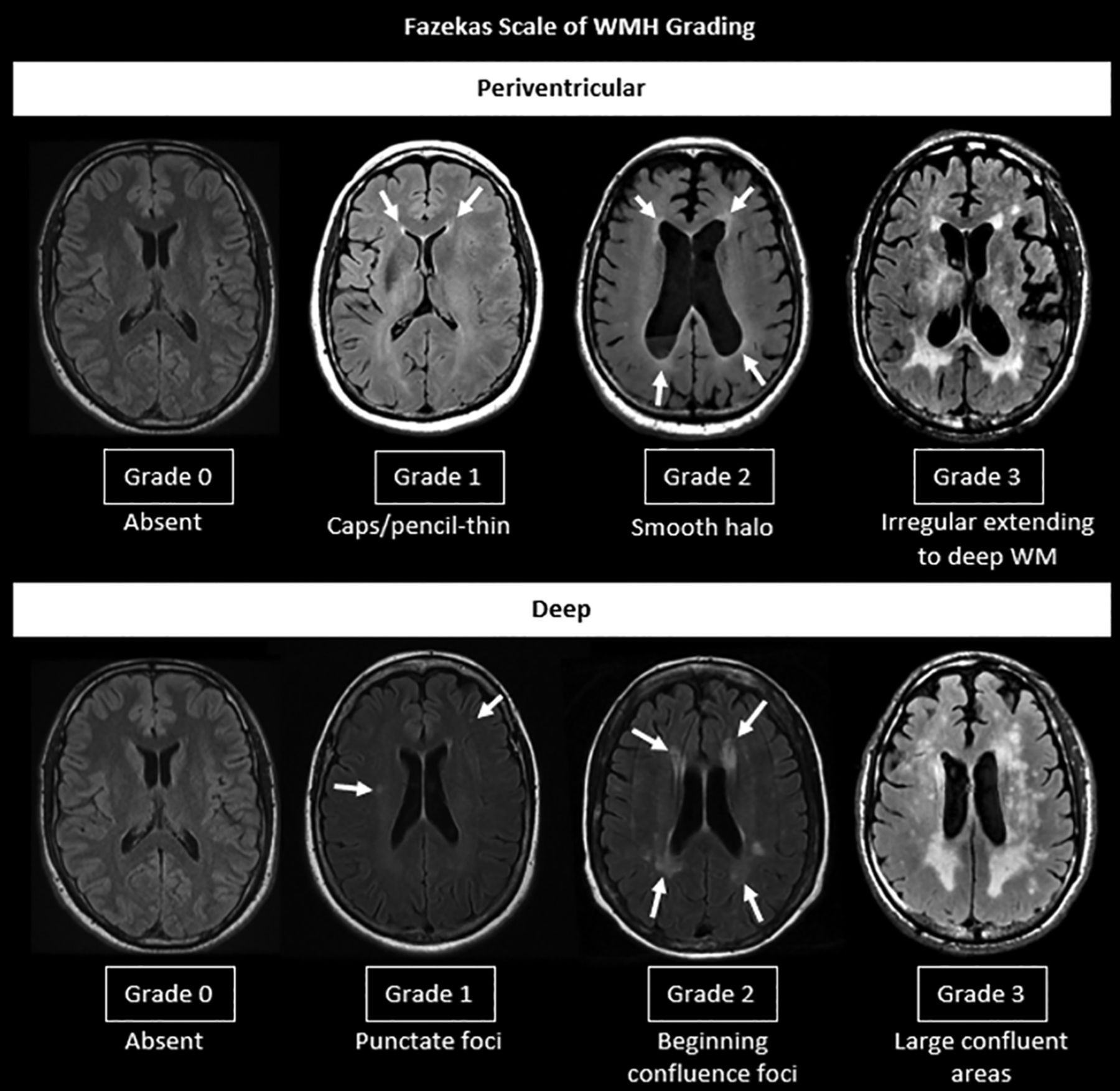
- FIG 3.
Fazekas scale of WMH grading.
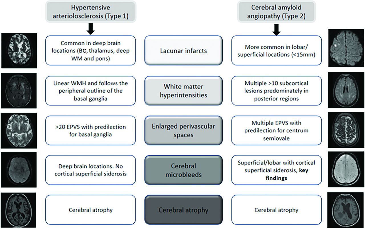
- FIG 4.
SVD MR imaging markers of the two most common etiopathogenic types of cerebral SVD in older adults, HA and CAA. BG indicates basal ganglia; EPVS: enlarged prominent perivascular spaces.
- FIG 5.
High SVD burden in a 55-year-old man with a history of arterial hypertension. Note multiple microbleeds in the left thalamus and occipital lobe (dotted arrows) on SWI (A), remote lacunar infarcts in the right centrum semiovale (white arrows, B), and WMH Fazekas 2 on FLAIR (B). More than 20 dilated perivascular spaces are seen on axial T2WI (circle, C) and remote lacunar infarct in right lentiform nucleus (white arrow, C). The total SVD score is 4. The patient developed an acute lacunar infarct (black arrow) on DWI (D) 10 months later.
- FIG 6.
Sample pipeline for studying brain connectivity for assessing global brain health. A, Data are processed to generate structural (tractography) or functional (fMRI) connectivity measures. B, A parcellation scheme is applied to obtain connectivity between different regions. C, The connectivity data are represented as an adjacency matrix. D, Network properties are calculated on the adjacency matrix. In the sample image, regions in the network are sized by importance (ie, centrality) and edges are sized by the strength of the connection.


Tables
Classification Type 1: HAFibrinoid necrosis, lipohyalinosis, microatheroma, microaneurysms Type 2: Sporadic and hereditary CAA Type 3: Inherited or genetic SVD (distinct from CAA)For example, CADASIL, CARASIL, MELAS, Fabry disease, COL4A1 mutations, and so forth Type 4: Inflammatory- and immunologically-mediated SVDExamples: Wegener granulomatosis, Churg-Strauss syndrome, microscopic polyangiitis, Henoch-Schönlein purpura, cryoglobulinemic vasculitis, cutaneous leukocytoclastic angiitis, primary angiitis of the CNS, Sjögren syndrome, rheumatoid vasculitis, scleroderma, dermatomyositis, and so forth Type 5: Venous collagenosis Type 6: Other SVDExamples: postradiation angiopathy and nonamyloid microvessel degeneration in Alzheimer disease Note:—MELAS indicates Mitochondrial Encephalopathy, Lactic Acidosis, and Stroke-like episodes; CARASIL, cerebral autosomal recessive arteriopathy with subcortical infarcts and leukoencephalopathy.
MR Imaging Sequence and Visual Rating Total SVD Scorea Type 1: Hypertensive42 Type 2: CAA33 Classic markers Lacune T2WI/FLAIR Any lacunes: 1 point WMH FLAIR/Fazekas scale (0–3):Periventricular WMH: 0 = absent; 1 = caps or pencil-thin; 2 = smooth halo; 3 = irregular extending to deep WMDeep WMH: 0 = absent; 1 = punctate foci; 2 = beginning confluence; 3 = large confluent areas Deep WMH (Fazekas 2 or 3) or periventricular WMH (Fazekas 3): 1 point Deep WMH (Fazekas 2 or 3) or
periventricular WMH (Fazekas 3): 1 pointPVS T2WI 4-point visual rating scale:0: no PVS; 1 (mild): ≤10 PVS; 2 (moderate): 11–20 PVS; 3 (moderate to severe): 21–40 PVS; and 4 (severe): >40 PVS >20 PVS in the basal ganglia: 1 point >20 PVS in CSO: 1 point Microbleeds T2* GRE/SWI; several visual scores are available including MARS and BOMBS Any microbleed: 1 point 2–4 Cortical CMB: 1 point;≥5 cortical CMB: 2 points New markers cSS T2* GRE/SWI, categorized as focal or disseminated Focal cSS: 1 point; disseminated cSS: 2 points Cortical microinfarct High-field MR imaging (>3T)3D T1WI3D DIR Note:—GRE indicates gradient recalled-echo; MARS, Microbleed Anatomical Rating Scale; BOMBS, Brain Observer MicroBleed Scales; CSO, centrum semiovale; cSS, cortical superficial siderosis; BG, basal ganglia; DIR, double inversion recovery.
↵a Type 1: Hypertensive; type 2: CAA.


