Advertisement

Main menu

  • Home
  • Content
    • Current Issue
    • Accepted Manuscripts
    • Article Preview
    • Past Issue Archive
    • Video Articles
    • AJNR Case Collection
    • Case of the Week Archive
    • Case of the Month Archive
    • Classic Case Archive
  • Special Collections
    • AJNR Awards
    • Low-Field MRI
    • Alzheimer Disease
    • ASNR Foundation Special Collection
    • Photon-Counting CT
    • View All
  • Multimedia
    • AJNR Podcasts
    • AJNR SCANtastic
    • Trainee Corner
    • MRI Safety Corner
    • Imaging Protocols
  • For Authors
    • Submit a Manuscript
    • Submit a Video Article
    • Submit an eLetter to the Editor/Response
    • Manuscript Submission Guidelines
    • Statistical Tips
    • Fast Publishing of Accepted Manuscripts
    • Graphical Abstract Preparation
    • Imaging Protocol Submission
    • Author Policies
  • About Us
    • About AJNR
    • Editorial Board
    • Editorial Board Alumni
  • More
    • Become a Reviewer/Academy of Reviewers
    • Subscribers
    • Permissions
    • Alerts
    • Feedback
    • Advertisers
    • ASNR Home

User menu

  • Alerts
  • Log in

Search

  • Advanced search
American Journal of Neuroradiology

American Journal of Neuroradiology

ASHNR American Society of Functional Neuroradiology ASHNR American Society of Pediatric Neuroradiology ASSR
  • Alerts
  • Log in

Advanced Search

  • Home
  • Content
    • Current Issue
    • Accepted Manuscripts
    • Article Preview
    • Past Issue Archive
    • Video Articles
    • AJNR Case Collection
    • Case of the Week Archive
    • Case of the Month Archive
    • Classic Case Archive
  • Special Collections
    • AJNR Awards
    • Low-Field MRI
    • Alzheimer Disease
    • ASNR Foundation Special Collection
    • Photon-Counting CT
    • View All
  • Multimedia
    • AJNR Podcasts
    • AJNR SCANtastic
    • Trainee Corner
    • MRI Safety Corner
    • Imaging Protocols
  • For Authors
    • Submit a Manuscript
    • Submit a Video Article
    • Submit an eLetter to the Editor/Response
    • Manuscript Submission Guidelines
    • Statistical Tips
    • Fast Publishing of Accepted Manuscripts
    • Graphical Abstract Preparation
    • Imaging Protocol Submission
    • Author Policies
  • About Us
    • About AJNR
    • Editorial Board
    • Editorial Board Alumni
  • More
    • Become a Reviewer/Academy of Reviewers
    • Subscribers
    • Permissions
    • Alerts
    • Feedback
    • Advertisers
    • ASNR Home
  • Follow AJNR on Twitter
  • Visit AJNR on Facebook
  • Follow AJNR on Instagram
  • Join AJNR on LinkedIn
  • RSS Feeds

AJNR Awards, New Junior Editors, and more. Read the latest AJNR updates

Research ArticleAdult Brain
Open Access

Decreased CSF Dynamics in Treatment-Naive Patients with Essential Hypertension: A Study with Phase-Contrast Cine MR Imaging

L. Ma, W. He, X. Li, X. Liu, H. Cao, L. Guo, X. Xiao, Y. Xu and Y. Wu
American Journal of Neuroradiology October 2021, DOI: https://doi.org/10.3174/ajnr.A7284
L. Ma
aFrom the Department of Medical Imaging (L.M., X. Li, X. Liu, H.C., L.G., X.X., Y.X., Y.W.), Nanfang Hospital, Southern Medical University, Guangzhou, China
  • Find this author on Google Scholar
  • Find this author on PubMed
  • Search for this author on this site
  • ORCID record for L. Ma
W. He
bDepartment of Radiology (W.H.), Guangdong 999 Brain Hospital, Guangzhou, China
  • Find this author on Google Scholar
  • Find this author on PubMed
  • Search for this author on this site
  • ORCID record for W. He
X. Li
aFrom the Department of Medical Imaging (L.M., X. Li, X. Liu, H.C., L.G., X.X., Y.X., Y.W.), Nanfang Hospital, Southern Medical University, Guangzhou, China
  • Find this author on Google Scholar
  • Find this author on PubMed
  • Search for this author on this site
  • ORCID record for X. Li
X. Liu
aFrom the Department of Medical Imaging (L.M., X. Li, X. Liu, H.C., L.G., X.X., Y.X., Y.W.), Nanfang Hospital, Southern Medical University, Guangzhou, China
  • Find this author on Google Scholar
  • Find this author on PubMed
  • Search for this author on this site
  • ORCID record for X. Liu
H. Cao
aFrom the Department of Medical Imaging (L.M., X. Li, X. Liu, H.C., L.G., X.X., Y.X., Y.W.), Nanfang Hospital, Southern Medical University, Guangzhou, China
  • Find this author on Google Scholar
  • Find this author on PubMed
  • Search for this author on this site
  • ORCID record for H. Cao
L. Guo
aFrom the Department of Medical Imaging (L.M., X. Li, X. Liu, H.C., L.G., X.X., Y.X., Y.W.), Nanfang Hospital, Southern Medical University, Guangzhou, China
  • Find this author on Google Scholar
  • Find this author on PubMed
  • Search for this author on this site
  • ORCID record for L. Guo
X. Xiao
aFrom the Department of Medical Imaging (L.M., X. Li, X. Liu, H.C., L.G., X.X., Y.X., Y.W.), Nanfang Hospital, Southern Medical University, Guangzhou, China
  • Find this author on Google Scholar
  • Find this author on PubMed
  • Search for this author on this site
  • ORCID record for X. Xiao
Y. Xu
aFrom the Department of Medical Imaging (L.M., X. Li, X. Liu, H.C., L.G., X.X., Y.X., Y.W.), Nanfang Hospital, Southern Medical University, Guangzhou, China
  • Find this author on Google Scholar
  • Find this author on PubMed
  • Search for this author on this site
  • ORCID record for Y. Xu
Y. Wu
aFrom the Department of Medical Imaging (L.M., X. Li, X. Liu, H.C., L.G., X.X., Y.X., Y.W.), Nanfang Hospital, Southern Medical University, Guangzhou, China
  • Find this author on Google Scholar
  • Find this author on PubMed
  • Search for this author on this site
  • ORCID record for Y. Wu
  • Article
  • Figures & Data
  • Info & Metrics
  • Responses
  • References
  • PDF
Loading

Article Figures & Data

Figures

  • Tables
  • FIG 1.
    • Download figure
    • Open in new tab
    • Download powerpoint
    FIG 1.

    Flow chart of the study population.
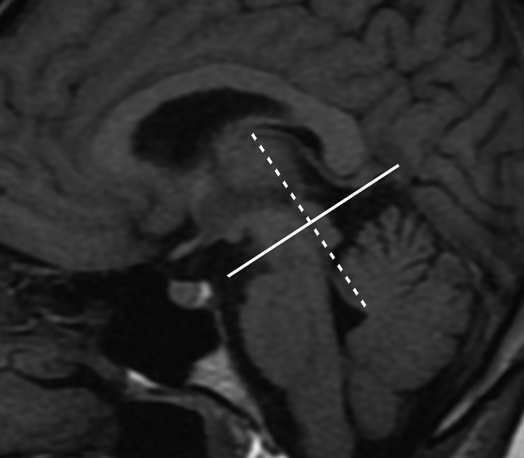
  • FIG 2.
    • Download figure
    • Open in new tab
    • Download powerpoint
    FIG 2.

    Illustration of the location of phase-contrast MR imaging planes. The midline sagittal T1WI MR image shows the phase-contrast MR imaging planes (solid line) located at the ampulla level of the cerebral aqueduct and perpendicular to its long axis (dotted line).
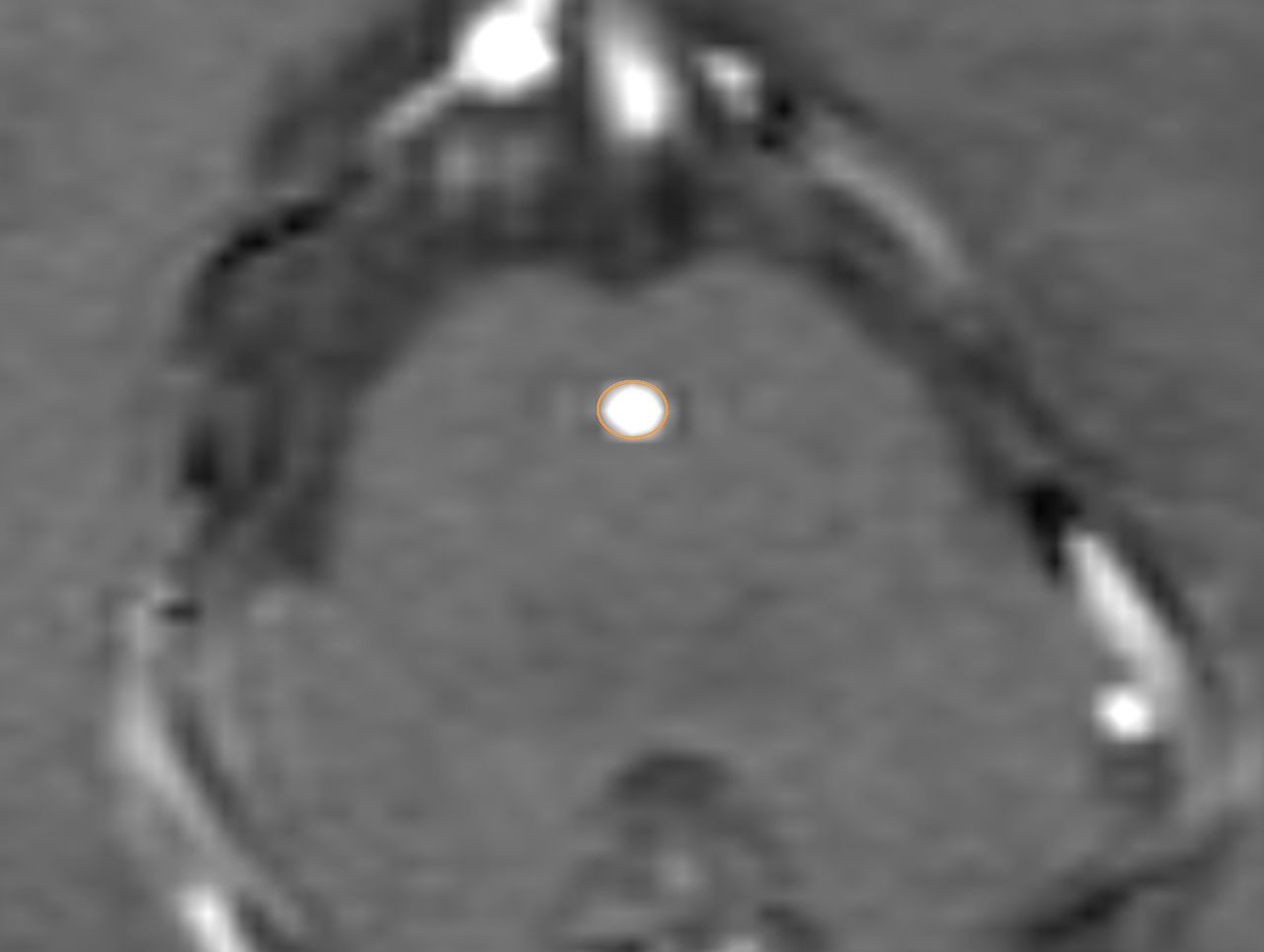
  • FIG 3.
    • Download figure
    • Open in new tab
    • Download powerpoint
    FIG 3.

    Placement of ROI to contain the whole aqueduct.

  • FIG 4.
    • Download figure
    • Open in new tab
    • Download powerpoint
    FIG 4.

    Flow curves of the essential hypertension and control groups. Flows are plotted during the successive phases of a cardiac cycle time (millisecond) and expressed in milliliter/minute. Each flow curve for both groups represents average flow values. Positive deflections represent craniocaudal flow (CSF systole), and negative deflections represent caudocranial flow (CSF diastole).

Tables

  • Figures
    • View popup
    Table 1:

    Clinical characteristics of patients with essential hypertension and healthy subjects

    CharacteristicsPatientsControlsP Value
    No.6060
    Age (mean) (yr)61.24 (SD, 8.9)60.38 (SD, 7.54).84
    Male3133.71
    PP (mean) (mm Hg)53.78 (SD, 14.86)42.43 (SD, 4.36)<.001
    SBP (mean) (mm Hg)148.54 (SD, 13.55)123.40 (SD, 6.06).001
    DBP (mean) (mm Hg)94.76 (SD, 7.72)80.97 (SD, 2.42)<.001
    Disease duration (mean) (yr)4.92 (SD, 2.72)0<.001
    • View popup
    Table 2:

    Comparison of mean CSF flow parameters in the hypertension and control groups

    GroupFFV (mL)FPV (cm/s)RFV (mL)RPV (cm/s)AF (mL/min)NFV (mL)
    Patients0.054 (SD, 0.021)5.338 (SD, 2.024)0.042 (SD, 0.019)4.837 (SD, 2.196)7.480 (SD, 3.221)0.012 (SD, 0.005)
    Controls0.080 (SD, 0.040)6.197 (SD, 2.394)0.070 (SD, 0.042)5.823 (SD, 2.821)9.824 (SD, 5.435)0.010 (SD, 0.011)
    P value<.001.04.005.04.006.12
    • View popup
    Table 3:

    Univariate linear regression coefficients for relationships between hypertension parameters and CSF flow parametersa

    VariablesFFV (mL)bFPV (cm/s)bRFV (mL)bRPV (cm/s)bAF (mL/min)b
    Duration−0.81 (−0.98 to −0.64)c−0.83 (−0.99 to −0.67)c−0.84 (−0.99 to −0.68)c−0.83 (−0.99 to −0.67)c−0.77 (−0.95 to −0.58)c
    PP−0.76 (−0.95 to −0.57)c−0.73 (−1.01 to −0.71)c−0.74 (−0.94 to −0.55)c−0.25 (−1.01−0.03)c−0.74 (−0.94 to −0.54)c
    SBP−0.86 (−1.01 to −0.71)c−0.86 (−0.97 to −0.62)c−0.88 (−1.02 to −0.75)c−0.87 (−0.75 to −0.72)c−0.83 (−0.99 to −0.67)c
    DBP0.05 (−0.33−0.25)−0.10 (−0.39−0.19)−0.12 (−0.41−0.17)−0.11 (−0.40−0.18)0.04 (−0.33−0.25)
    Age−0.13 (−0.42−0.16)−0.05 (−0.34−0.24)−0.11 (−0.40−0.18)−0.09 (−0.38−0.20)−0.07 (−0.36−0.22)
    Sex−0.09 (−0.38−0.20)−0.10 (−0.38−0.19)−0.12 (−0.41−0.17)−0.11 (−0.40−0.18)−0.14 (−0.43−0.15)
    • ↵a Numbers in parentheses are 95% CI.

    • ↵b Crude regression coefficients indicative of increase (positive value) or decrease (negative value) in CSF flow parameters.

    • ↵c P < .05.

    • View popup
    Table 4:

    Multiple linear regression coefficients for relationships between hypertension parameters and CSF flow parametersa

    VariablesFFV (mL)bFPV (cm/s)bRFV (mL)bRPV (cm/s)bAF (mL/min)b
    Duration−0.34 (−0.61 to −0.07)c−0.37 (−0.64 to −0.10)c−0.31 (−0.56 to −0.07)c−0.35 (−0.61 to −0.09)c−0.25 (−0.55−0.05)c
    SBP−0.44 (−0.83 to −0.06)c−0.50 (−0.88 to −0.12)c−0.61 (−0.97 to −0.26)c−0.55 (−0.91 to −0.18)c−0.50 (−0.93 to −0.08)c
    PP−0.25 (−0.43−0.13)c−0.16 (−0.33−0.22)c−0.11 (−0.26−0.25)c−0.23 (−0.30−0.24)c−0.34 (−0.45−0.17)c
    • ↵a Numbers in parentheses are 95% CI.

    • ↵b β, adjusted regression coefficients indicative of an increase (positive value) or decrease (negative value) in CSF flow parameters.

    • ↵c P < .05.

PreviousNext
Back to top
Advertisement
Print
Download PDF
Email Article

Thank you for your interest in spreading the word on American Journal of Neuroradiology.

NOTE: We only request your email address so that the person you are recommending the page to knows that you wanted them to see it, and that it is not junk mail. We do not capture any email address.

Enter multiple addresses on separate lines or separate them with commas.
Decreased CSF Dynamics in Treatment-Naive Patients with Essential Hypertension: A Study with Phase-Contrast Cine MR Imaging
(Your Name) has sent you a message from American Journal of Neuroradiology
(Your Name) thought you would like to see the American Journal of Neuroradiology web site.
CAPTCHA
This question is for testing whether or not you are a human visitor and to prevent automated spam submissions.
Cite this article
L. Ma, W. He, X. Li, X. Liu, H. Cao, L. Guo, X. Xiao, Y. Xu, Y. Wu
Decreased CSF Dynamics in Treatment-Naive Patients with Essential Hypertension: A Study with Phase-Contrast Cine MR Imaging
American Journal of Neuroradiology Oct 2021, DOI: 10.3174/ajnr.A7284

Citation Manager Formats

  • BibTeX
  • Bookends
  • EasyBib
  • EndNote (tagged)
  • EndNote 8 (xml)
  • Medlars
  • Mendeley
  • Papers
  • RefWorks Tagged
  • Ref Manager
  • RIS
  • Zotero
0 Responses
Respond to this article
Share
Bookmark this article
Decreased CSF Dynamics in Treatment-Naive Patients with Essential Hypertension: A Study with Phase-Contrast Cine MR Imaging
L. Ma, W. He, X. Li, X. Liu, H. Cao, L. Guo, X. Xiao, Y. Xu, Y. Wu
American Journal of Neuroradiology Oct 2021, DOI: 10.3174/ajnr.A7284
del.icio.us logo Twitter logo Facebook logo Mendeley logo
  • Tweet Widget
  • Facebook Like
  • Google Plus One
Purchase

Jump to section

  • Article
    • Abstract
    • ABBREVIATIONS:
    • MATERIALS AND METHODS
    • RESULTS
    • DISCUSSION
    • CONCLUSIONS
    • Footnotes
    • References
  • Figures & Data
  • Info & Metrics
  • Responses
  • References
  • PDF

Related Articles

  • No related articles found.
  • PubMed
  • Google Scholar

Cited By...

  • No citing articles found.
  • Crossref
  • Google Scholar

This article has not yet been cited by articles in journals that are participating in Crossref Cited-by Linking.

More in this TOC Section

  • Diagnostic Neuroradiology of Monoclonal Antibodies
  • Clinical Outcomes After Chiari I Decompression
  • Segmentation of Brain Metastases with BLAST
Show more Adult Brain

Similar Articles

Advertisement

Indexed Content

  • Current Issue
  • Accepted Manuscripts
  • Article Preview
  • Past Issues
  • Editorials
  • Editor's Choice
  • Fellows' Journal Club
  • Letters to the Editor
  • Video Articles

Cases

  • Case Collection
  • Archive - Case of the Week
  • Archive - Case of the Month
  • Archive - Classic Case

More from AJNR

  • Trainee Corner
  • Imaging Protocols
  • MRI Safety Corner
  • Book Reviews

Multimedia

  • AJNR Podcasts
  • AJNR Scantastics

Resources

  • Turnaround Time
  • Submit a Manuscript
  • Submit a Video Article
  • Submit an eLetter to the Editor/Response
  • Manuscript Submission Guidelines
  • Statistical Tips
  • Fast Publishing of Accepted Manuscripts
  • Graphical Abstract Preparation
  • Imaging Protocol Submission
  • Evidence-Based Medicine Level Guide
  • Publishing Checklists
  • Author Policies
  • Become a Reviewer/Academy of Reviewers
  • News and Updates

About Us

  • About AJNR
  • Editorial Board
  • Editorial Board Alumni
  • Alerts
  • Permissions
  • Not an AJNR Subscriber? Join Now
  • Advertise with Us
  • Librarian Resources
  • Feedback
  • Terms and Conditions
  • AJNR Editorial Board Alumni

American Society of Neuroradiology

  • Not an ASNR Member? Join Now

© 2025 by the American Society of Neuroradiology All rights, including for text and data mining, AI training, and similar technologies, are reserved.
Print ISSN: 0195-6108 Online ISSN: 1936-959X

Powered by HighWire