
Article Figures & Data
Figures
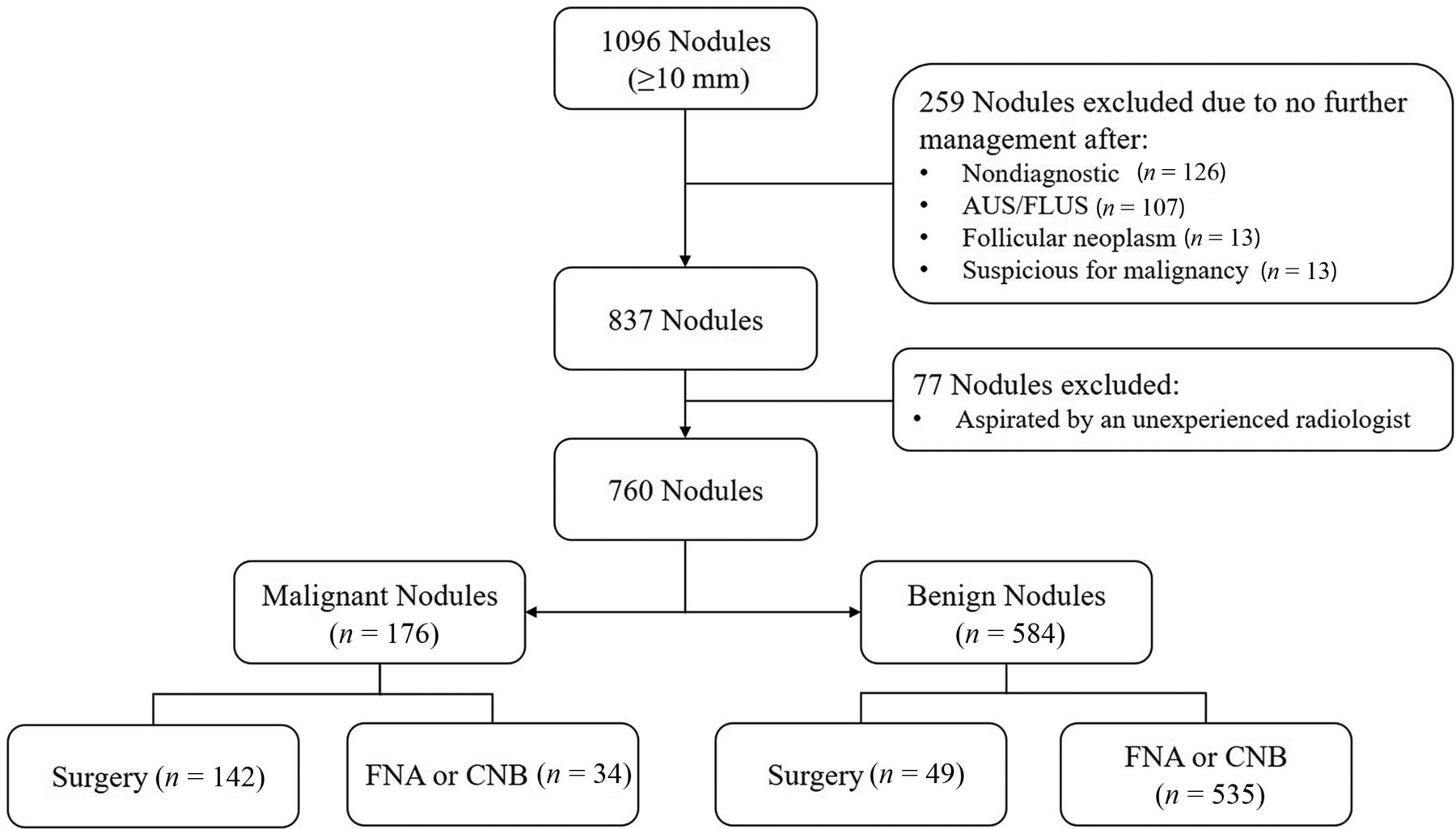
- FIG 1.
Flowchart of the study population. AUS/FLUS indicates atypia of undetermined significance/follicular lesion of undetermined significance.
- FIG 2.
Comparison of ROC curves between the CNN (solid line) and ACR TI-RADS categories (dotted line). The area under the ROC curve of the CNN (0.917; 95% confidence interval, 0.895–0.936) was higher than that in the ACR TI-RADS categories (0.891; 95% confidence interval, 0.867–0.912) (P = .017). The areas under the ROC curve of the CNN using a malignancy risk percentage between 0 and 100 and ACR TI-RADS categories using a TR category from 1 to 5 were compared as continuous values.


Tables
- Table 1:
Patient demographics and distribution of ACR TI-RADS features in benign and malignant thyroid nodules (n = 760)a
Characteristics All (n = 760) Benign Nodules (n = 584) Malignant Nodules (n = 176) P Value Sex .035 Women 587 462 (79.4%) 125 (71.4%) Men 170 120 (20.6%) 50 (28.6%) Age (median) (interquartile range) (yr) 51 (39–61) 52 (41–61) 45 (34–60) <.001 Nodule size (median) (interquartile range) (mm) 20 (14–30) 23 (15–32) 14 (11–20) <.001 Nodule features Composition <.001 Cystic or almost completely cystic 50 47 (8.0%) 3 (1.7%) Spongiform 1 1 (0.2%) 0 Mixed cystic and solid 234 222 (38.0%) 12 (6.8%) Solid or almost completely solid 475 314 (53.8%) 161 (91.5%) Echogenicity <.001 Anechoic 0 0 Hyperechoic or isoechoic 410 387 (66.3%) 23 (13.1%) Hypoechoic 329 191 (32.7%) 138 (78.4%) Very hypoechoic 21 6 (1.0%) 15 (8.5%) Shape <.001 Wider-than-tall 671 554 (94.9%) 117 (66.5%) Taller-than-wide 89 30 (5.1%) 59 (33.5%) Margin <.001 Smooth 579 535 (91.6%) 44 (25.0%) Ill-defined 0 0 0 Lobulated or irregular 181 49 (8.4%) 132 (75.0%) Extrathyroidal extension 0 0 0 Echogenic foci <.001 None or large comet-tail artifacts 536 477 (81.7%) 59 (33.5%) Macrocalcifications 91 69 (11.8%) 22 (12.5%) Peripheral (rim) calcifications 10 10 (1.7%) 0 Punctate echogenic foci 123 28 (4.8%) 95 (54.0%) ↵a Data are numbers of nodules, with percentages in parentheses.
- Table 2:
Calculated malignancy risk of each category according to the risk stratification of ACR TI-RADS
TR1 TR2 TR3 TR4 TR5 Total Suggested risk of malignancy (%)20,24 ≤2 ≤2 2< and ≤5 5< and ≤20 >20 ACR TI-RADS category No. of malignant nodules 0 4 9 33 130 176 Assigned total nodules 41 158 185 209 167 760 Calculated risk of malignancy (%) 0 2.5 4.9 15.8 77.8 23.2 CNNa No. of malignant nodules 0 0 0 9 167 176 Assigned total nodules 0 5 45 307 403 760 Calculated risk of malignancy (%) 0 0 0 2.9 41.4 23.2 ↵a Malignancy percentages provided by the CNN were re-categorized according to the suggested cancer risk levels of ACR TI-RADS.
CNN (95% CI) ACR TI-RADS (95% CI) P Value Sensitivity 81.8% (76.1–87.5) 73.9% (67.4–80.4) .009 Specificity 86.1% (83.3–88.9) 93.7% (91.7–95.6) <.001 Accuracy 85.1% (82.6–87.7) 89.1% (86.9–91.3) .003 Positive predictive value 64.0% (57.7–70.3) 77.8% (71.6–84.1) <.001 Negative predictive value 94.0% (92–96) 92.2% (90.1–94.4) .046 AUCa 0.917 (0.895-0.936) 0.891 (0.867-0.912) .017 ↵a The AUCs of the CNN using a malignancy risk percentage between 0 and 100 and ACR TI-RADS categories using a TR category from 1 to 5 were compared as continuous values.


Jump to section
Related Articles
Cited By...
- No citing articles found.