Advertisement
  • Main menu
  • User menu
  • Search
  • English ▼
    • English
    • Afrikaans
    • Albanian
    • Amharic
    • Arabic
    • Armenian
    • Azerbaijani
    • Basque
    • Belarusian
    • Bengali
    • Bosnian
    • Bulgarian
    • Catalan
    • Cebuano
    • Chichewa
    • Chinese (Simplified)
    • Chinese (Traditional)
    • Corsican
    • Croatian
    • Czech
    • Danish
    • Dutch
    • Esperanto
    • Estonian
    • Filipino
    • Finnish
    • French
    • Frisian
    • Galician
    • Georgian
    • German
    • Greek
    • Gujarati
    • Haitian Creole
    • Hausa
    • Hawaiian
    • Hebrew
    • Hindi
    • Hmong
    • Hungarian
    • Icelandic
    • Igbo
    • Indonesian
    • Irish
    • Italian
    • Japanese
    • Javanese
    • Kannada
    • Kazakh
    • Khmer
    • Korean
    • Kurdish (Kurmanji)
    • Kyrgyz
    • Lao
    • Latin
    • Latvian
    • Lithuanian
    • Luxembourgish
    • Macedonian
    • Malagasy
    • Malay
    • Malayalam
    • Maltese
    • Maori
    • Marathi
    • Mongolian
    • Myanmar (Burmese)
    • Nepali
    • Norwegian
    • Pashto
    • Persian
    • Polish
    • Portuguese
    • Punjabi
    • Romanian
    • Russian
    • Samoan
    • Scottish Gaelic
    • Serbian
    • Sesotho
    • Shona
    • Sindhi
    • Sinhala
    • Slovak
    • Slovenian
    • Somali
    • Spanish
    • Sudanese
    • Swahili
    • Swedish
    • Tajik
    • Tamil
    • Telugu
    • Thai
    • Turkish
    • Ukrainian
    • Urdu
    • Uzbek
    • Vietnamese
    • Welsh
    • Xhosa
    • Yiddish
    • Yoruba
    • Zulu

Main menu

  • Home
  • Content
    • Current Issue
    • Accepted Manuscripts
    • Article Preview
    • Past Issue Archive
    • Video Articles
    • AJNR Case Collection
    • Case of the Week Archive
    • Case of the Month Archive
    • Classic Case Archive
  • Special Collections
    • Advancing NeuroMRI with High-Relaxivity Contrast Agents
    • AJNR Awards
    • Low-Field MRI
    • Alzheimer Disease
    • ASNR Foundation Special Collection
    • Photon-Counting CT
    • View All
  • Multimedia
    • AJNR Podcasts
    • AJNR SCANtastic
    • Trainee Corner
    • MRI Safety Corner
    • Imaging Protocols
  • For Authors
    • Submit a Manuscript
    • Submit a Video Article
    • Submit an eLetter to the Editor/Response
    • Manuscript Submission Guidelines
    • Statistical Tips
    • Fast Publishing of Accepted Manuscripts
    • Graphical Abstract Preparation
    • Imaging Protocol Submission
    • Author Policies
  • About Us
    • About AJNR
    • Editorial Board
    • Editorial Board Alumni
  • More
    • Become a Reviewer/Academy of Reviewers
    • Subscribers
    • Permissions
    • Alerts
    • Feedback
    • Advertisers
    • ASNR Home

User menu

  • Alerts
  • Log in

Search

  • Advanced search
American Journal of Neuroradiology

American Journal of Neuroradiology

ASHNR American Society of Functional Neuroradiology ASHNR American Society of Pediatric Neuroradiology ASSR
  • Alerts
  • Log in

Advanced Search

English ▼
  • English
  • Afrikaans
  • Albanian
  • Amharic
  • Arabic
  • Armenian
  • Azerbaijani
  • Basque
  • Belarusian
  • Bengali
  • Bosnian
  • Bulgarian
  • Catalan
  • Cebuano
  • Chichewa
  • Chinese (Simplified)
  • Chinese (Traditional)
  • Corsican
  • Croatian
  • Czech
  • Danish
  • Dutch
  • Esperanto
  • Estonian
  • Filipino
  • Finnish
  • French
  • Frisian
  • Galician
  • Georgian
  • German
  • Greek
  • Gujarati
  • Haitian Creole
  • Hausa
  • Hawaiian
  • Hebrew
  • Hindi
  • Hmong
  • Hungarian
  • Icelandic
  • Igbo
  • Indonesian
  • Irish
  • Italian
  • Japanese
  • Javanese
  • Kannada
  • Kazakh
  • Khmer
  • Korean
  • Kurdish (Kurmanji)
  • Kyrgyz
  • Lao
  • Latin
  • Latvian
  • Lithuanian
  • Luxembourgish
  • Macedonian
  • Malagasy
  • Malay
  • Malayalam
  • Maltese
  • Maori
  • Marathi
  • Mongolian
  • Myanmar (Burmese)
  • Nepali
  • Norwegian
  • Pashto
  • Persian
  • Polish
  • Portuguese
  • Punjabi
  • Romanian
  • Russian
  • Samoan
  • Scottish Gaelic
  • Serbian
  • Sesotho
  • Shona
  • Sindhi
  • Sinhala
  • Slovak
  • Slovenian
  • Somali
  • Spanish
  • Sudanese
  • Swahili
  • Swedish
  • Tajik
  • Tamil
  • Telugu
  • Thai
  • Turkish
  • Ukrainian
  • Urdu
  • Uzbek
  • Vietnamese
  • Welsh
  • Xhosa
  • Yiddish
  • Yoruba
  • Zulu
  • Home
  • Content
    • Current Issue
    • Accepted Manuscripts
    • Article Preview
    • Past Issue Archive
    • Video Articles
    • AJNR Case Collection
    • Case of the Week Archive
    • Case of the Month Archive
    • Classic Case Archive
  • Special Collections
    • Advancing NeuroMRI with High-Relaxivity Contrast Agents
    • AJNR Awards
    • Low-Field MRI
    • Alzheimer Disease
    • ASNR Foundation Special Collection
    • Photon-Counting CT
    • View All
  • Multimedia
    • AJNR Podcasts
    • AJNR SCANtastic
    • Trainee Corner
    • MRI Safety Corner
    • Imaging Protocols
  • For Authors
    • Submit a Manuscript
    • Submit a Video Article
    • Submit an eLetter to the Editor/Response
    • Manuscript Submission Guidelines
    • Statistical Tips
    • Fast Publishing of Accepted Manuscripts
    • Graphical Abstract Preparation
    • Imaging Protocol Submission
    • Author Policies
  • About Us
    • About AJNR
    • Editorial Board
    • Editorial Board Alumni
  • More
    • Become a Reviewer/Academy of Reviewers
    • Subscribers
    • Permissions
    • Alerts
    • Feedback
    • Advertisers
    • ASNR Home
  • Follow AJNR on Twitter
  • Visit AJNR on Facebook
  • Follow AJNR on Instagram
  • Join AJNR on LinkedIn
  • RSS Feeds

AJNR Awards, New Junior Editors, and more. Read the latest AJNR updates


Improved Turnaround Times | Median time to first decision: 12 days

Research ArticlePediatric Neuroimaging

Interobserver Reliability of a MR Imaging Scoring System in Infants with Hypoxic-Ischemic Encephalopathy

E. Szakmar, H. Meunier, M. El-Dib, E. Yang and T.E. Inder
American Journal of Neuroradiology March 2021, DOI: https://doi.org/10.3174/ajnr.A7048
E. Szakmar
aFrom the Department of Pediatric Newborn Medicine (E.S., H.M., M.E.-D., T.E.I.), Brigham and Women’s Hospital, Harvard Medical School, Boston, Massachusetts
bFirst Department of Pediatrics (E.S.), Semmelweis University, Budapest, Hungary
  • Find this author on Google Scholar
  • Find this author on PubMed
  • Search for this author on this site
  • ORCID record for E. Szakmar
H. Meunier
aFrom the Department of Pediatric Newborn Medicine (E.S., H.M., M.E.-D., T.E.I.), Brigham and Women’s Hospital, Harvard Medical School, Boston, Massachusetts
cDepartment of Neonatal Intensive Care Unit, (H.M.), Hôpital Alix de Champagne, Reims, France
  • Find this author on Google Scholar
  • Find this author on PubMed
  • Search for this author on this site
  • ORCID record for H. Meunier
M. El-Dib
aFrom the Department of Pediatric Newborn Medicine (E.S., H.M., M.E.-D., T.E.I.), Brigham and Women’s Hospital, Harvard Medical School, Boston, Massachusetts
  • Find this author on Google Scholar
  • Find this author on PubMed
  • Search for this author on this site
  • ORCID record for M. El-Dib
E. Yang
dDepartment of Radiology (E.Y.), Boston Children’s Hospital, Harvard Medical School, Boston, Massachusetts
  • Find this author on Google Scholar
  • Find this author on PubMed
  • Search for this author on this site
  • ORCID record for E. Yang
T.E. Inder
aFrom the Department of Pediatric Newborn Medicine (E.S., H.M., M.E.-D., T.E.I.), Brigham and Women’s Hospital, Harvard Medical School, Boston, Massachusetts
  • Find this author on Google Scholar
  • Find this author on PubMed
  • Search for this author on this site
  • ORCID record for T.E. Inder
  • Article
  • Figures & Data
  • Info & Metrics
  • Responses
  • References
  • PDF
Loading

Article Figures & Data

Figures

  • Tables
  • FIG 1.
    • Download figure
    • Open in new tab
    • Download powerpoint
    FIG 1.

    A, The gestational age of the infants considered when evaluating the myelination of the PLIC. In the coronal T1-weighted image, the myelination of the PLIC was considered as age-appropriate for a near-term infant (35 weeks of gestation) and was scored as normal. B, The gestational age of the infants was considered when evaluating the peak of the NAA. In 1H-MR spectroscopy (TE = 30 ms), the peak NAA was considered as age-appropriate for a near-term infant (35 weeks of gestation) and was scored as normal. C, The lesion that had involvement of both the WM and cortex was scored individually only for the principal area. In axial DWI, the diffusion restriction in the cortex and its location were scored as focal (1 lobe) and unilateral (score of 2). The WM involvement was scored individually as focal and unilateral (score of 2). D, The extension of signal abnormality (involving 1 lobe or >1 lobe) was scored on the basis of the primary area of injury. In axial ADC mapping, the diffusion restriction in the WM was scored as focal (score of 1) because only the frontal lobe was involved and the location was scored as bilateral (score of 2).

  • FIG 2.
    • Download figure
    • Open in new tab
    • Download powerpoint
    FIG 2.

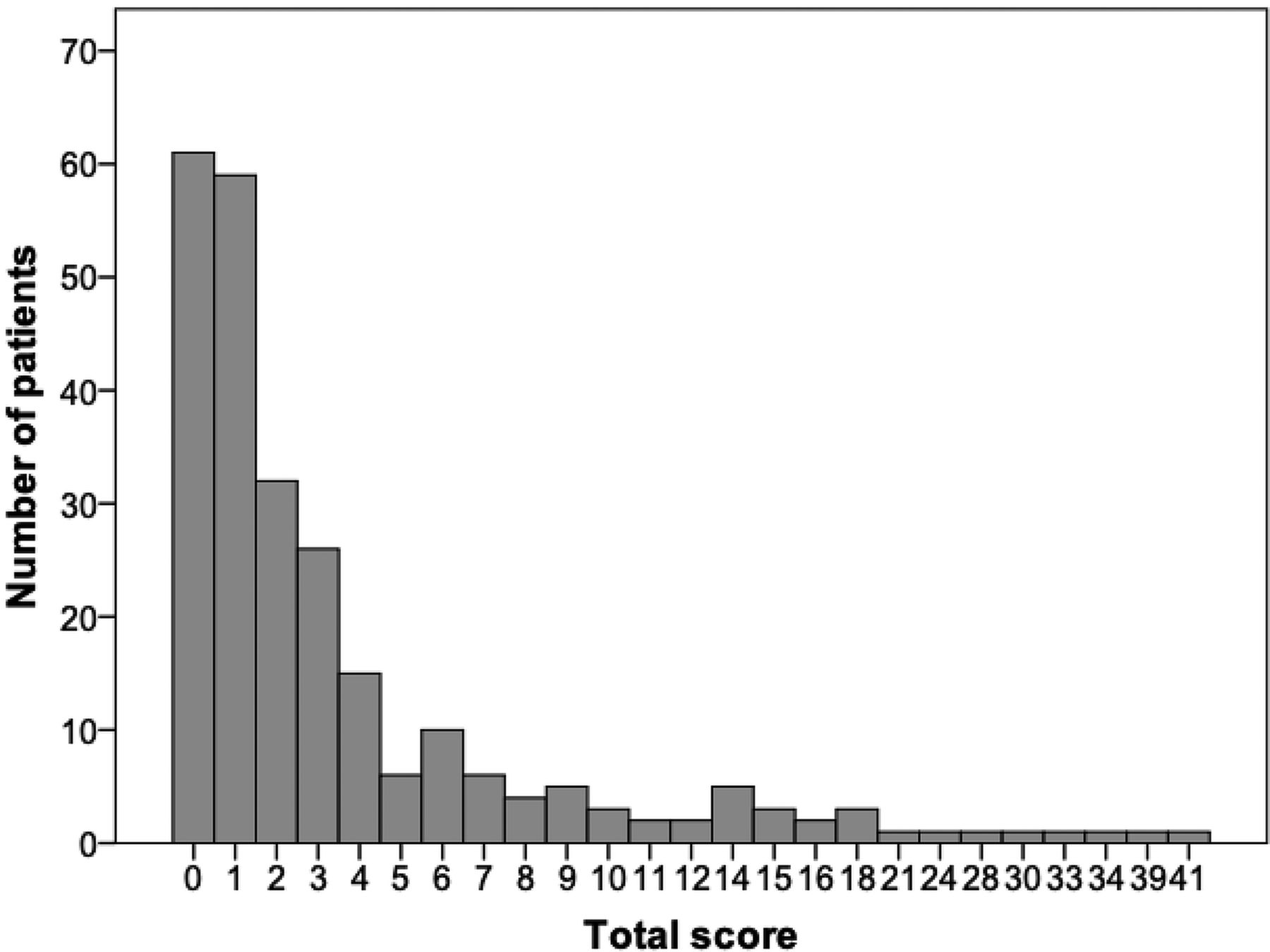
    The frequency distribution of the total score in the full cohort.
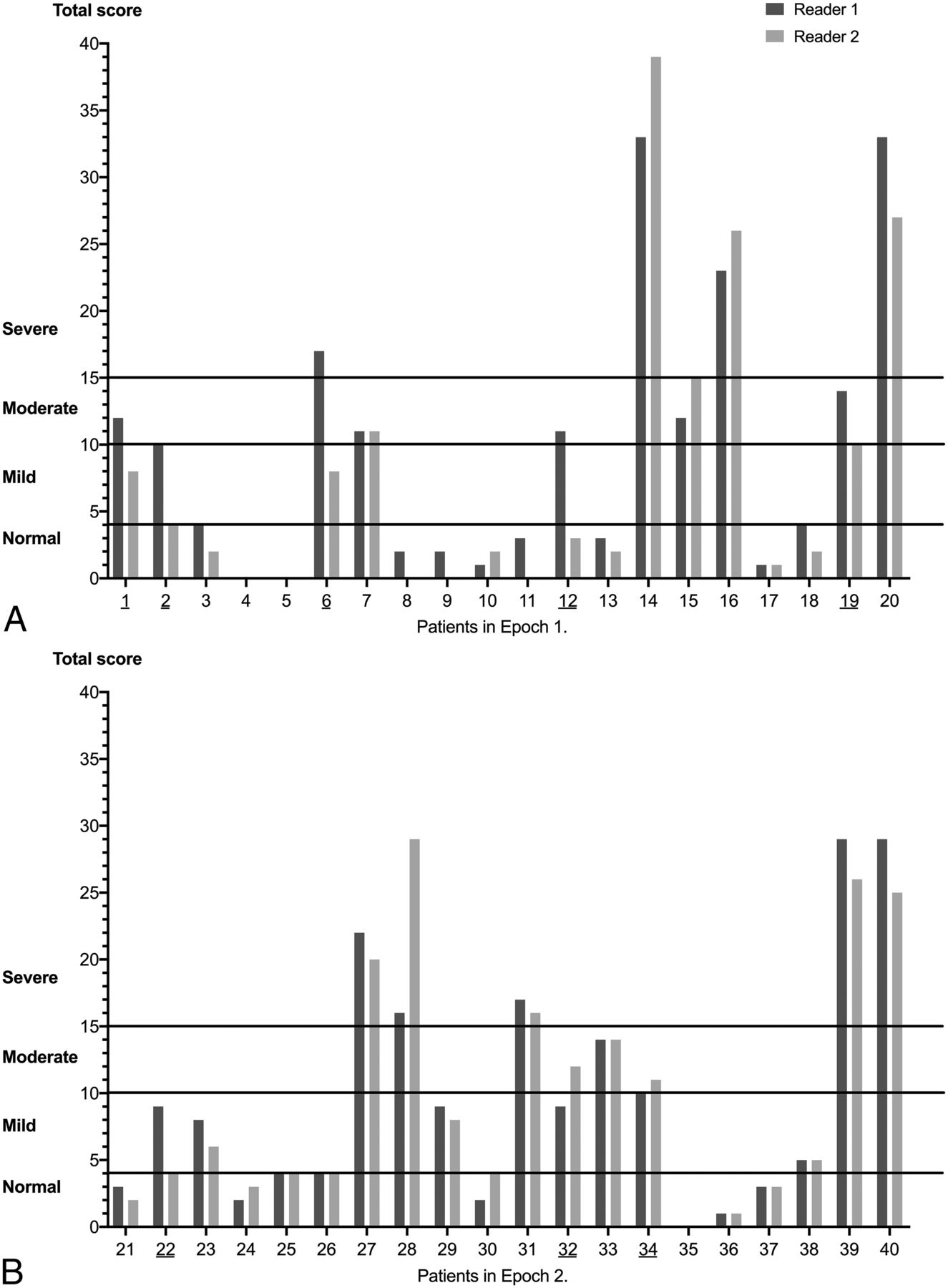
  • FIG 3.
    • Download figure
    • Open in new tab
    • Download powerpoint
    FIG 3.

    The severity of brain injury based on the total score for each subject in the first (A) and second epochs (B).

Tables

  • Figures
    • View popup
    Table 1:

    Demographics and prenatal data of the full cohorta

    Full Cohort (n = 252)
    Gestational age (wk)39 (38–40)
    Birth weight (g)3180 (2830–3544)
    Sex (% of males)145 (57.5%)
    Inborn166 (65.9%)
    Apgar at 1 min2 (1–4)
    Apgar at 5 min6 (5–7)
    Apgar at 10 min7 (6–8)
    UA pH7.04 (6.94–7.12)
    UA BD (mmol/L)11.7 (8.5–14.3)
    UA lactate (mmol/L)8.9 (6.4–10.4)
    UV pH7.13 (7.02–7.23)
    UV BD (mmol/L)10.1 (6.9–12.9)
    UV lactate (mmol/L)7.3 (5.6–9.2)
    Postnatal pH7.24 (7.15–7.30)
    Postnatal BD (mmol/L)9.2 (6.2–12.9)
    Postnatal lactate (mmol/L)7.9 (4.9–11.2)
    Stage of HIE
     Mild HIE124/234 (53.0%)
     Moderate HIE105/234 (44.9%)
     Severe HIE5/234 (2.1%)
    • Note:—BD indicates base deficit; UA, umbilical artery; UV, umbilical vein.

    • ↵a Nonparametric data are presented as median with interquartile range. The stage of HIE was based on the modified Sarnat stage. The Sarnat stage was available for 234 infants.

    • View popup
    Table 2:

    Interrater reliability for subscores and for total scorea

    Before Adjustment (n = 20)After Adjustment (n = 20)
    ICC95% CIICC95% CI
    Gray matter0.950.86–0.980.950.88–0.98
    GM with 1H-MR spectroscopy0.950.82–0.980.960.89–0.98
    WM/cortex0.970.91–0.990.980.96–0.99
    Cerebellum0.950.88–0.981.00–
    Additional0.860.62–0.940.660.14–0.87
    Total score0.960.89–0.990.960.89–0.98
    • Note:— GM indicates Gray Matter; ICC, intraclass correlation coefficient; WM, white matter; ‐, not applicable (NA).

    • ↵a The ICC with a 2-way random-effects model was calculated to assess the interrater variability between 2 experienced readers.

    • View popup
    Table 3:

    Severity of injury based on the total scoresa

    Severity of Brain InjuryBefore Adjustment (n = 20)After Adjustment (n = 20)
    Reader 1Reader 2P ValueReader 1Reader 2P Value
    Normal (≤4) (No.) (%)10 (50%)12 (60%).508 (40%)9 (45%) 1.00
    Mild (5–10) (No.) (%)1 (5%)3 (15%).636 (30%)3 (15%) .25
    Moderate (11–15) (No.) (%)5 (25%)2 (10%).251 (5%)3 (15%).50
    Severe (>15) (No.) (%)4 (20%)3 (15%)1.005 (25%)5 (25%) 1.00
    • ↵a A McNemar test was administered to determine whether there was a difference in the proportion of the severity of the brain injury between the 2 readers.

PreviousNext
Back to top
Advertisement
Print
Download PDF
Email Article
Cite this article
0 Responses
Respond to this article
Share
Bookmark this article
Interobserver Reliability of a MR Imaging Scoring System in Infants with Hypoxic-Ischemic Encephalopathy
E. Szakmar, H. Meunier, M. El-Dib, E. Yang, T.E. Inder
American Journal of Neuroradiology Mar 2021, DOI: 10.3174/ajnr.A7048
del.icio.us logo Twitter logo Facebook logo Mendeley logo
  • Tweet Widget
Purchase

Jump to section

  • Article
    • Abstract
    • ABBREVIATIONS:
    • MATERIALS AND METHODS
    • RESULTS
    • DISCUSSION
    • CONCLUSIONS
    • Footnotes
    • References
  • Figures & Data
  • Info & Metrics
  • Responses
  • References
  • PDF

Related Articles

  • No related articles found.
  • PubMed
  • Google Scholar

Cited By...

  • No citing articles found.
  • Crossref
  • Google Scholar

This article has not yet been cited by articles in journals that are participating in Crossref Cited-by Linking.

More in this TOC Section

  • Pediatric Glioma:Imaging Clusters, Molecular Traits
  • Clinical SVR of Fetal Brain MRI
  • Subpial Hemorrhage & Motor Outcomes in Neonates
Show more Pediatric Neuroimaging

Similar Articles

Advertisement

Indexed Content

  • Current Issue
  • Accepted Manuscripts
  • Article Preview
  • Past Issues
  • Editorials
  • Editor's Choice
  • Fellows' Journal Club
  • Letters to the Editor
  • Video Articles

Cases

  • Case Collection
  • Archive - Case of the Week
  • Archive - Case of the Month
  • Archive - Classic Case

More from AJNR

  • Trainee Corner
  • Imaging Protocols
  • MRI Safety Corner
  • Book Reviews

Multimedia

  • AJNR Podcasts
  • AJNR Scantastics

Resources

  • Turnaround Time
  • Submit a Manuscript
  • Submit a Video Article
  • Submit an eLetter to the Editor/Response
  • Manuscript Submission Guidelines
  • Statistical Tips
  • Fast Publishing of Accepted Manuscripts
  • Graphical Abstract Preparation
  • Imaging Protocol Submission
  • Evidence-Based Medicine Level Guide
  • Publishing Checklists
  • Author Policies
  • Become a Reviewer/Academy of Reviewers
  • News and Updates

About Us

  • About AJNR
  • Editorial Board
  • Editorial Board Alumni
  • Alerts
  • Permissions
  • Not an AJNR Subscriber? Join Now
  • Advertise with Us
  • Librarian Resources
  • Feedback
  • Terms and Conditions
  • AJNR Editorial Board Alumni

American Society of Neuroradiology

  • Not an ASNR Member? Join Now

© 2025 by the American Society of Neuroradiology All rights, including for text and data mining, AI training, and similar technologies, are reserved.
Print ISSN: 0195-6108 Online ISSN: 1936-959X

Powered by HighWire
Email this Article

Thank you for your interest in spreading the word on American Journal of Neuroradiology.

NOTE: We only request your email address so that the person you are recommending the page to knows that you wanted them to see it, and that it is not junk mail. We do not capture any email address.

Enter multiple addresses on separate lines or separate them with commas.
Interobserver Reliability of a MR Imaging Scoring System in Infants with Hypoxic-Ischemic Encephalopathy
(Your Name) has sent you a message from American Journal of Neuroradiology
(Your Name) thought you would like to see the American Journal of Neuroradiology web site.
CAPTCHA
This question is for testing whether or not you are a human visitor and to prevent automated spam submissions.
Cite this article
E. Szakmar, H. Meunier, M. El-Dib, E. Yang, T.E. Inder
Interobserver Reliability of a MR Imaging Scoring System in Infants with Hypoxic-Ischemic Encephalopathy
American Journal of Neuroradiology Mar 2021, DOI: 10.3174/ajnr.A7048

Citation Manager Formats

  • BibTeX
  • Bookends
  • EasyBib
  • EndNote (tagged)
  • EndNote 8 (xml)
  • Medlars
  • Mendeley
  • Papers
  • RefWorks Tagged
  • Ref Manager
  • RIS
  • Zotero

We use cookies on this site to enhance your user experience. By clicking any link on this page you are giving your consent for us to set cookies.