Advertisement

Main menu

  • Home
  • Content
    • Current Issue
    • Accepted Manuscripts
    • Article Preview
    • Past Issue Archive
    • Video Articles
    • AJNR Case Collection
    • Case of the Week Archive
    • Case of the Month Archive
    • Classic Case Archive
  • Special Collections
    • AJNR Awards
    • Low-Field MRI
    • Alzheimer Disease
    • ASNR Foundation Special Collection
    • Photon-Counting CT
    • View All
  • Multimedia
    • AJNR Podcasts
    • AJNR SCANtastic
    • Trainee Corner
    • MRI Safety Corner
    • Imaging Protocols
  • For Authors
    • Submit a Manuscript
    • Submit a Video Article
    • Submit an eLetter to the Editor/Response
    • Manuscript Submission Guidelines
    • Statistical Tips
    • Fast Publishing of Accepted Manuscripts
    • Graphical Abstract Preparation
    • Imaging Protocol Submission
    • Author Policies
  • About Us
    • About AJNR
    • Editorial Board
    • Editorial Board Alumni
  • More
    • Become a Reviewer/Academy of Reviewers
    • Subscribers
    • Permissions
    • Alerts
    • Feedback
    • Advertisers
    • ASNR Home

User menu

  • Alerts
  • Log in

Search

  • Advanced search
American Journal of Neuroradiology

American Journal of Neuroradiology

ASHNR American Society of Functional Neuroradiology ASHNR American Society of Pediatric Neuroradiology ASSR
  • Alerts
  • Log in

Advanced Search

  • Home
  • Content
    • Current Issue
    • Accepted Manuscripts
    • Article Preview
    • Past Issue Archive
    • Video Articles
    • AJNR Case Collection
    • Case of the Week Archive
    • Case of the Month Archive
    • Classic Case Archive
  • Special Collections
    • AJNR Awards
    • Low-Field MRI
    • Alzheimer Disease
    • ASNR Foundation Special Collection
    • Photon-Counting CT
    • View All
  • Multimedia
    • AJNR Podcasts
    • AJNR SCANtastic
    • Trainee Corner
    • MRI Safety Corner
    • Imaging Protocols
  • For Authors
    • Submit a Manuscript
    • Submit a Video Article
    • Submit an eLetter to the Editor/Response
    • Manuscript Submission Guidelines
    • Statistical Tips
    • Fast Publishing of Accepted Manuscripts
    • Graphical Abstract Preparation
    • Imaging Protocol Submission
    • Author Policies
  • About Us
    • About AJNR
    • Editorial Board
    • Editorial Board Alumni
  • More
    • Become a Reviewer/Academy of Reviewers
    • Subscribers
    • Permissions
    • Alerts
    • Feedback
    • Advertisers
    • ASNR Home
  • Follow AJNR on Twitter
  • Visit AJNR on Facebook
  • Follow AJNR on Instagram
  • Join AJNR on LinkedIn
  • RSS Feeds

AJNR Awards, New Junior Editors, and more. Read the latest AJNR updates

Research ArticleAdult Brain
Open Access

Brain and Lung Imaging Correlation in Patients with COVID-19: Could the Severity of Lung Disease Reflect the Prevalence of Acute Abnormalities on Neuroimaging? A Global Multicenter Observational Study

A. Mahammedi, A. Ramos, N. Bargalló, M. Gaskill, S. Kapur, L. Saba, H. Carrete, S. Sengupta, E. Salvador, A. Hilario, Y. Revilla, M. Sanchez, M. Perez-Nuñez, S. Bachir, B. Zhang, L. Oleaga, J. Sergio, L. Koren, P. Martin-Medina, L. Wang, M. Benegas, F. Ostos, G. Gonzalez-Ortega, P. Calleja, G. Udstuen, B. Williamson, V. Khandwala, S. Chadalavada, D. Woo and A. Vagal
American Journal of Neuroradiology March 2021, DOI: https://doi.org/10.3174/ajnr.A7072
A. Mahammedi
aFrom the Departments of Neuroradiology, (A.M., A.V., M.G., L.W., G.U., B.W., V.K.), Cardiopulmonary Imaging, (S.K.), Neurology (S.S., D.W.), and Department of Informatics and Interventional Radiology, (S.C.), University of Cincinnati Medical Center, Cincinnati, Ohio
  • Find this author on Google Scholar
  • Find this author on PubMed
  • Search for this author on this site
  • ORCID record for A. Mahammedi
A. Ramos
bDepartments of Epidemiology and Biostatistics (B.Z.) and Pediatrics, (S.B.), Cincinnati Children’s Hospital Medical Center, Cincinnati, Ohio
  • Find this author on Google Scholar
  • Find this author on PubMed
  • Search for this author on this site
  • ORCID record for A. Ramos
N. Bargalló
dDepartments of Neuroradiology (L.O., N.B.) and Cardiopulmonary Imaging (M.S., M.B., J.S.), Hospital Clínic de Barcelona, Sunyer Biomedical Research Institute, Barcelona, Spain
  • Find this author on Google Scholar
  • Find this author on PubMed
  • Search for this author on this site
  • ORCID record for N. Bargalló
M. Gaskill
aFrom the Departments of Neuroradiology, (A.M., A.V., M.G., L.W., G.U., B.W., V.K.), Cardiopulmonary Imaging, (S.K.), Neurology (S.S., D.W.), and Department of Informatics and Interventional Radiology, (S.C.), University of Cincinnati Medical Center, Cincinnati, Ohio
  • Find this author on Google Scholar
  • Find this author on PubMed
  • Search for this author on this site
  • ORCID record for M. Gaskill
S. Kapur
aFrom the Departments of Neuroradiology, (A.M., A.V., M.G., L.W., G.U., B.W., V.K.), Cardiopulmonary Imaging, (S.K.), Neurology (S.S., D.W.), and Department of Informatics and Interventional Radiology, (S.C.), University of Cincinnati Medical Center, Cincinnati, Ohio
  • Find this author on Google Scholar
  • Find this author on PubMed
  • Search for this author on this site
  • ORCID record for S. Kapur
L. Saba
eDepartment of Neuroradiology (L.S.), Azienda Ospedaliero Universitaria di Cagliari, Monserrato (Cagliari), Italy
  • Find this author on Google Scholar
  • Find this author on PubMed
  • Search for this author on this site
  • ORCID record for L. Saba
H. Carrete Jr
fDepartment of Neuroradiology (H.C.), Escola Paulista de Medicina, Federal University of São Paulo, São Paulo, Brazil
  • Find this author on Google Scholar
  • Find this author on PubMed
  • Search for this author on this site
  • ORCID record for H. Carrete Jr
S. Sengupta
aFrom the Departments of Neuroradiology, (A.M., A.V., M.G., L.W., G.U., B.W., V.K.), Cardiopulmonary Imaging, (S.K.), Neurology (S.S., D.W.), and Department of Informatics and Interventional Radiology, (S.C.), University of Cincinnati Medical Center, Cincinnati, Ohio
  • Find this author on Google Scholar
  • Find this author on PubMed
  • Search for this author on this site
  • ORCID record for S. Sengupta
E. Salvador
cDepartments of Neuroradiology (A.R., E.S., A.H., L.K., P.M.-M.), Cardiopulmonary Imaging (Y.R., M.P.-N.) and Neurology (F.O., G.G.-O., P.C.), Hospital Universitario 12 de Octubre, Madrid, Spain
  • Find this author on Google Scholar
  • Find this author on PubMed
  • Search for this author on this site
  • ORCID record for E. Salvador
A. Hilario
cDepartments of Neuroradiology (A.R., E.S., A.H., L.K., P.M.-M.), Cardiopulmonary Imaging (Y.R., M.P.-N.) and Neurology (F.O., G.G.-O., P.C.), Hospital Universitario 12 de Octubre, Madrid, Spain
  • Find this author on Google Scholar
  • Find this author on PubMed
  • Search for this author on this site
  • ORCID record for A. Hilario
Y. Revilla
cDepartments of Neuroradiology (A.R., E.S., A.H., L.K., P.M.-M.), Cardiopulmonary Imaging (Y.R., M.P.-N.) and Neurology (F.O., G.G.-O., P.C.), Hospital Universitario 12 de Octubre, Madrid, Spain
  • Find this author on Google Scholar
  • Find this author on PubMed
  • Search for this author on this site
  • ORCID record for Y. Revilla
M. Sanchez
dDepartments of Neuroradiology (L.O., N.B.) and Cardiopulmonary Imaging (M.S., M.B., J.S.), Hospital Clínic de Barcelona, Sunyer Biomedical Research Institute, Barcelona, Spain
  • Find this author on Google Scholar
  • Find this author on PubMed
  • Search for this author on this site
  • ORCID record for M. Sanchez
M. Perez-Nuñez
cDepartments of Neuroradiology (A.R., E.S., A.H., L.K., P.M.-M.), Cardiopulmonary Imaging (Y.R., M.P.-N.) and Neurology (F.O., G.G.-O., P.C.), Hospital Universitario 12 de Octubre, Madrid, Spain
  • Find this author on Google Scholar
  • Find this author on PubMed
  • Search for this author on this site
  • ORCID record for M. Perez-Nuñez
S. Bachir
fDepartment of Neuroradiology (H.C.), Escola Paulista de Medicina, Federal University of São Paulo, São Paulo, Brazil
  • Find this author on Google Scholar
  • Find this author on PubMed
  • Search for this author on this site
  • ORCID record for S. Bachir
B. Zhang
bDepartments of Epidemiology and Biostatistics (B.Z.) and Pediatrics, (S.B.), Cincinnati Children’s Hospital Medical Center, Cincinnati, Ohio
  • Find this author on Google Scholar
  • Find this author on PubMed
  • Search for this author on this site
  • ORCID record for B. Zhang
L. Oleaga
dDepartments of Neuroradiology (L.O., N.B.) and Cardiopulmonary Imaging (M.S., M.B., J.S.), Hospital Clínic de Barcelona, Sunyer Biomedical Research Institute, Barcelona, Spain
  • Find this author on Google Scholar
  • Find this author on PubMed
  • Search for this author on this site
  • ORCID record for L. Oleaga
J. Sergio
dDepartments of Neuroradiology (L.O., N.B.) and Cardiopulmonary Imaging (M.S., M.B., J.S.), Hospital Clínic de Barcelona, Sunyer Biomedical Research Institute, Barcelona, Spain
  • Find this author on Google Scholar
  • Find this author on PubMed
  • Search for this author on this site
  • ORCID record for J. Sergio
L. Koren
cDepartments of Neuroradiology (A.R., E.S., A.H., L.K., P.M.-M.), Cardiopulmonary Imaging (Y.R., M.P.-N.) and Neurology (F.O., G.G.-O., P.C.), Hospital Universitario 12 de Octubre, Madrid, Spain
  • Find this author on Google Scholar
  • Find this author on PubMed
  • Search for this author on this site
  • ORCID record for L. Koren
P. Martin-Medina
cDepartments of Neuroradiology (A.R., E.S., A.H., L.K., P.M.-M.), Cardiopulmonary Imaging (Y.R., M.P.-N.) and Neurology (F.O., G.G.-O., P.C.), Hospital Universitario 12 de Octubre, Madrid, Spain
  • Find this author on Google Scholar
  • Find this author on PubMed
  • Search for this author on this site
  • ORCID record for P. Martin-Medina
L. Wang
aFrom the Departments of Neuroradiology, (A.M., A.V., M.G., L.W., G.U., B.W., V.K.), Cardiopulmonary Imaging, (S.K.), Neurology (S.S., D.W.), and Department of Informatics and Interventional Radiology, (S.C.), University of Cincinnati Medical Center, Cincinnati, Ohio
  • Find this author on Google Scholar
  • Find this author on PubMed
  • Search for this author on this site
  • ORCID record for L. Wang
M. Benegas
dDepartments of Neuroradiology (L.O., N.B.) and Cardiopulmonary Imaging (M.S., M.B., J.S.), Hospital Clínic de Barcelona, Sunyer Biomedical Research Institute, Barcelona, Spain
  • Find this author on Google Scholar
  • Find this author on PubMed
  • Search for this author on this site
  • ORCID record for M. Benegas
F. Ostos
cDepartments of Neuroradiology (A.R., E.S., A.H., L.K., P.M.-M.), Cardiopulmonary Imaging (Y.R., M.P.-N.) and Neurology (F.O., G.G.-O., P.C.), Hospital Universitario 12 de Octubre, Madrid, Spain
  • Find this author on Google Scholar
  • Find this author on PubMed
  • Search for this author on this site
  • ORCID record for F. Ostos
G. Gonzalez-Ortega
cDepartments of Neuroradiology (A.R., E.S., A.H., L.K., P.M.-M.), Cardiopulmonary Imaging (Y.R., M.P.-N.) and Neurology (F.O., G.G.-O., P.C.), Hospital Universitario 12 de Octubre, Madrid, Spain
  • Find this author on Google Scholar
  • Find this author on PubMed
  • Search for this author on this site
  • ORCID record for G. Gonzalez-Ortega
P. Calleja
cDepartments of Neuroradiology (A.R., E.S., A.H., L.K., P.M.-M.), Cardiopulmonary Imaging (Y.R., M.P.-N.) and Neurology (F.O., G.G.-O., P.C.), Hospital Universitario 12 de Octubre, Madrid, Spain
  • Find this author on Google Scholar
  • Find this author on PubMed
  • Search for this author on this site
  • ORCID record for P. Calleja
G. Udstuen
aFrom the Departments of Neuroradiology, (A.M., A.V., M.G., L.W., G.U., B.W., V.K.), Cardiopulmonary Imaging, (S.K.), Neurology (S.S., D.W.), and Department of Informatics and Interventional Radiology, (S.C.), University of Cincinnati Medical Center, Cincinnati, Ohio
  • Find this author on Google Scholar
  • Find this author on PubMed
  • Search for this author on this site
  • ORCID record for G. Udstuen
B. Williamson
aFrom the Departments of Neuroradiology, (A.M., A.V., M.G., L.W., G.U., B.W., V.K.), Cardiopulmonary Imaging, (S.K.), Neurology (S.S., D.W.), and Department of Informatics and Interventional Radiology, (S.C.), University of Cincinnati Medical Center, Cincinnati, Ohio
  • Find this author on Google Scholar
  • Find this author on PubMed
  • Search for this author on this site
  • ORCID record for B. Williamson
V. Khandwala
aFrom the Departments of Neuroradiology, (A.M., A.V., M.G., L.W., G.U., B.W., V.K.), Cardiopulmonary Imaging, (S.K.), Neurology (S.S., D.W.), and Department of Informatics and Interventional Radiology, (S.C.), University of Cincinnati Medical Center, Cincinnati, Ohio
  • Find this author on Google Scholar
  • Find this author on PubMed
  • Search for this author on this site
  • ORCID record for V. Khandwala
S. Chadalavada
aFrom the Departments of Neuroradiology, (A.M., A.V., M.G., L.W., G.U., B.W., V.K.), Cardiopulmonary Imaging, (S.K.), Neurology (S.S., D.W.), and Department of Informatics and Interventional Radiology, (S.C.), University of Cincinnati Medical Center, Cincinnati, Ohio
  • Find this author on Google Scholar
  • Find this author on PubMed
  • Search for this author on this site
  • ORCID record for S. Chadalavada
D. Woo
aFrom the Departments of Neuroradiology, (A.M., A.V., M.G., L.W., G.U., B.W., V.K.), Cardiopulmonary Imaging, (S.K.), Neurology (S.S., D.W.), and Department of Informatics and Interventional Radiology, (S.C.), University of Cincinnati Medical Center, Cincinnati, Ohio
  • Find this author on Google Scholar
  • Find this author on PubMed
  • Search for this author on this site
  • ORCID record for D. Woo
A. Vagal
aFrom the Departments of Neuroradiology, (A.M., A.V., M.G., L.W., G.U., B.W., V.K.), Cardiopulmonary Imaging, (S.K.), Neurology (S.S., D.W.), and Department of Informatics and Interventional Radiology, (S.C.), University of Cincinnati Medical Center, Cincinnati, Ohio
  • Find this author on Google Scholar
  • Find this author on PubMed
  • Search for this author on this site
  • ORCID record for A. Vagal
  • Article
  • Figures & Data
  • Supplemental
  • Info & Metrics
  • Responses
  • References
  • PDF
Loading

Article Figures & Data

Figures

  • Tables
  • FIG 1.
    • Download figure
    • Open in new tab
    • Download powerpoint
    FIG 1.

    Flow chart of the study.
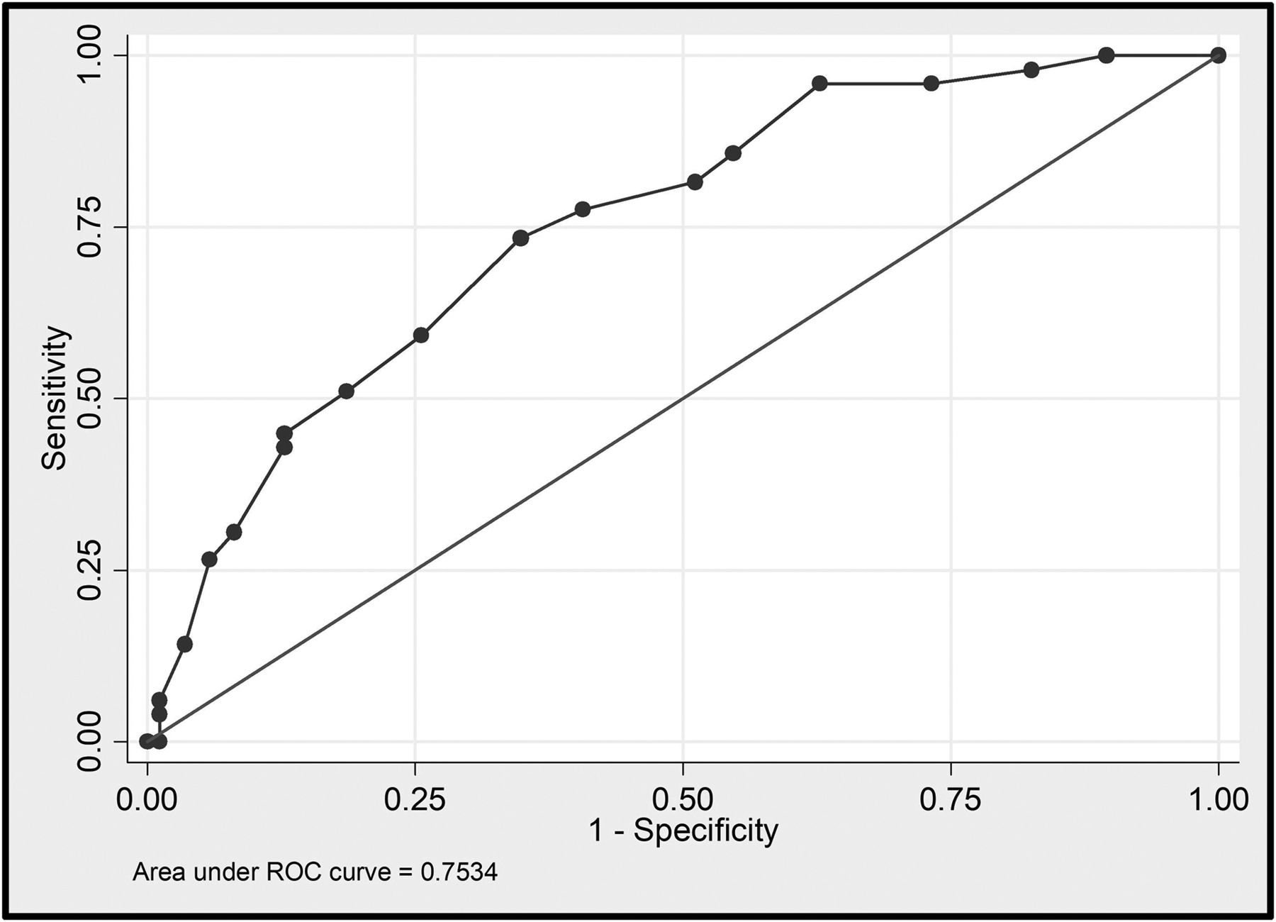
  • FIG 2.
    • Download figure
    • Open in new tab
    • Download powerpoint
    FIG 2.

    Receiver operating characteristic curve showing the sensitivity and specificity of the CT lung severity score as a predictor of acute neuroimaging abnormalities in patients with COVID-19 with neurologic symptoms. The area under the curve corresponds to the accuracy. ROC indicates receiver operating characteristic.
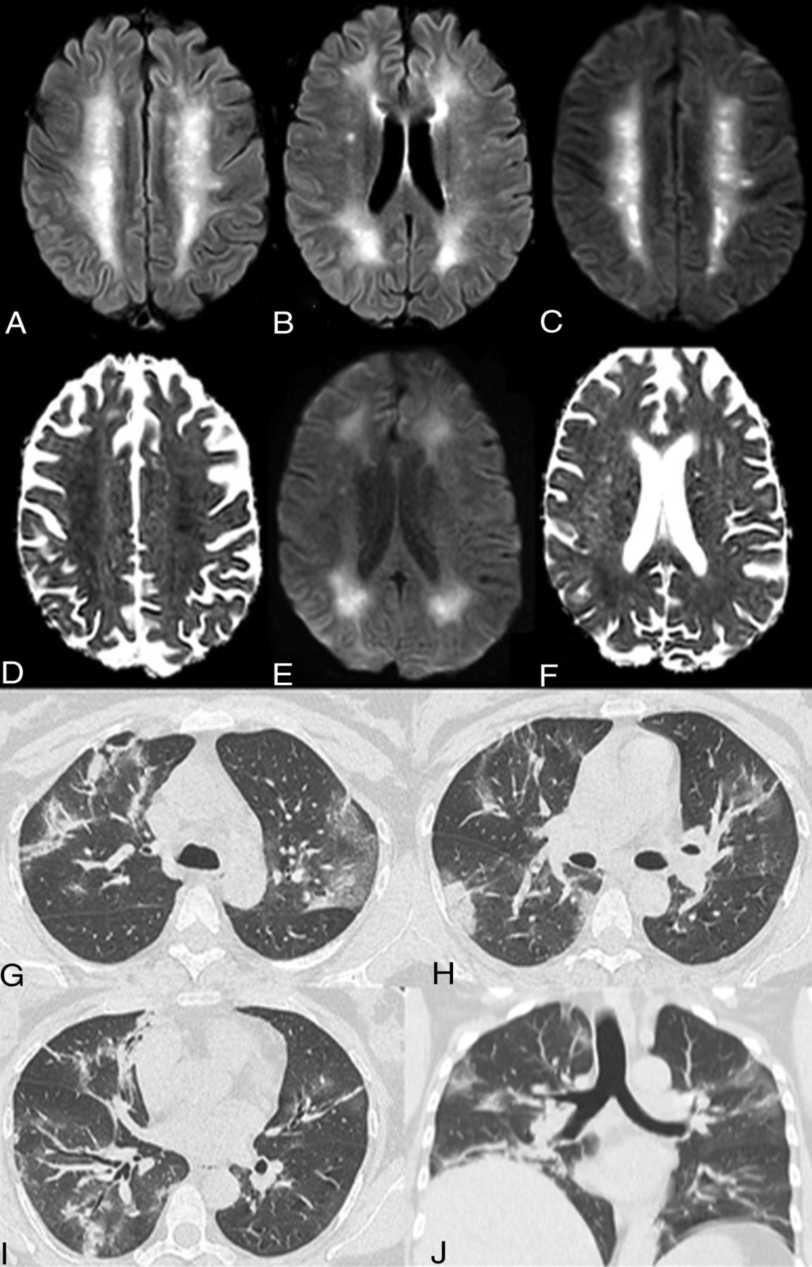
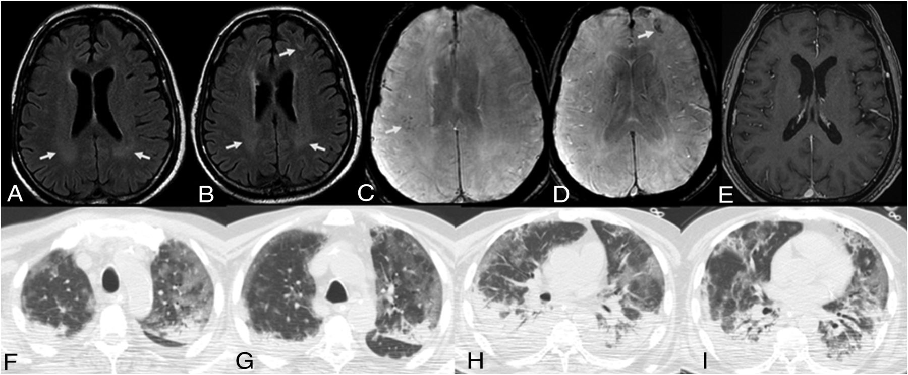
  • FIG 3.
    • Download figure
    • Open in new tab
    • Download powerpoint
    FIG 3.

    Acute leukoencephalopathy. A 48-year-old man without a history of seizures presented with convulsions and altered mental status. Extensive and confluent symmetric deep and subcortical white matter FLAIR hyperintensities in the bilateral centra semiovale (A) and periventricular frontal and parietal regions (B) with associated mild restricted diffusion on DWI/ADC images, most prominent in the centra semiovale (C and D) and peritrigonal regions (E and F). No associated enhancement or microbleed was seen on the T1 postcontrast and SWI (not shown). Coronal MPR and axial noncontrast images in lung windows demonstrate mixed ground-glass and consolidative opacities in all lobes with a lower lung and peripheral predominance (G–J). The CT lung severity score was 16 (right upper lobe, right lower lobe, left upper lobe, left lower lobe, 25%–49% and right middle lobe 50%–75%). The chest CT scan was obtained 8 days after the initial onset of respiratory symptoms.
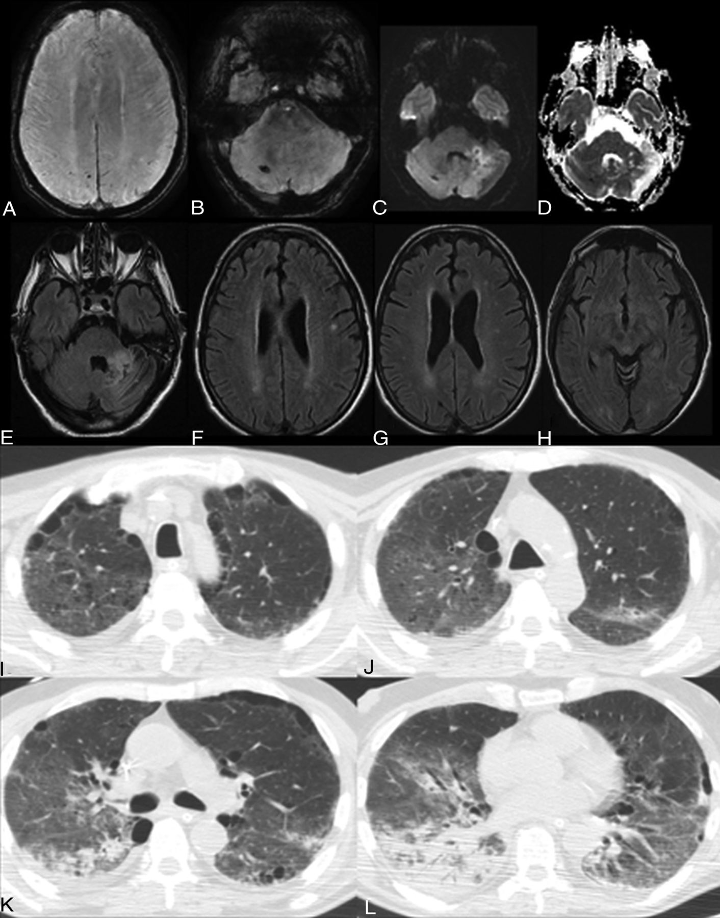
  • FIG 4.
    • Download figure
    • Open in new tab
    • Download powerpoint
    FIG 4.

    Acute infarct, multifocal microbleeds, and subcortical white matter FLAIR hyperintensity. A 65-year-old man presented with altered mental status and ischemic stroke. Available prior brain MR imaging findings from 1 month ago were normal. A and B, New punctate microbleeds on gradient-echo sequences in the right superior parietal lobule, left inferior parietal lobule, and right cerebellum. Note a small area of restricted diffusion in the anterior left cerebellum on DWI/ADC images (C–D) with FLAIR hyperintensities (E), consistent with acute infarct. New punctate deep and subcortical white matter FLAIR hyperintensities without associated hemorrhage or restricted diffusion in the left-greater-than-right frontal (F), parietal (G), and occipital lobes (H). Axial chest CT images show right apical GGOs and biapical paraseptal emphysema (I). Bilateral GGOs are right greater than left (J). Patchy consolidations in the apical segment of the lower lobe of the right lung (K). Extensive consolidation with an air bronchogram in the right lower lobe (L). The CT lung severity score was 14. The chest CT scan was obtained 10 days after initial onset of respiratory symptoms.
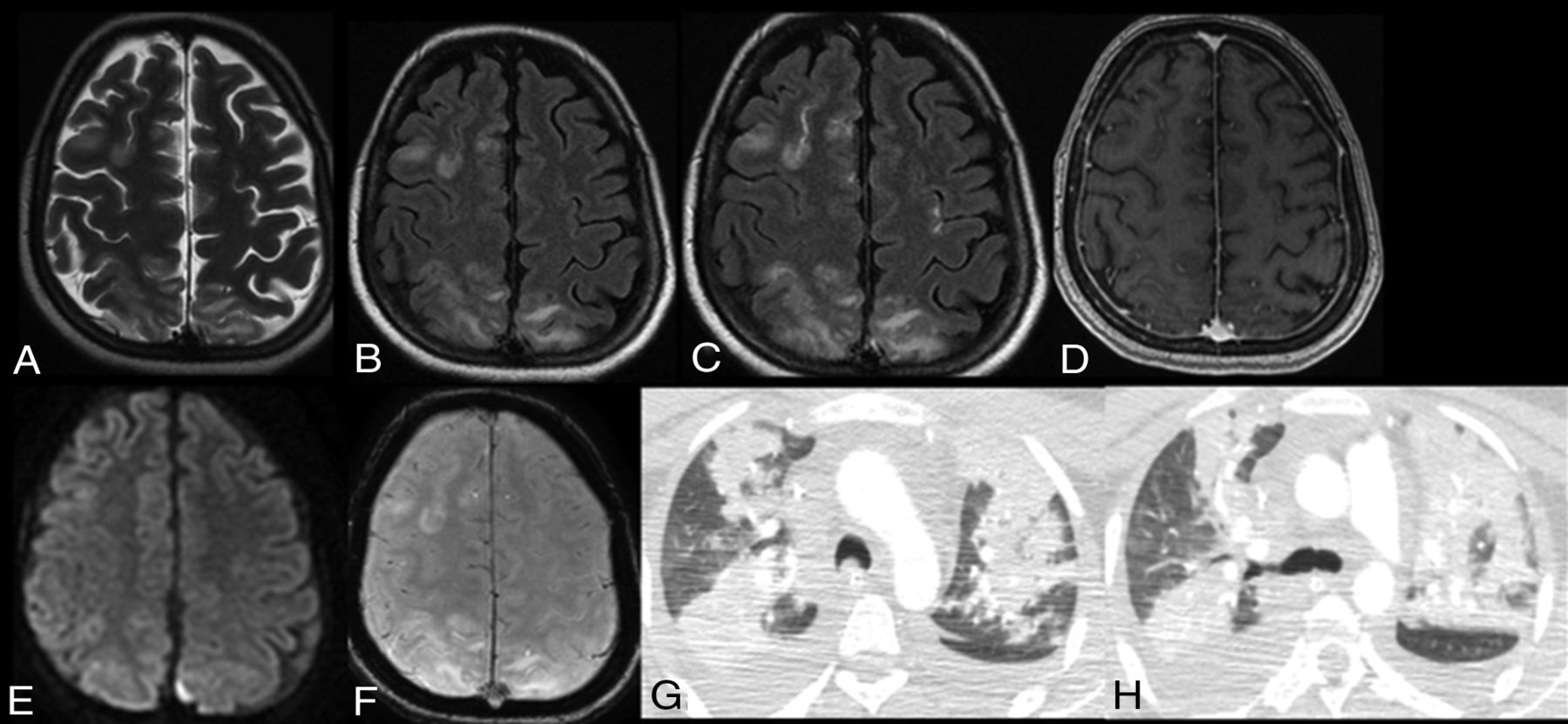
  • FIG 5.
    • Download figure
    • Open in new tab
    • Download powerpoint
    FIG 5.

    PRES. A 25-year-old woman presented with altered mental status and ischemic stroke. A and B, Extensive cortical and subcortical areas of T2/FLAIR hyperintensity in the right frontal and bilateral parietal lobes. Associated patchy and confluent leptomeningeal enhancement is seen only on FLAIR postcontrast image (C) but not on T1 postcontrast image (D). Note no restriction diffusion or hemosiderin on DWI and gradient-echo sequences (E and F). Axial chest CT scans show new extensive bilateral patchy consolidations in the upper lobes with bilateral right-more-than-left pleural effusions (G and H). The CT lung severity score was 12. The chest CT scan was obtained 7 days after the initial onset of respiratory symptoms.

  • FIG 6.
    • Download figure
    • Open in new tab
    • Download powerpoint
    FIG 6.

    Multifocal microbleeds and deep white matter FLAIR hyperintensities. A 63-year-old man presented with altered mental status. Available prior brain MR imaging findings from 3 months ago were normal. Note confluent periventricular white matter T2/FLAIR hyperintensities, most prominent in the peritrigonal regions (A and B), with new punctate microbleeds on gradient-echo sequences in the right frontoparietal regions (C) and left frontal lobe (D). No associated enhancement is seen on the T1 postcontrast image. Axial chest CT images in lung windows show bilateral, peripheral, predominant ground-glass opacities and dependent consolidations in the upper and lower lobes (F–I). Note air bronchograms in both lower lobes (H and I). The CT lung severity score was 18. The chest CT scan was obtained 11 days after the initial onset of respiratory symptoms.
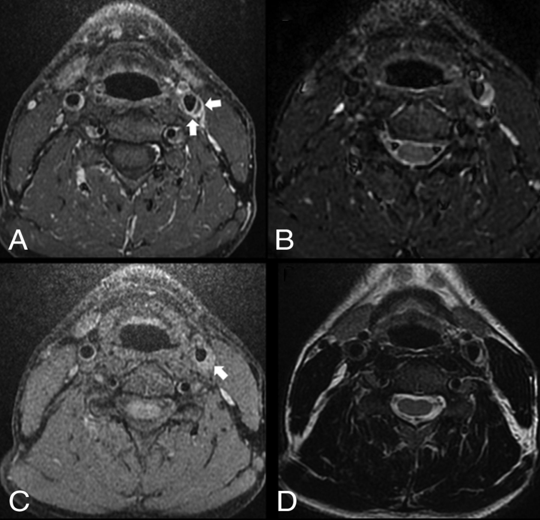
  • FIG 7.
    • Download figure
    • Open in new tab
    • Download powerpoint
    FIG 7.

    Left TIPIC (carotidynia). A 40-year-old man without significant medical history or known trauma presented with myoclonus and acute tenderness overlying the left carotid artery with increased pulsation. Axial T1 fat-saturated postcontrast image shows asymmetric enhancement and nonstenotic thickening of the left common carotid wall (A, arrows), with T1 precontrast hyperintensity of a crescent-shaped subintimal focus (C, arrow), which is also hyperintense on T2 fat-saturated and T2-weighted (B and D) images. Differential considerations include carotidynia or dissection. However, given the significantly improved symptoms after anti-inflammatory treatment, the lack of luminal stenosis, and the patient’s neck pain characteristics, findings are most consistent with carotidynia. No follow-up imaging was available.

Tables

  • Figures
  • Neuroimaging characteristics of hospitalized patients with COVID-19 with new onset of neurologic symptomsa

    Neuroimaging CharacteristicsAll Patients (n = 135), CT (n = 132) or MR imaging (n = 36)
    T2/FLAIR white matter signal abnormality
     Nonconfluent punctate deep and subcortical white matter disease22/36 (61)
     Isolated, nonspecific9/36 (25)
     Associated restricted diffusion onlyb6/36 (17)
     Associated microhemorrhage onlyb3/36 (8)
     Associated microhemorrhage and restricted diffusionb4/36 (11)
    Confluent symmetric T2 hyperintensity without restricted diffusion or hemorrhage2/36 (5)
    Confluent symmetric T2 hyperintensity with mild restricted diffusionc2/36 (5)
    Enhancement (MR imaging with and without IV contrast)LeptomeningealdParenchymale Cranial nerves2/17 (12)2/17 (12)0/17 (0)
    Acute ischemic infarctsVascular territorySmall/watershed infarctsCardioembolic36/135 (27)21/135 (15)10/135 (7)5/135 (4)
    Intracranial hemorrhagesParenchymalSubarachnoidMicrohemorrhage14/135 (10)3/135 (2)4/135 (3)7/36 (19)
    Acute leukoencephalopathyc4/36 (11)
    PRES3/36 (8)
    Hypoxic-ischemic encephalopathy2/36 (5)
    TIPICf2/7 (28)
    • ↵a Numbers in parentheses are percentages.

    • bThought to be most consistent with acute lacunar infarcts with a few associated microhemorrhages (Figs 4 and 6).

    • cAcute leukoencephalopathy. A 48-year-old man without a history of seizures presented with convulsion and altered mental status (Fig 3).

    • dSeen on FLAIR postcontrast only and likely related to PRES (Fig 5).

    • eSeptic emboli with atypical left parietal abscess. A 70-year-old woman with high blood pressure, chronic kidney disease, and type 2 diabetes mellitus. Long admission in the intensive care unit with intubation for COVID-19 and bilateral pneumonia. She presented with alteration of mental state and difficulty to progress in the weaning process. No history of malignancy (Online Supplemental Data).

    • fLeft TIPIC (carotidynia). A 40 -year-old man without significant medical history or known trauma presented with myoclonus and acute tenderness overlying the left carotid artery with increased pulsation. His symptoms significantly improved after steroid therapy (Fig 7).

PreviousNext
Back to top
Advertisement
Print
Download PDF
Email Article

Thank you for your interest in spreading the word on American Journal of Neuroradiology.

NOTE: We only request your email address so that the person you are recommending the page to knows that you wanted them to see it, and that it is not junk mail. We do not capture any email address.

Enter multiple addresses on separate lines or separate them with commas.
Brain and Lung Imaging Correlation in Patients with COVID-19: Could the Severity of Lung Disease Reflect the Prevalence of Acute Abnormalities on Neuroimaging? A Global Multicenter Observational Study
(Your Name) has sent you a message from American Journal of Neuroradiology
(Your Name) thought you would like to see the American Journal of Neuroradiology web site.
CAPTCHA
This question is for testing whether or not you are a human visitor and to prevent automated spam submissions.
Cite this article
A. Mahammedi, A. Ramos, N. Bargalló, M. Gaskill, S. Kapur, L. Saba, H. Carrete, S. Sengupta, E. Salvador, A. Hilario, Y. Revilla, M. Sanchez, M. Perez-Nuñez, S. Bachir, B. Zhang, L. Oleaga, J. Sergio, L. Koren, P. Martin-Medina, L. Wang, M. Benegas, F. Ostos, G. Gonzalez-Ortega, P. Calleja, G. Udstuen, B. Williamson, V. Khandwala, S. Chadalavada, D. Woo, A. Vagal
Brain and Lung Imaging Correlation in Patients with COVID-19: Could the Severity of Lung Disease Reflect the Prevalence of Acute Abnormalities on Neuroimaging? A Global Multicenter Observational Study
American Journal of Neuroradiology Mar 2021, DOI: 10.3174/ajnr.A7072

Citation Manager Formats

  • BibTeX
  • Bookends
  • EasyBib
  • EndNote (tagged)
  • EndNote 8 (xml)
  • Medlars
  • Mendeley
  • Papers
  • RefWorks Tagged
  • Ref Manager
  • RIS
  • Zotero
0 Responses
Respond to this article
Share
Bookmark this article
Brain and Lung Imaging Correlation in Patients with COVID-19: Could the Severity of Lung Disease Reflect the Prevalence of Acute Abnormalities on Neuroimaging? A Global Multicenter Observational Study
A. Mahammedi, A. Ramos, N. Bargalló, M. Gaskill, S. Kapur, L. Saba, H. Carrete, S. Sengupta, E. Salvador, A. Hilario, Y. Revilla, M. Sanchez, M. Perez-Nuñez, S. Bachir, B. Zhang, L. Oleaga, J. Sergio, L. Koren, P. Martin-Medina, L. Wang, M. Benegas, F. Ostos, G. Gonzalez-Ortega, P. Calleja, G. Udstuen, B. Williamson, V. Khandwala, S. Chadalavada, D. Woo, A. Vagal
American Journal of Neuroradiology Mar 2021, DOI: 10.3174/ajnr.A7072
del.icio.us logo Twitter logo Facebook logo Mendeley logo
  • Tweet Widget
  • Facebook Like
  • Google Plus One
Purchase

Jump to section

  • Article
    • Abstract
    • ABBREVIATIONS:
    • MATERIALS AND METHODS
    • RESULTS
    • DISCUSSION
    • CONCLUSIONS
    • Acknowledgments
    • Footnotes
    • References
  • Figures & Data
  • Supplemental
  • Info & Metrics
  • Responses
  • References
  • PDF

Related Articles

  • PubMed
  • Google Scholar

Cited By...

  • No citing articles found.
  • Crossref
  • Google Scholar

This article has not yet been cited by articles in journals that are participating in Crossref Cited-by Linking.

More in this TOC Section

  • Diagnostic Neuroradiology of Monoclonal Antibodies
  • Clinical Outcomes After Chiari I Decompression
  • Segmentation of Brain Metastases with BLAST
Show more Adult Brain

Similar Articles

Advertisement

Indexed Content

  • Current Issue
  • Accepted Manuscripts
  • Article Preview
  • Past Issues
  • Editorials
  • Editor's Choice
  • Fellows' Journal Club
  • Letters to the Editor
  • Video Articles

Cases

  • Case Collection
  • Archive - Case of the Week
  • Archive - Case of the Month
  • Archive - Classic Case

More from AJNR

  • Trainee Corner
  • Imaging Protocols
  • MRI Safety Corner
  • Book Reviews

Multimedia

  • AJNR Podcasts
  • AJNR Scantastics

Resources

  • Turnaround Time
  • Submit a Manuscript
  • Submit a Video Article
  • Submit an eLetter to the Editor/Response
  • Manuscript Submission Guidelines
  • Statistical Tips
  • Fast Publishing of Accepted Manuscripts
  • Graphical Abstract Preparation
  • Imaging Protocol Submission
  • Evidence-Based Medicine Level Guide
  • Publishing Checklists
  • Author Policies
  • Become a Reviewer/Academy of Reviewers
  • News and Updates

About Us

  • About AJNR
  • Editorial Board
  • Editorial Board Alumni
  • Alerts
  • Permissions
  • Not an AJNR Subscriber? Join Now
  • Advertise with Us
  • Librarian Resources
  • Feedback
  • Terms and Conditions
  • AJNR Editorial Board Alumni

American Society of Neuroradiology

  • Not an ASNR Member? Join Now

© 2025 by the American Society of Neuroradiology All rights, including for text and data mining, AI training, and similar technologies, are reserved.
Print ISSN: 0195-6108 Online ISSN: 1936-959X

Powered by HighWire