
Article Figures & Data
Figures
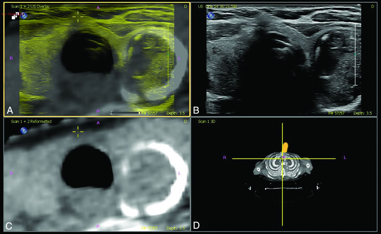
- FIG 1.
Matched plane fusion, manual correction. A, Overlay US (yellow) and CT (gray). B, US image. C, Reformatted CT image. D, Volume representation of CT image and probe location.
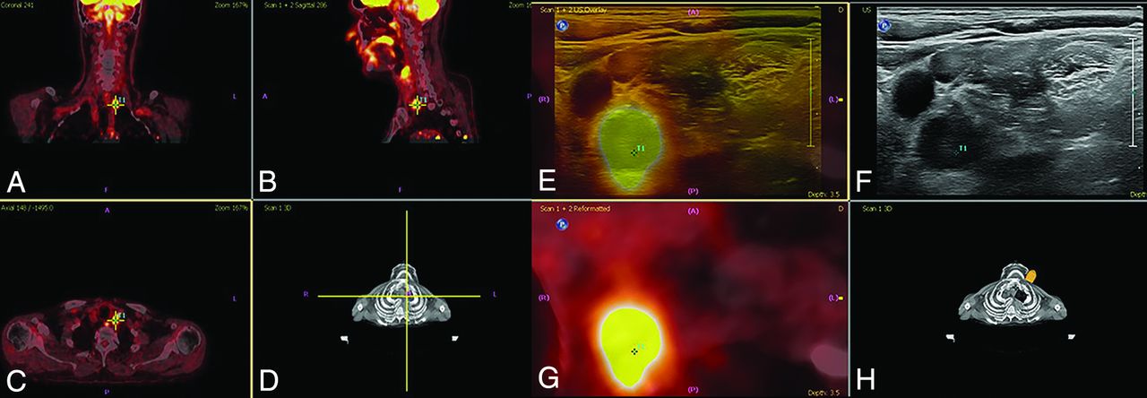
- FIG 2.
Target planning and real-time fusion of FDG-PET-positive lymph nodes to identify PET-positive nodes. A, Coronal view. B, Sagittal view. C, Axial view. D, Volume representation of FDG-PET/CT image and probe location, PET-positive nodes where targeted, and real-time image fused with ultrasound. E, Overlay US (yellow) and PET/CT (gray). F, US image. G, Reformatted PET/CT image. H, Volume representation of the CT image and probe location. Fusion of PET and CT and target planning took place using the electromagnetic navigation system PercuNav. First, routine ultrasound and routine USgFNAC were performed. Second, ultrasound and FDG-PET-positive nodes were real-time fused. USgFNAC in PET-positive nodes was confirmed, and additional fused-USgFNAC of missed PET-positive nodes was performed.
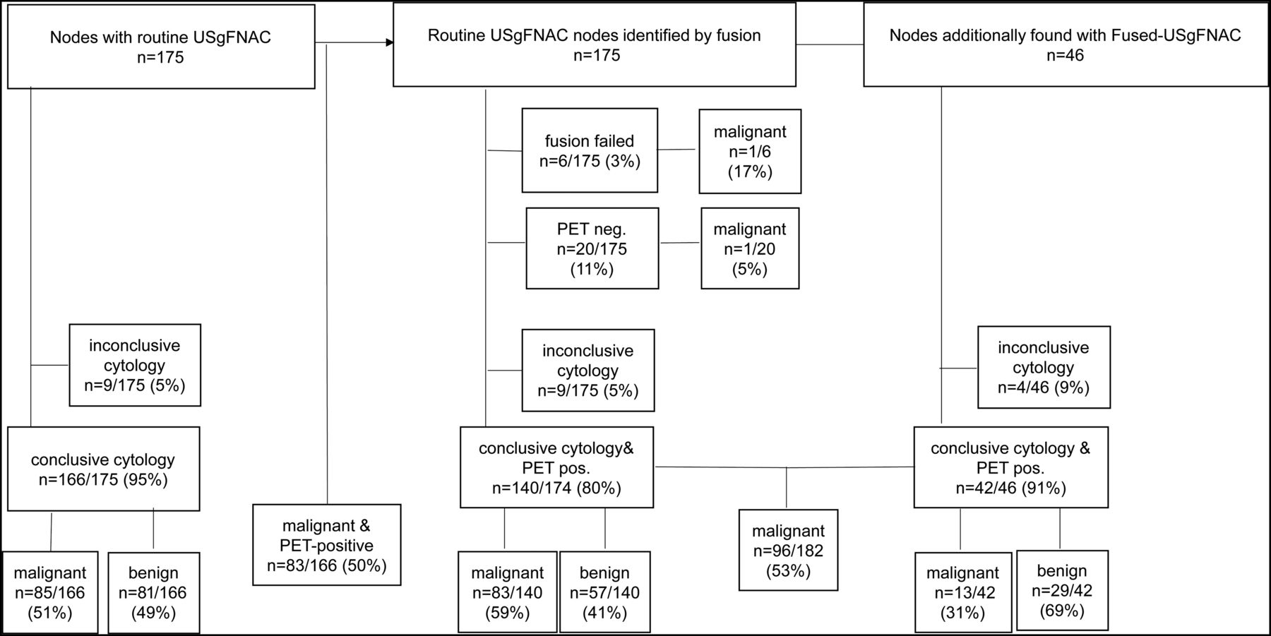
- FIG 3.
Flow chart results of routine USgFNAC and fused-USgFNAC. Pos. Indicates positive.
- FIG 4.
Change of N stage after additional fused-USgFNAC. The patient presented with cT3N0 oropharyngeal squamous cell carcinoma. A, Results of routine USgFNAC N1. B and C, PET/CT of the same node, controlled by image fusion. D–F, Additional nodes on PET/CT; all nodes have been fused, and fused-USgFNAC was performed. G, The deep parapharyngeal node was missed at routine ultrasound and only recognized after fusion. H, A PET-positive node with a normal appearance on routine ultrasound. I, Fused-USgFNAC-proved benign PET-positive contralateral node. Cytologically proved pN stage after fused-USgFNAC was pN2b, while it was N1 with USgFNAC and N2c on PET/CT. The green arrows point to the PET-positive nodes.


Tables
Diagnosis No. Percentage Adeno ca parotid gland 1 1% Angiosarcoma 1 1% B-cell lymphoma 1 1% Lung carcinoma 2 2% Melanoma 6 6% Merkel cell carcinoma 2 2% Rhabdomyosarcoma 1 1% SCC hypopharyngeal 7 7% SCC laryngeal 16 17% SCC nasal cavity sinus 4 4% SCC nasopharyngeal 1 1% SCC oral cavity 19 20% SCC oropharyngeal 25 26% SCC skin 1 1% SCC unknown primary 6 6% Second branchial cleft 1 1% Tuberculosis 1 1% Unknown primary 1 1% Total 96 100% Note:—Aneno ca indicates adenocarcinoma; SCC, squamous cell carcinoma.
↵a In total, 82% of all patients had SCC.
Treatment RT CRT BRT PDT Chemo Surgery, no ND 11 5 1 0 0 0 SND/SNB 20 8 1 0 0 0 No surgery 63 31 21 6 1 4 Noa treatment 2 0 0 0 0 0 Total 96 44 23 6 1 4 Note:—RT indicates radiation therapy; CRT, radiochemotherapy; BRT, bioradiation therapy; PDT, photodynamic therapy; Chemo, chemotherapy; SND, selective neck dissection; SNB, sentinel node biopsy.
↵a Two patients did not have treatment because of benign lesions: second branchial cleft cyst and tuberculosis.
Level Total Malignant Benign Insufficient No. No. Sizes (mm) No. Sizes (mm) No. Sizes (mm) 1 2 0 1 3 1 8 1a 1 0 1 4 0 1b 4 1 9 3 4, 3, 6 0 2 12 3 7, 4, 6 8 9, 6, 11, 8, 5, 6, 8, 5 1 8 2b 1 1 6 0 0 3 13 4 9, 8, 6, 6 8 5, 4, 3, 5, 4, 4, 4, 6 1 5 4 7 2 6, 12 4 1 4 5 3 1 9 2 5, 6 0 Parot.gl. 2 0 2 5, 6 0 Cheek 1 1 6 0 0 Total 46 13 29 4 Note:—1 to 5 indicates the neck levels; Parot.gl., parotid gland.


Jump to section
Related Articles
Cited By...
- No citing articles found.