Advertisement

Main menu

  • Home
  • Content
    • Current Issue
    • Accepted Manuscripts
    • Article Preview
    • Past Issue Archive
    • Video Articles
    • AJNR Case Collection
    • Case of the Week Archive
    • Case of the Month Archive
    • Classic Case Archive
  • Special Collections
    • Advancing NeuroMRI with High-Relaxivity Contrast Agents
    • AJNR Awards
    • Low-Field MRI
    • Alzheimer Disease
    • ASNR Foundation Special Collection
    • Photon-Counting CT
    • View All
  • Multimedia
    • AJNR Podcasts
    • AJNR SCANtastic
    • Trainee Corner
    • MRI Safety Corner
    • Imaging Protocols
  • For Authors
    • Submit a Manuscript
    • Submit a Video Article
    • Submit an eLetter to the Editor/Response
    • Manuscript Submission Guidelines
    • Statistical Tips
    • Fast Publishing of Accepted Manuscripts
    • Graphical Abstract Preparation
    • Imaging Protocol Submission
    • Author Policies
  • About Us
    • About AJNR
    • Editorial Board
    • Editorial Board Alumni
  • More
    • Become a Reviewer/Academy of Reviewers
    • Subscribers
    • Permissions
    • Alerts
    • Feedback
    • Advertisers
    • ASNR Home

User menu

  • Alerts
  • Log in

Search

  • Advanced search
American Journal of Neuroradiology

American Journal of Neuroradiology

ASHNR American Society of Functional Neuroradiology ASHNR American Society of Pediatric Neuroradiology ASSR
  • Alerts
  • Log in

Advanced Search

  • Home
  • Content
    • Current Issue
    • Accepted Manuscripts
    • Article Preview
    • Past Issue Archive
    • Video Articles
    • AJNR Case Collection
    • Case of the Week Archive
    • Case of the Month Archive
    • Classic Case Archive
  • Special Collections
    • Advancing NeuroMRI with High-Relaxivity Contrast Agents
    • AJNR Awards
    • Low-Field MRI
    • Alzheimer Disease
    • ASNR Foundation Special Collection
    • Photon-Counting CT
    • View All
  • Multimedia
    • AJNR Podcasts
    • AJNR SCANtastic
    • Trainee Corner
    • MRI Safety Corner
    • Imaging Protocols
  • For Authors
    • Submit a Manuscript
    • Submit a Video Article
    • Submit an eLetter to the Editor/Response
    • Manuscript Submission Guidelines
    • Statistical Tips
    • Fast Publishing of Accepted Manuscripts
    • Graphical Abstract Preparation
    • Imaging Protocol Submission
    • Author Policies
  • About Us
    • About AJNR
    • Editorial Board
    • Editorial Board Alumni
  • More
    • Become a Reviewer/Academy of Reviewers
    • Subscribers
    • Permissions
    • Alerts
    • Feedback
    • Advertisers
    • ASNR Home
  • Follow AJNR on Twitter
  • Visit AJNR on Facebook
  • Follow AJNR on Instagram
  • Join AJNR on LinkedIn
  • RSS Feeds

AJNR Awards, New Junior Editors, and more. Read the latest AJNR updates


Improved Turnaround Times | Median time to first decision: 12 days

Research ArticleAdult Brain
Open Access

Development and Validation of a Deep Learning–Based Automatic Brain Segmentation and Classification Algorithm for Alzheimer Disease Using 3D T1-Weighted Volumetric Images

C.H. Suh, W.H. Shim, S.J. Kim, J.H. Roh, J.-H. Lee, M.-J. Kim, S. Park, W. Jung, J. Sung, G.-H. Jahng, and for the Alzheimer’s Disease Neuroimaging Initiative
American Journal of Neuroradiology November 2020, DOI: https://doi.org/10.3174/ajnr.A6848
C.H. Suh
aFrom the Department of Radiology and Research Institute of Radiology (C.H.S., W.H.S., S.J.K.)
  • Find this author on Google Scholar
  • Find this author on PubMed
  • Search for this author on this site
  • ORCID record for C.H. Suh
W.H. Shim
aFrom the Department of Radiology and Research Institute of Radiology (C.H.S., W.H.S., S.J.K.)
  • Find this author on Google Scholar
  • Find this author on PubMed
  • Search for this author on this site
  • ORCID record for W.H. Shim
S.J. Kim
aFrom the Department of Radiology and Research Institute of Radiology (C.H.S., W.H.S., S.J.K.)
  • Find this author on Google Scholar
  • Find this author on PubMed
  • Search for this author on this site
  • ORCID record for S.J. Kim
J.H. Roh
bDepartment of Neurology (J.H.R., J.-H.L.)
dDepartment of Physiology (J.H.R.), Korea University College of Medicine, Seoul, Republic of Korea
  • Find this author on Google Scholar
  • Find this author on PubMed
  • Search for this author on this site
  • ORCID record for J.H. Roh
J.-H. Lee
bDepartment of Neurology (J.H.R., J.-H.L.)
  • Find this author on Google Scholar
  • Find this author on PubMed
  • Search for this author on this site
  • ORCID record for J.-H. Lee
M.-J. Kim
cHealth Screening and Promotion Center (M.-J.K.), Asan Medical Center, Seoul, Republic of Korea
  • Find this author on Google Scholar
  • Find this author on PubMed
  • Search for this author on this site
  • ORCID record for M.-J. Kim
S. Park
eVUNO Inc (S.P., W.J., J.S.), Seoul, Republic of Korea
  • Find this author on Google Scholar
  • Find this author on PubMed
  • Search for this author on this site
  • ORCID record for S. Park
W. Jung
eVUNO Inc (S.P., W.J., J.S.), Seoul, Republic of Korea
  • Find this author on Google Scholar
  • Find this author on PubMed
  • Search for this author on this site
  • ORCID record for W. Jung
J. Sung
eVUNO Inc (S.P., W.J., J.S.), Seoul, Republic of Korea
  • Find this author on Google Scholar
  • Find this author on PubMed
  • Search for this author on this site
  • ORCID record for J. Sung
G.-H. Jahng,
fDepartment of Radiology (G.-H.J.), Kyung Hee University Hospital at Gangdong, College of Medicine, Kyung Hee University, Seoul, Republic of Korea
  • Find this author on Google Scholar
  • Find this author on PubMed
  • Search for this author on this site
  • ORCID record for G.-H. Jahng,
  • Article
  • Figures & Data
  • Supplemental
  • Info & Metrics
  • Responses
  • References
  • PDF
Loading

Article Figures & Data

Figures

  • Tables
  • FIG 1.
    • Download figure
    • Open in new tab
    • Download powerpoint
    FIG 1.

    Network architecture of the brain parcellation and classification model. CONV indicates convolution.
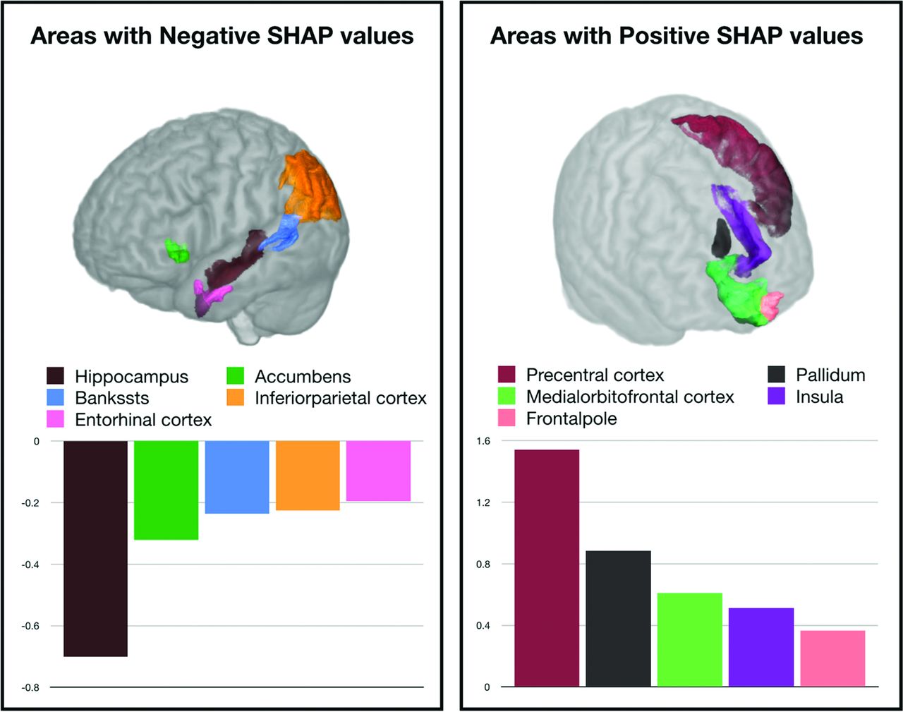
  • FIG 2.
    • Download figure
    • Open in new tab
    • Download powerpoint
    FIG 2.

    The impact of feature (volume of each brain region) on the AD prediction model, as represented by Shapley values, in which the impact of a feature is defined as the change in the expected output of the model when a feature is observed versus unknown. A, Visualization of the top 5 brain regions representing feature impacts pushing the decision of the model to AD, along with average feature impact. B, Visualization of the top 5 brain regions representing feature impacts pushing the decision of the model to healthy controls, along with average feature impact. Bankssts indicates banks of the superior temporal sulcus; SHAP, Shapley Additive Explanations (https://pbiecek.github.io/ema/shapley.html).

Tables

  • Figures
    • View popup
    Table 1:

    Characteristics of the development and datasetsa

    Asan Medical CenterKyung Hee University Hospital in GangdongADNIOASIS
    No. of patients1099212711705
    Age (mean) (yr)65 ± 1370 ± 976 ± 768 ± 10
    No. of male patients500 (45)52 (25)412 (58)403 (57)
    No. of female patients599 (55)160 (75)299 (42)302 (43)
    Classification
    AD161 (15)68 (32)178 (25)145 (21)
     Education (yr)9.9 (4.8)NA14.5 (3.4)14.0 (3.2)
     MMSE score18.5 (4.7)17.4 (5.3)22.8 (3.1)24.4 (5.1)
     Clinical Dementia Rating1.00 (0.49)1.10 (0.47)0.73 (0.34)0.68 (0.28)
     Global Deterioration ScaleNANA1.7 (1.4)3.2 (7.3)
    MCI363 (33)63 (30)317 (45)0
     Education (yr)10.1 (5.0)NA15.9 (2.5)
     MMSE score24.9 (3.6)25.7 (3.7)26.4 (2.1)
     Clinical Dementia Rating0.51 (0.09)0.61 (1.16)0.5
     Global Deterioration ScaleNANA1.5 (1.3)
    Healthy control575 (52)81 (38)216 (30)560 (79)
     Education (yr)NANA16.2 (2.8)15.2 (2.7)
     MMSE score29.5 (0.5)27.7 (2.5)29.1 (1.0)28.8 (3.2)
     Clinical Dementia RatingNA0.24 (0.26)00
     Global Deterioration ScaleNANA0.8 (1.1)1.3 (4.0)
    • Note:—MMSE indicates Mini-Mental State Examination; NA, not available.

    • ↵a Unless otherwise indicated, data are reported as number (%).

    • View popup
    Table 2:

    Diagnostic performance of logistic regression, the linear Support Vector Machine, and the deep learning–based automatic classification algorithm in the datasetsa

    Logistic RegressionLinear SVMXGBoostP ValuebP Valuec
    AD vs MCI
     Asan Medical Center0.770 (0.761–0.779)0.772 (0.761–0.782)0.803 (0.802–0.805)<.001<.001
     Kyung Hee University Hospital at Gangdong0.798 (0.775–0.822)0.804 (0.783–0.824)0.825 (0.810–0.840).018.030
     ADNI0.706 (0.702–0.710)0.700 (0.695–0.704)0.758 (0.755–0.760)<.001<.001
    MCI vs healthy control
     Asan Medical Center0.812 (0.806–0.817)0.830 (0.821–0.840)0.870 (0.868–0.872)<.001<.001
     Kyung Hee University Hospital at Gangdong0.692 (0.678–0.706)0.687 (0.669–0.706)0.705 (0.699–0.712).029.023
     ADNI0.698 (0.686–0.710)0.702 (0.697–0.708)0.668 (0.664–0.671)<.001<.001
    AD vs healthy controls
     Asan Medical Center0.953 (0.949–0.958)0.960 (0.958–0.963)0.982 (0.980–0.985)<.001<.001
     Kyung Hee University Hospital at Gangdong0.905 (0.889–0.921)0.911 (0.903–0.920)0.940 (0.933–0.947)<.001<.001
     ADNI0.863 (0.856–0.870)0.860 (0.857–0.863)0.885 (0.879–0.891)<.001<.001
     OASISd0.826 (0.817–0.835)0.820 (0.809–0.832)0.840 (0.837–0.844).001<.001
    • ↵a Data are AUC (95% CI).

    • ↵b P values: between logistic regression and XGBoost.

    • ↵c P values: between linear SVM and XGBosst.

    • ↵d OASIS dataset included only AD and healthy controls.

PreviousNext
Back to top
Advertisement
Print
Download PDF
Email Article

Thank you for your interest in spreading the word on American Journal of Neuroradiology.

NOTE: We only request your email address so that the person you are recommending the page to knows that you wanted them to see it, and that it is not junk mail. We do not capture any email address.

Enter multiple addresses on separate lines or separate them with commas.
Development and Validation of a Deep Learning–Based Automatic Brain Segmentation and Classification Algorithm for Alzheimer Disease Using 3D T1-Weighted Volumetric Images
(Your Name) has sent you a message from American Journal of Neuroradiology
(Your Name) thought you would like to see the American Journal of Neuroradiology web site.
CAPTCHA
This question is for testing whether or not you are a human visitor and to prevent automated spam submissions.
Cite this article
C.H. Suh, W.H. Shim, S.J. Kim, J.H. Roh, J.-H. Lee, M.-J. Kim, S. Park, W. Jung, J. Sung, G.-H. Jahng,, for the Alzheimer’s Disease Neuroimaging Initiative
Development and Validation of a Deep Learning–Based Automatic Brain Segmentation and Classification Algorithm for Alzheimer Disease Using 3D T1-Weighted Volumetric Images
American Journal of Neuroradiology Nov 2020, DOI: 10.3174/ajnr.A6848

Citation Manager Formats

  • BibTeX
  • Bookends
  • EasyBib
  • EndNote (tagged)
  • EndNote 8 (xml)
  • Medlars
  • Mendeley
  • Papers
  • RefWorks Tagged
  • Ref Manager
  • RIS
  • Zotero
0 Responses
Respond to this article
Share
Bookmark this article
Development and Validation of a Deep Learning–Based Automatic Brain Segmentation and Classification Algorithm for Alzheimer Disease Using 3D T1-Weighted Volumetric Images
C.H. Suh, W.H. Shim, S.J. Kim, J.H. Roh, J.-H. Lee, M.-J. Kim, S. Park, W. Jung, J. Sung, G.-H. Jahng,, for the Alzheimer’s Disease Neuroimaging Initiative
American Journal of Neuroradiology Nov 2020, DOI: 10.3174/ajnr.A6848
del.icio.us logo Twitter logo Facebook logo Mendeley logo
  • Tweet Widget
  • Facebook Like
  • Google Plus One
Purchase

Jump to section

  • Article
    • Abstract
    • ABBREVIATIONS:
    • MATERIALS AND METHODS
    • RESULTS
    • DISCUSSION
    • CONCLUSIONS
    • Footnotes
    • References
  • Figures & Data
  • Supplemental
  • Info & Metrics
  • Responses
  • References
  • PDF

Related Articles

  • PubMed
  • Google Scholar

Cited By...

  • Sex Differences in the Clinical and Imaging Characteristics of Korean CADASIL Patients: A Korean Nationwide Retrospective Study
  • Deep Learning-Based Algorithm for Automatic Quantification of Nigrosome-1 and Parkinsonism Classification Using Susceptibility Map-Weighted MRI
  • Simple Words over Rich Imaging: Accurate Brain Disease Classification via Language Model Analysis of Radiological Reports
  • Crossref
  • Google Scholar

This article has not yet been cited by articles in journals that are participating in Crossref Cited-by Linking.

More in this TOC Section

Adult Brain

  • Diagnostic Neuroradiology of Monoclonal Antibodies
  • Clinical Outcomes After Chiari I Decompression
  • Segmentation of Brain Metastases with BLAST
Show more Adult Brain

Functional

  • Kurtosis and Epileptogenic Tubers: A Pilot Study
  • Glutaric Aciduria Type 1: DK vs. Conventional MRI
  • WM Neuroaxonal Loss in Type 1 Diabetes
Show more Functional

Similar Articles

Advertisement

Indexed Content

  • Current Issue
  • Accepted Manuscripts
  • Article Preview
  • Past Issues
  • Editorials
  • Editor's Choice
  • Fellows' Journal Club
  • Letters to the Editor
  • Video Articles

Cases

  • Case Collection
  • Archive - Case of the Week
  • Archive - Case of the Month
  • Archive - Classic Case

More from AJNR

  • Trainee Corner
  • Imaging Protocols
  • MRI Safety Corner
  • Book Reviews

Multimedia

  • AJNR Podcasts
  • AJNR Scantastics

Resources

  • Turnaround Time
  • Submit a Manuscript
  • Submit a Video Article
  • Submit an eLetter to the Editor/Response
  • Manuscript Submission Guidelines
  • Statistical Tips
  • Fast Publishing of Accepted Manuscripts
  • Graphical Abstract Preparation
  • Imaging Protocol Submission
  • Evidence-Based Medicine Level Guide
  • Publishing Checklists
  • Author Policies
  • Become a Reviewer/Academy of Reviewers
  • News and Updates

About Us

  • About AJNR
  • Editorial Board
  • Editorial Board Alumni
  • Alerts
  • Permissions
  • Not an AJNR Subscriber? Join Now
  • Advertise with Us
  • Librarian Resources
  • Feedback
  • Terms and Conditions
  • AJNR Editorial Board Alumni

American Society of Neuroradiology

  • Not an ASNR Member? Join Now

© 2025 by the American Society of Neuroradiology All rights, including for text and data mining, AI training, and similar technologies, are reserved.
Print ISSN: 0195-6108 Online ISSN: 1936-959X

Powered by HighWire