
Article Figures & Data
Figures
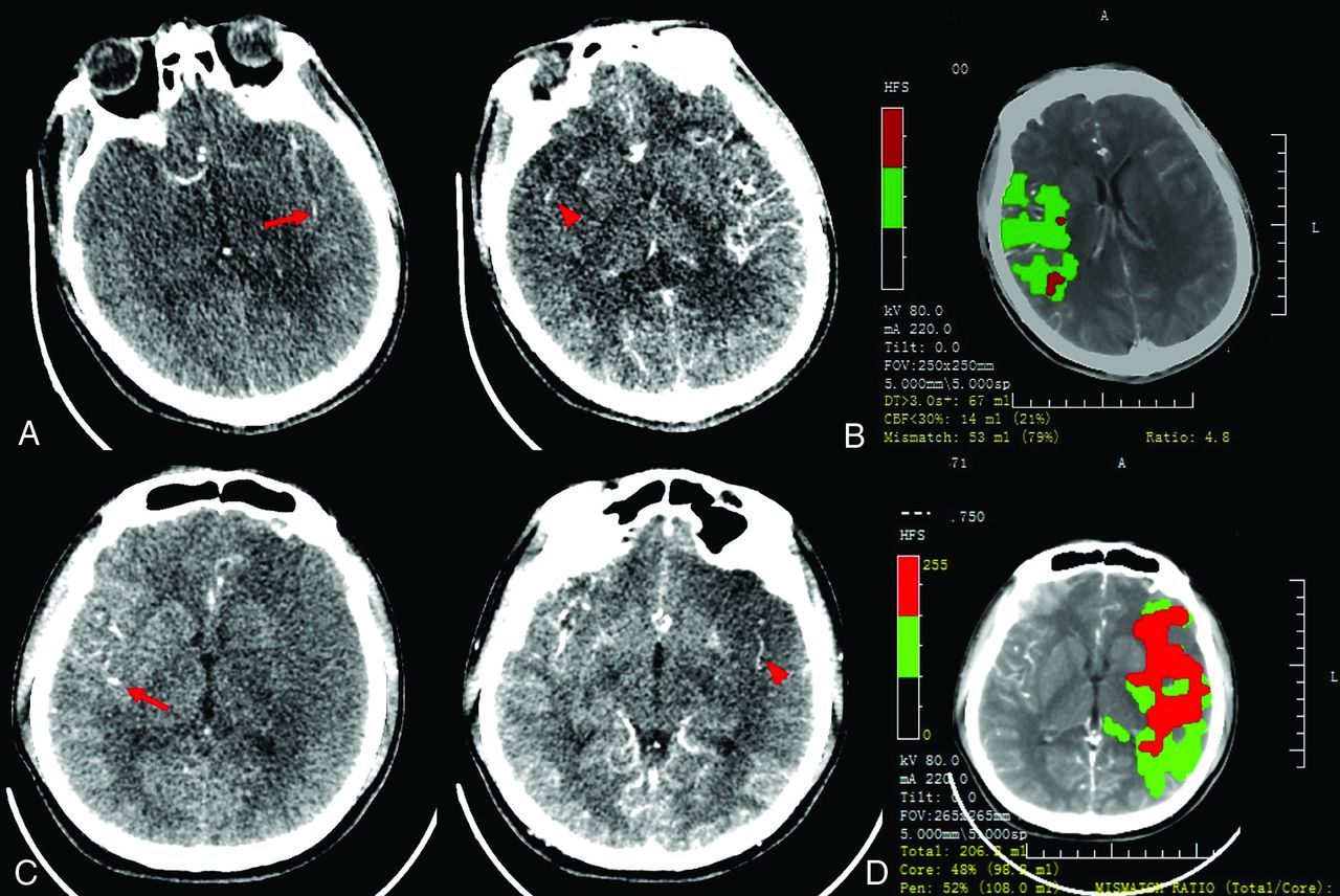
- FIG 1.
Case 1. A 46-year-old man with acute occlusion of the right MCA. CTP source imaging (SI) from a 256-section CT scanner showed that the first detection of contrast in the affected right hemisphere (red arrowhead) was delayed by 1 cycle (4 seconds for 1 cycle) after the first detection of contrast in the left Sylvian fissure (red arrow). The relative filling time delay (rFTD) was 4 seconds, which corresponds to a grade of 1 (A). Mismatch imaging calculated by software demonstrated a target mismatch between hypoperfusion volume (67 mL) and core volume (14 mL) (B). Case 2. A 24-year-old man with acute occlusion of the left MCA. CTP-SI from a 64-section CT scanner showed that the first detection of contrast in the affected left hemisphere (red arrowhead) was delayed by 5 cycles (1.3 seconds for 1 cycle) after the first detection of contrast in the right Sylvian fissure (red arrow). The rFTD was 6.5 seconds, which corresponded to a grade of 2 (C). Mismatch imaging showed a large core volume of 98 mL and no TMM (D).
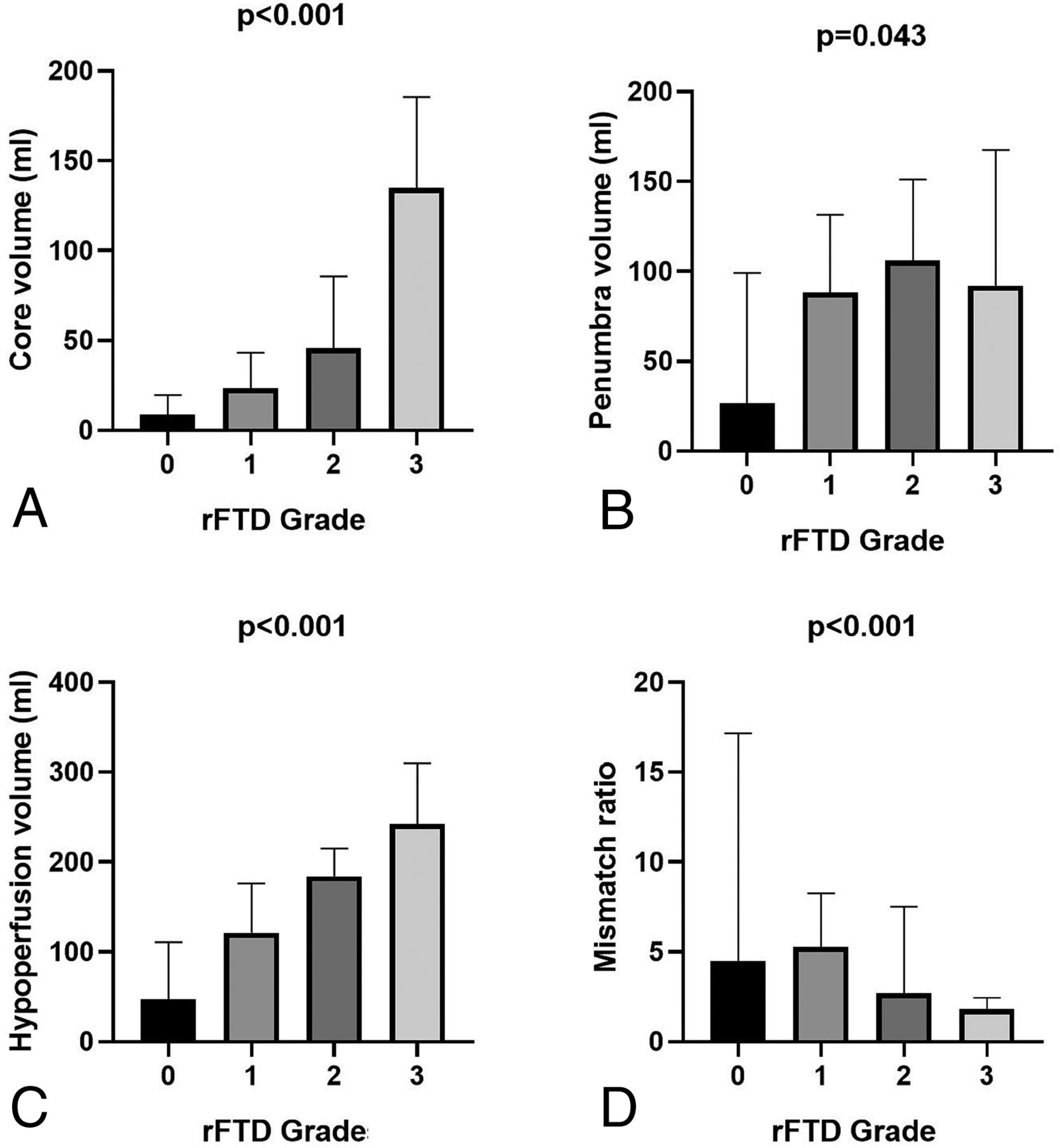
- FIG 2.
Comparison of ischemic volume parameters, including infarct core (A), penumbra (B), and hypoperfusion (C), and the mismatch ratio of hypoperfusion to core volume (D) among relative filling time delay grades.
- FIG 3.
Percentages of target mismatch (A), infarct core volume < 70 mL (B), and ICV < 51 mL (C) among relative filling time delay grades.


Tables
Characteristic Total Grade 0 Grade 1 Grade 2 Grade 3 P Patients, n 138 14 78 33 13 Age, (median) (IQR) (yr) 69 (59, 76) 63.5 (55.25, 72.75) 66 (57, 75) 71 (59, 76.5) 75 (64, 79) .169 Males, n (%) 86 (62.3) 7 (50) 48 (61.5) 21 (63.6) 10 (76.9) .546 Clinical characteristics Onset-to-CT time (median) (IQR) (min) 119 (63, 201) 115.5 (70.25, 161.75) 120 (71.5, 224) 105 (52.75, 203) 123 (48.5, 174) .845 Hypertension, n (%) 90 (65.9) 9 (64.3) 49 (62.8) 23 (69.7) 10 (76.9) .738 Diabetes, n (%) 45 (32.6) 6 (42.9) 18 (23.1) 15 (45.5) 6 (46.2) .059 Smoking, n (%) 53 (38.4) 6 (42.9) 30 (38.5) 11 (33.3) 6 (46.2) .848 Previous stroke and/or TIA, n (%) 26 (18.8) 2 (14.3) 13 (16.7) 5 (15.2) 6 (46.2) .07 Atrial fibrillation, n (%) 54 (39.1) 5 (35.7) 28 (35.9) 14 (42.4) 7 (53.8) .627 Dyslipidemia, n (%) 24 (17.4) 2 (14.3) 14 (17.9) 6 (18.2) 2 (15.4) .984 SBP (median) (IQR) (mm Hg) 147 (130, 162) 137 (124.5, 166.5) 145 (134.5, 160) 158 (128.5, 169.75) 147 (131, 172.5) .508 DBP (median) (IQR) (mm Hg) 83 (80, 91) 82.5 (82.25, 89.5) 85 (80, 91.5) 85.5 (79.25, 99.75) 80 (69, 90.5) .276 ABG (median) (IQR) (mol/L) 7.4 (6.1, 9.6) 7.15 (6.33, 8.89) 7 (5.7, 8.69) 8.05 (6.29, 10.2) 7.9 (7.2, 9.45) .206 Baseline NIHSS score (median) (IQR) 16 (11, 19) 10.5 (4.75, 18.5) 14 (9, 17) 17.5 (12, 20) 19 (16.5, 22) <.001 Occluded site, n (%) .067 MCA 87 (63) 10 (71.4) 50 (64.1) 21 (63.6) 6 (46.2) ICA 44 (32.1) 3 (21.4) 25 (32.1) 12 (36.4) 4 (30.8) MCA + ICA 7 (5.1) 1 (7.1) 3 (3.8) 0 (0) 3 (23.1) Perfusion parameters Core (median) (IQR) (mL) 30.5 (10, 87, 61.25) 7.35 (0.13, 15.6) 23.3 (8.65, 43.8) 45.75 (25.17, 90.25) 135 (77.85, 185.5) <.001 Penumbra (median) (IQR) (mL) 88.3 (52.75, 138.7) 47.65 (5, 113.37) 90 (54, 136) 110.05 (62.42, 155.02) 92 (50, 167.45) .043 Low perfusion (median) (IQR) (mL) 132.3 (83.32, 213.25) 55.55 (14.5, 116.55) 121.6 (80.5, 195.5) 186.55 (141.55, 216.15) 242 (154, 309.85) <.001 Mismatch ratioa (median) (IQR) 3.9 (2.4, 7.1) 5 (3.22, 21.85) 5.45 (2.24, 8.42) 2.6 (1.74, 7.5) 1.8 (1.4, 2.44) <.001 TMM, n (%) 99 (71.7) 9 (64.3) 70 (89.7) 18 (54.5) 2 (15.4) <.001 ICV <70 mL, n (%) 108 (78.3) 14 (100) 71 (91) 20 (60.6) 3 (23.1) <.001 ICV <51 mL, n (%) 93 (67.4) 13 (92.9) 62 (79.5) 18 (54.5) 0 (0) <.001 Note:—SBP indicates systolic blood pressure; DBP, diastolic blood pressure; ABG, admission blood glucose.
↵a Calculated by low perfusion volume to infarct core volume.
- Table 2:
Multivariate regression analysis of the independent association between rFTD grade ≤ 1 and TMM, ICV < 70 mL, and ICV < 51 mL
Variable adOR 95% CI P Sensitivity Specificity Prediction of TMM 6.25a 2.48–15.72 <.001 0.79 0.66 Prediction of ICV <70 mL 9.73b 3.11–30.35 <.001 0.78 0.76 Prediction of ICV <51 mL 5.84c 2.3–14.83 <.001 0.8 0.62


Jump to section
Related Articles
Cited By...
- No citing articles found.