Advertisement

Main menu

  • Home
  • Content
    • Current Issue
    • Accepted Manuscripts
    • Article Preview
    • Past Issue Archive
    • Video Articles
    • AJNR Case Collection
    • Case of the Week Archive
    • Case of the Month Archive
    • Classic Case Archive
  • Special Collections
    • AJNR Awards
    • Low-Field MRI
    • Alzheimer Disease
    • ASNR Foundation Special Collection
    • Photon-Counting CT
    • View All
  • Multimedia
    • AJNR Podcasts
    • AJNR SCANtastic
    • Trainee Corner
    • MRI Safety Corner
    • Imaging Protocols
  • For Authors
    • Submit a Manuscript
    • Submit a Video Article
    • Submit an eLetter to the Editor/Response
    • Manuscript Submission Guidelines
    • Statistical Tips
    • Fast Publishing of Accepted Manuscripts
    • Graphical Abstract Preparation
    • Imaging Protocol Submission
    • Author Policies
  • About Us
    • About AJNR
    • Editorial Board
    • Editorial Board Alumni
  • More
    • Become a Reviewer/Academy of Reviewers
    • Subscribers
    • Permissions
    • Alerts
    • Feedback
    • Advertisers
    • ASNR Home

User menu

  • Alerts
  • Log in

Search

  • Advanced search
American Journal of Neuroradiology

American Journal of Neuroradiology

ASHNR American Society of Functional Neuroradiology ASHNR American Society of Pediatric Neuroradiology ASSR
  • Alerts
  • Log in

Advanced Search

  • Home
  • Content
    • Current Issue
    • Accepted Manuscripts
    • Article Preview
    • Past Issue Archive
    • Video Articles
    • AJNR Case Collection
    • Case of the Week Archive
    • Case of the Month Archive
    • Classic Case Archive
  • Special Collections
    • AJNR Awards
    • Low-Field MRI
    • Alzheimer Disease
    • ASNR Foundation Special Collection
    • Photon-Counting CT
    • View All
  • Multimedia
    • AJNR Podcasts
    • AJNR SCANtastic
    • Trainee Corner
    • MRI Safety Corner
    • Imaging Protocols
  • For Authors
    • Submit a Manuscript
    • Submit a Video Article
    • Submit an eLetter to the Editor/Response
    • Manuscript Submission Guidelines
    • Statistical Tips
    • Fast Publishing of Accepted Manuscripts
    • Graphical Abstract Preparation
    • Imaging Protocol Submission
    • Author Policies
  • About Us
    • About AJNR
    • Editorial Board
    • Editorial Board Alumni
  • More
    • Become a Reviewer/Academy of Reviewers
    • Subscribers
    • Permissions
    • Alerts
    • Feedback
    • Advertisers
    • ASNR Home
  • Follow AJNR on Twitter
  • Visit AJNR on Facebook
  • Follow AJNR on Instagram
  • Join AJNR on LinkedIn
  • RSS Feeds

AJNR Awards, New Junior Editors, and more. Read the latest AJNR updates

Research ArticleSpine Imaging and Spine Image-Guided Interventions
Open Access

MRI Predictors of Recurrence and Outcome after Acute Transverse Myelitis of Unidentified Etiology

E. Bulut, T. Shoemaker, J. Karakaya, D.M. Ray, M.A. Mealy, M. Levy and I. Izbudak
American Journal of Neuroradiology July 2019, DOI: https://doi.org/10.3174/ajnr.A6121
E. Bulut
aFrom the Departments of Radiology (E.B.)
  • Find this author on Google Scholar
  • Find this author on PubMed
  • Search for this author on this site
  • ORCID record for E. Bulut
T. Shoemaker
cDepartment of Neurology (T.S., M.A.M., M.L.)
eDepartment of Neurological Sciences (T.S.), Rush University Medical Center, Chicago, Illinois
  • Find this author on Google Scholar
  • Find this author on PubMed
  • Search for this author on this site
  • ORCID record for T. Shoemaker
J. Karakaya
bStatistics (J.K.), Hacettepe University Faculty of Medicine, Ankara, Turkey
  • Find this author on Google Scholar
  • Find this author on PubMed
  • Search for this author on this site
  • ORCID record for J. Karakaya
D.M. Ray
dDivision of Neuroradiology (D.M.R., I.I.), The Russell H. Morgan Department of Radiology and Radiological Science, Johns Hopkins University School of Medicine, Baltimore, Maryland
  • Find this author on Google Scholar
  • Find this author on PubMed
  • Search for this author on this site
  • ORCID record for D.M. Ray
M.A. Mealy
cDepartment of Neurology (T.S., M.A.M., M.L.)
  • Find this author on Google Scholar
  • Find this author on PubMed
  • Search for this author on this site
  • ORCID record for M.A. Mealy
M. Levy
cDepartment of Neurology (T.S., M.A.M., M.L.)
fDepartment of Neurology (M.L.), Massachusetts General Hospital and Harvard Medical School, Boston, Massachusetts.
  • Find this author on Google Scholar
  • Find this author on PubMed
  • Search for this author on this site
  • ORCID record for M. Levy
I. Izbudak
dDivision of Neuroradiology (D.M.R., I.I.), The Russell H. Morgan Department of Radiology and Radiological Science, Johns Hopkins University School of Medicine, Baltimore, Maryland
  • Find this author on Google Scholar
  • Find this author on PubMed
  • Search for this author on this site
  • ORCID record for I. Izbudak
  • Article
  • Figures & Data
  • Info & Metrics
  • Responses
  • References
  • PDF
Loading

Article Figures & Data

Figures

  • Tables
  • Fig 1.
    • Download figure
    • Open in new tab
    • Download powerpoint
    Fig 1.

    Flow chart of patients who met the inclusion/exclusion criteria for the study population.
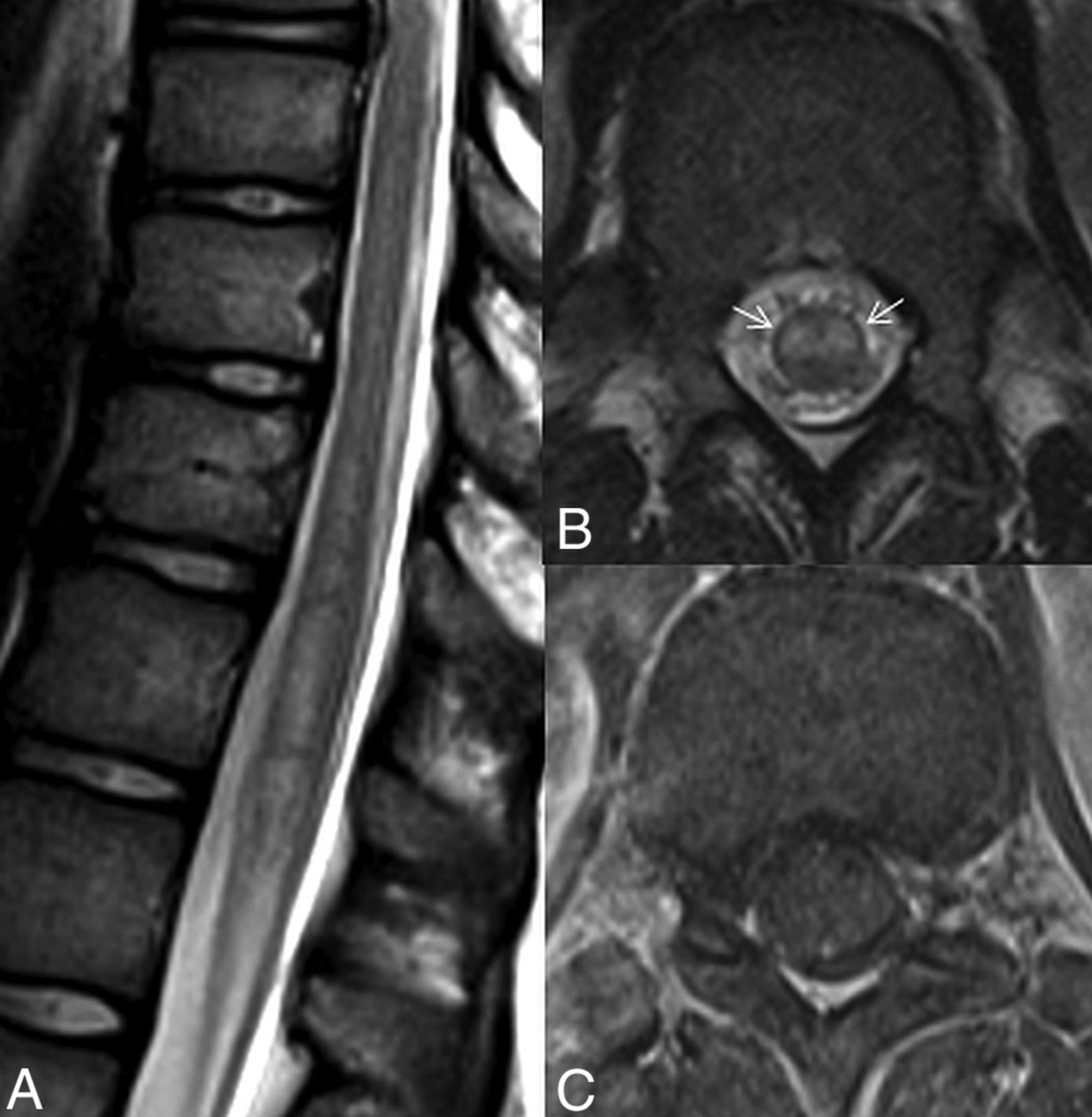
  • Fig 2.
    • Download figure
    • Open in new tab
    • Download powerpoint
    Fig 2.

    Spine MR images of a 13-year-old female patient with monophasic idiopathic transverse myelitis. The sagittal T2-weighted image (A) shows a longitudinally extensive, minimally expansile hyperintense lesion at the distal spinal cord. The axial T2-weighted (B) and postcontrast T1-weighted (C) images show central involvement with the owl's eyes sign (arrows in B) and no discernible contrast enhancement of the lesion.
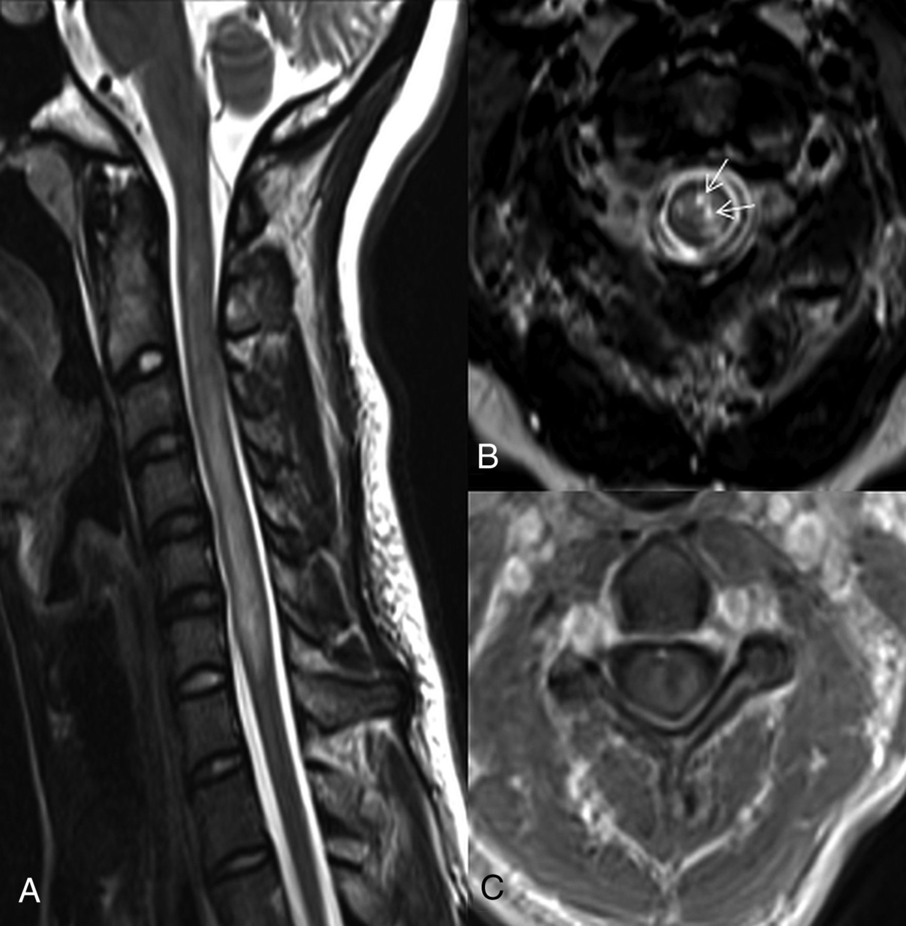
  • Fig 3.
    • Download figure
    • Open in new tab
    • Download powerpoint
    Fig 3.

    Spine MR images of a 29-year-old female patient with recurrent transverse myelitis. The sagittal T2-weighted image (A) shows a longitudinally extensive, expansile hyperintense lesion at the cervical spine. The axial T2-weighted (B) and postcontrast T1-weighted (C) images show gray and white matter involvement with bright spotty lesions (arrows in B) and ring enhancement of the lesion. The patient was diagnosed with NMOSD later at follow-up.

Tables

  • Figures
    • View popup
    Table 1:

    Imaging parameters used in spinal MRI

    SequenceTR/TI (ms)TE (ms)Matrix SizeFOV (mm)Section Thickness/Spacing (mm)
    T1WI sagittal397–5628.7–11256–384 × 256–28825–32 × 35.3–45.23–3.5/3.3–4.3
    T2WI sagittal2340–407096–109256–384 × 256–28825–32 × 35.3–45.23–3.5/3.3–4.3
    STIR sagittal3210–4610/150–22042–79256–384 × 256–28825–32 × 35.3–45.23–3.5/3.3–4.3
    T2WI axial2874–410098–109256–320 × 192–28018–20 × 25.4–28.33–4/3.3–5
    T1WI axial400–6548.4–9.7256–320 × 192–28018–20 × 25.4–28.33–4/3.3–5
    • View popup
    Table 2:

    The clinical characteristics of 77 patients who presented with acute transverse myelitis

    CharacteristicsMonophasic (n = 50)Recurrent (n = 27)P Values
    Age at initial manifestation (mean) (yr)34.2 ± 20.640.4 ± 18.5.196
    Female (n) (%)25 (50%)21 (77.8%).033
    Ethnicity.020
        African American (n) (%)10 (20%)10 (37%)
        Caucasian (n) (%)38 (76%)12 (44.4%)
        Asian American (n) (%)1 (2%)3 (11%)
        Hispanic (n) (%)1 (2%)2 (7.4%)
    Seropositivity for anti-AQP4 Ab1 (2%)8 (29.6%).003
    Mean follow-up time (yr)2.4 ± 1.73.8 ± 3.8.023
    Mean EDSS score4.2 ± 2.13.3 ± 1.8.064
    Patients with follow-up spine MRIs (n) (%)16 (32%)27 (100%)<.001
    Use of immunosuppressive/immunomodulatory treatment (n) (%)9 (18%)18 (66.7%)<.001
    • Note:—AQP4 Ab indicates aquaporin 4 antibody.

    • View popup
    Table 3:

    The frequencies of spinal MRI findings and associations with monophasic/relapsing disease

    MRI FindingsMonophasic Disease (n = 50) (n) (%)Recurrent Disease (n = 27) (n) (%)P ValueUnadjusted OR (95% CI)
    LETM (n = 42) (54.5%)22 (44%)20 (74.1%).022a3.63 (1.30–10.14)
    Multifocal lesions (n = 16) (22.5%)9/44 (20.5%)7 (25.9%).8081.36 (0.44–4.21)
    Distribution.304
        Cervical (n = 21) (27.3%)14 (28%)7 (25.9%)
        Cervicothoracic (n = 14) (18.2%)6 (12%)8 (29.6%)
        Thoracic (n = 35) (45.5%)25 (50%)10 (37%)
        Holocord (n = 7) (9.1%)5 (10%)2 (7.4%)
    Brain stem extension (n = 5) (6.5%)1/46 (2.2%)4/25 (16%).049a8.57 (0.90–81.46)
    Location.521
        Gray matter (n = 3) (3.9%)3 (6%)0
        Gray + white matter (n = 65) (84.4%)42 (84%)23 (85.2%)
        White matter (n = 9) (11.7%)5 (10%)4 (14.8%)
    >1/2 of the cord area (n = 55) (71.4%)33 (66%)22 (81.5%).2422.26 (0.73–7.04)
    Cord expansion (n = 48) (62.3%)26 (52%)22 (81.5%).021a4.06 (1.32–12.42)
    T1 hypointensity (n = 23) (30.3%)11/49 (22.4%)12 (44.4%).0892.76 (1.01–7.61)
    BSLs (n = 27) (35.1%)12 (24%)15 (55.6%).012a3.95 (1.45–10.74)
    Owl's eyes sign (n = 2) (2.6%)2 (4%)0.5390.58 (0.06–5.88)
    Enhancement (n = 48) (62.3%)26 (52%)22/26 (84.6%).011a5.07 (1.52–16.87)
    Brain involvement (n = 17) (27%)11/43 (25.6%)6/20 (30%).9501.24 (0.38–4.04)
    • ↵a Significant.

    • View popup
    Table 4:

    Adjusted ORs for associations of imaging variables/age

    VariableAdjusted ORsP Value95% Cl
    Cord expansion5.30.0181.33–21.11
    BSLs3.63.0401.06–12.43
    Enhancement5.05.0231.25–20.34
    Age1.03.0840.99–1.061
    Constant0.01.00
PreviousNext
Back to top
Advertisement
Print
Download PDF
Email Article

Thank you for your interest in spreading the word on American Journal of Neuroradiology.

NOTE: We only request your email address so that the person you are recommending the page to knows that you wanted them to see it, and that it is not junk mail. We do not capture any email address.

Enter multiple addresses on separate lines or separate them with commas.
MRI Predictors of Recurrence and Outcome after Acute Transverse Myelitis of Unidentified Etiology
(Your Name) has sent you a message from American Journal of Neuroradiology
(Your Name) thought you would like to see the American Journal of Neuroradiology web site.
CAPTCHA
This question is for testing whether or not you are a human visitor and to prevent automated spam submissions.
Cite this article
E. Bulut, T. Shoemaker, J. Karakaya, D.M. Ray, M.A. Mealy, M. Levy, I. Izbudak
MRI Predictors of Recurrence and Outcome after Acute Transverse Myelitis of Unidentified Etiology
American Journal of Neuroradiology Jul 2019, DOI: 10.3174/ajnr.A6121

Citation Manager Formats

  • BibTeX
  • Bookends
  • EasyBib
  • EndNote (tagged)
  • EndNote 8 (xml)
  • Medlars
  • Mendeley
  • Papers
  • RefWorks Tagged
  • Ref Manager
  • RIS
  • Zotero
0 Responses
Respond to this article
Share
Bookmark this article
MRI Predictors of Recurrence and Outcome after Acute Transverse Myelitis of Unidentified Etiology
E. Bulut, T. Shoemaker, J. Karakaya, D.M. Ray, M.A. Mealy, M. Levy, I. Izbudak
American Journal of Neuroradiology Jul 2019, DOI: 10.3174/ajnr.A6121
del.icio.us logo Twitter logo Facebook logo Mendeley logo
  • Tweet Widget
  • Facebook Like
  • Google Plus One
Purchase

Jump to section

  • Article
    • Abstract
    • ABBREVIATIONS:
    • Materials and Methods
    • Results
    • Discussion
    • Conclusions
    • Footnotes
    • REFERENCES
  • Figures & Data
  • Info & Metrics
  • Responses
  • References
  • PDF

Related Articles

  • No related articles found.
  • PubMed
  • Google Scholar

Cited By...

  • Enhancing Lesion Detection in Inflammatory Myelopathies: A Deep Learning-Reconstructed Double Inversion Recovery MRI Approach
  • Crossref
  • Google Scholar

This article has not yet been cited by articles in journals that are participating in Crossref Cited-by Linking.

More in this TOC Section

  • Management Outcomes For VO Spine Biopsy
  • Characteristics of SIH Type I Culprit Lesions
  • Advanced Imaging of Type 2 Spinal CSF Leaks
Show more Spine Imaging and Spine Image-Guided Interventions

Similar Articles

Advertisement

Indexed Content

  • Current Issue
  • Accepted Manuscripts
  • Article Preview
  • Past Issues
  • Editorials
  • Editor's Choice
  • Fellows' Journal Club
  • Letters to the Editor
  • Video Articles

Cases

  • Case Collection
  • Archive - Case of the Week
  • Archive - Case of the Month
  • Archive - Classic Case

More from AJNR

  • Trainee Corner
  • Imaging Protocols
  • MRI Safety Corner
  • Book Reviews

Multimedia

  • AJNR Podcasts
  • AJNR Scantastics

Resources

  • Turnaround Time
  • Submit a Manuscript
  • Submit a Video Article
  • Submit an eLetter to the Editor/Response
  • Manuscript Submission Guidelines
  • Statistical Tips
  • Fast Publishing of Accepted Manuscripts
  • Graphical Abstract Preparation
  • Imaging Protocol Submission
  • Evidence-Based Medicine Level Guide
  • Publishing Checklists
  • Author Policies
  • Become a Reviewer/Academy of Reviewers
  • News and Updates

About Us

  • About AJNR
  • Editorial Board
  • Editorial Board Alumni
  • Alerts
  • Permissions
  • Not an AJNR Subscriber? Join Now
  • Advertise with Us
  • Librarian Resources
  • Feedback
  • Terms and Conditions
  • AJNR Editorial Board Alumni

American Society of Neuroradiology

  • Not an ASNR Member? Join Now

© 2025 by the American Society of Neuroradiology All rights, including for text and data mining, AI training, and similar technologies, are reserved.
Print ISSN: 0195-6108 Online ISSN: 1936-959X

Powered by HighWire