Advertisement

Main menu

  • Home
  • Content
    • Current Issue
    • Accepted Manuscripts
    • Article Preview
    • Past Issue Archive
    • Video Articles
    • AJNR Case Collection
    • Case of the Week Archive
    • Case of the Month Archive
    • Classic Case Archive
  • Special Collections
    • AJNR Awards
    • Low-Field MRI
    • Alzheimer Disease
    • ASNR Foundation Special Collection
    • Photon-Counting CT
    • View All
  • Multimedia
    • AJNR Podcasts
    • AJNR SCANtastic
    • Trainee Corner
    • MRI Safety Corner
    • Imaging Protocols
  • For Authors
    • Submit a Manuscript
    • Submit a Video Article
    • Submit an eLetter to the Editor/Response
    • Manuscript Submission Guidelines
    • Statistical Tips
    • Fast Publishing of Accepted Manuscripts
    • Graphical Abstract Preparation
    • Imaging Protocol Submission
    • Author Policies
  • About Us
    • About AJNR
    • Editorial Board
    • Editorial Board Alumni
  • More
    • Become a Reviewer/Academy of Reviewers
    • Subscribers
    • Permissions
    • Alerts
    • Feedback
    • Advertisers
    • ASNR Home

User menu

  • Alerts
  • Log in

Search

  • Advanced search
American Journal of Neuroradiology

American Journal of Neuroradiology

ASHNR American Society of Functional Neuroradiology ASHNR American Society of Pediatric Neuroradiology ASSR
  • Alerts
  • Log in

Advanced Search

  • Home
  • Content
    • Current Issue
    • Accepted Manuscripts
    • Article Preview
    • Past Issue Archive
    • Video Articles
    • AJNR Case Collection
    • Case of the Week Archive
    • Case of the Month Archive
    • Classic Case Archive
  • Special Collections
    • AJNR Awards
    • Low-Field MRI
    • Alzheimer Disease
    • ASNR Foundation Special Collection
    • Photon-Counting CT
    • View All
  • Multimedia
    • AJNR Podcasts
    • AJNR SCANtastic
    • Trainee Corner
    • MRI Safety Corner
    • Imaging Protocols
  • For Authors
    • Submit a Manuscript
    • Submit a Video Article
    • Submit an eLetter to the Editor/Response
    • Manuscript Submission Guidelines
    • Statistical Tips
    • Fast Publishing of Accepted Manuscripts
    • Graphical Abstract Preparation
    • Imaging Protocol Submission
    • Author Policies
  • About Us
    • About AJNR
    • Editorial Board
    • Editorial Board Alumni
  • More
    • Become a Reviewer/Academy of Reviewers
    • Subscribers
    • Permissions
    • Alerts
    • Feedback
    • Advertisers
    • ASNR Home
  • Follow AJNR on Twitter
  • Visit AJNR on Facebook
  • Follow AJNR on Instagram
  • Join AJNR on LinkedIn
  • RSS Feeds

AJNR Awards, New Junior Editors, and more. Read the latest AJNR updates

Research ArticleAdult Brain
Open Access

3T MRI Whole-Brain Microscopy Discrimination of Subcortical Anatomy, Part 2: Basal Forebrain

M.J. Hoch, M.T. Bruno, A. Faustin, N. Cruz, A.Y. Mogilner, L. Crandall, T. Wisniewski, O. Devinsky and T.M. Shepherd
American Journal of Neuroradiology June 2019, DOI: https://doi.org/10.3174/ajnr.A6088
M.J. Hoch
aFrom the Department of Radiology and Imaging Sciences, (M.J.H.), Emory University, Atlanta, Georgia
  • Find this author on Google Scholar
  • Find this author on PubMed
  • Search for this author on this site
  • ORCID record for M.J. Hoch
M.T. Bruno
bDepartments of Radiology (M.T.B., N.C., T.M.S.)
  • Find this author on Google Scholar
  • Find this author on PubMed
  • Search for this author on this site
  • ORCID record for M.T. Bruno
A. Faustin
cPathology (A.F., T.W.)
  • Find this author on Google Scholar
  • Find this author on PubMed
  • Search for this author on this site
  • ORCID record for A. Faustin
N. Cruz
bDepartments of Radiology (M.T.B., N.C., T.M.S.)
  • Find this author on Google Scholar
  • Find this author on PubMed
  • Search for this author on this site
  • ORCID record for N. Cruz
A.Y. Mogilner
dNeurosurgery (A.Y.M.)
  • Find this author on Google Scholar
  • Find this author on PubMed
  • Search for this author on this site
  • ORCID record for A.Y. Mogilner
L. Crandall
eNeurology (L.C., T.W., O.D.)
gSUDC Foundation (L.C., O.D.), New York, New York
  • Find this author on Google Scholar
  • Find this author on PubMed
  • Search for this author on this site
  • ORCID record for L. Crandall
T. Wisniewski
cPathology (A.F., T.W.)
eNeurology (L.C., T.W., O.D.)
fPsychiatry (T.W.), New York University, New York, New York
  • Find this author on Google Scholar
  • Find this author on PubMed
  • Search for this author on this site
  • ORCID record for T. Wisniewski
O. Devinsky
eNeurology (L.C., T.W., O.D.)
gSUDC Foundation (L.C., O.D.), New York, New York
  • Find this author on Google Scholar
  • Find this author on PubMed
  • Search for this author on this site
  • ORCID record for O. Devinsky
T.M. Shepherd
bDepartments of Radiology (M.T.B., N.C., T.M.S.)
hCenter for Advanced Imaging Innovation and Research (T.M.S.), New York, New York.
  • Find this author on Google Scholar
  • Find this author on PubMed
  • Search for this author on this site
  • ORCID record for T.M. Shepherd
  • Article
  • Figures & Data
  • Supplemental
  • Info & Metrics
  • Responses
  • References
  • PDF
Loading

Article Figures & Data

Figures

  • Tables
  • Fig 1.
    • Download figure
    • Open in new tab
    • Download powerpoint
    Fig 1.

    Selected coronal, sagittal, and axial images of the postmortem basal forebrain illustrating the serial imaging planes for Fig 2 and On-line Figs 1 and 2, respectively. Table 1 provides a complete list of labeled anatomy for all figures, indicated by the numbers in parentheses in the legends. The familiarity of T2 contrast and multiple imaging planes provided in this study should help facilitate learning the complex neuroanatomy of the basal forebrain.

  • Fig 2.
    • Download figure
    • Open in new tab
    • Download powerpoint
    Fig 2.

    Serial inferior-to-superior axial images of the postmortem basal forebrain parallel to the commissural plane (A–F, −4, −2, 0, 2, 4, and 8 mm relative to the intercommissural plane, respectively). The globus pallidus internus (17) is a therapeutic DBS target for Parkinson disease and dystonia.18 The globus pallidus internus is separated from the externus (16) by a thin hypointense band, the internal medullary lamina (55 in On-line Fig 1). Note the 2 divisions of the globus pallidus internus (medial and lateral) separated by the accessory medullary lamina in B and C. Contrast is less conspicuous in the more superior thalamus.
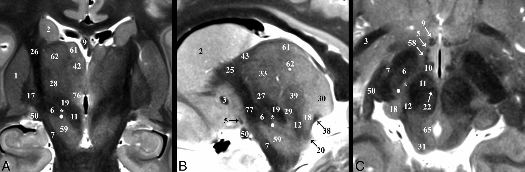
  • Fig 3.
    • Download figure
    • Open in new tab
    • Download powerpoint
    Fig 3.

    Selected images illustrating the subthalamic nucleus (6) in the basal forebrain. Coronal, sagittal, and axial images show the subthalamic nucleus as a biconvex hypointense structure nestled along the medial margin of the internal capsule (26). B, The darkest portion of the internal capsule just anterior to the subthalamic nucleus represents the Edinger comb system (77) containing the pallidosubthalamic, pallidonigral, and nigrostriatal tracts. The small white circle represents the potential DBS electrode tip placement site (Table 2) in the inferior portion of the zona incerta (asterisk), which corresponds with a better therapeutic profile according to Plaha et al.22

  • Fig 4.
    • Download figure
    • Open in new tab
    • Download powerpoint
    Fig 4.

    Selected images illustrating the hippocampal-thalamic pathways. Coronal, sagittal, oblique axial (the dashed line in B represents the oblique imaging plane for C), and magnified coronal images of the fornix (9) and mammillothalamic tract (10). A and B, The decreased size of the postcommissural fornix as it approaches the mammillary bodies (56) is likely due, in part, to the direct hippocampal pathway (36) in C, which bypasses the mammillary bodies to reach the anterior thalamic nuclei (41). D, Just medial to the Fields of Forel (78) and pallidofugal tracts, the principal mammillary tract (80) gives rise to the ascending mammillothalamic tract. Note the subthalamic fasciculus (81) and zona incerta (asterisk).

  • Fig 5.
    • Download figure
    • Open in new tab
    • Download powerpoint
    Fig 5.

    Selected images illustrating the pallidothalamic tracts. Sagittal, oblique axial (the dashed line in A represents the oblique imaging plane for B), and coronal images illustrating the complex 3D shapes and spatial relationships of the ansa lenticularis (5), lenticular fasciculus (58), and thalamic fasciculus (27). B, The ansa lenticularis originates from the inferomedial globus pallidus internus (17) and joins the lenticular fasciculus (H2 field of Forel) in the very hypointense prerubral H Fields of Forel (78). These pallidal efferents then ascend as the thalamic fasciculus (H1 Fields of Forel) to the ventral thalamus. The zona incerta (asterisk) is the bright signal intensity region in between lenticular and thalamic fasciculi in B and C. The subthalamic nucleus (6) can be seen in relationship to these structures in A. Note the dark structure just inferior to the 44 label and the dashed line is a thalamic perforating vessel.
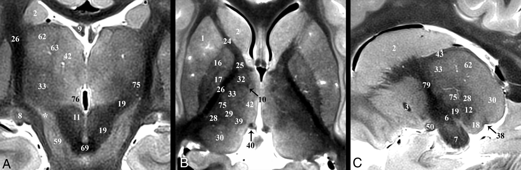
  • Fig 6.
    • Download figure
    • Open in new tab
    • Download powerpoint
    Fig 6.

    Coronal, axial, and sagittal images illustrating the superior ascent of the dentatorubrothalamic tract (19) to the Vim nucleus (75). A, Slight obliquity of the image allows depiction of the Vim and DRT on the left and the posterior aspect of the relatively more hyperintense nucleus ventrooralis (33) on the right (zona incerta labeled with asterisks). The relationship of the PLIC (26), nucleus ventrocaudalis anterior (28), and nucleus ventrocaudalis posterior (29) nuclei to the Vim is also illustrated. For completeness, the interested reader can find the proximal or prerubral component of the dentatorubrothalamic pathway also demonstrated in On-line Fig 3 of the previous report.57

Tables

  • Figures
    • View popup
    Table 1:

    Basal forebrain structures

    Labeling Key
    1) Putamen
    2) Caudate nucleus
    3) Anterior commissure
    4) Ansa peduncularis (inferior thalamic peduncle)
    5) Ansa lenticularis
    6) Subthalamic nucleus
    7) Cerebral peduncle
    8) Lateral geniculate nucleus
    9) Fornix
    10) Mammillothalamic tract
    11) Red nucleus
    12) Medial lemniscus
    13) Spinothalamic tract
    14) Central tegmental tract
    15) Inferior colliculus
    16) Globus pallidus externus
    17) Globus pallidus internus
    18) Medial geniculate nucleus
    19) Dentatorubrothalamic tract
    20) Brachium of the inferior colliculus
    21) Mesencephalic trigeminal nucleus
    22) Habenulopeduncular tract (fasiculus retroflexus)
    23) Posterior commissure
    24) Anterior limb of the internal capsule
    25) Genu of the internal capsule
    26) Posterior limb of the internal capsule
    27) Thalamic fasciculus (H1)
    28) Nucleus ventrocaudalis anterior
    29) Nucleus ventrocaudalis posterior
    30) Pulvinar
    31) Superior colliculus
    32) Nucleus lateropolaris
    33) Nucleus ventrooralis
    34) External capsule
    35) Retrolenticular internal capsule
    36) Direct hippocampal tract
    37) Habenular commissure
    38) Brachium of the superior colliculus
    39) Nucleus centralis
    40) Nucleus habenularis
    41) Anterior thalamic nuclear group
    42) Nucleus medialis
    43) External medullary lamina (thalamus)
    44) Extreme capsule
    45) Claustrum
    46) Caudolenticular gray bridges (pontes grisei caudatolenticulares)
    47) Olfactory tubercle
    48) Accumbens area
    49) Medial forebrain bundle
    50) Optic tract
    51) External medullary lamina (globus pallidus)
    52) Diagonal band of Broca
    53) Basal nucleus of Meynert
    54) Hypothalamic nuclei
    55) Internal medullary lamina (globus pallidus)
    56) Mammillary body
    57) Optic radiations
    58) Lenticular fasciculus (H2)
    59) Substantia nigra
    60) Massa intermedia
    61) Nucleus dorsalis superficialis
    62) Nucleus dorsalis oralis
    63) Internal medullary lamina (thalamus)
    64) Auditory radiations
    65) Periaqueductal gray matter
    66) Supraoptic decussation
    67) Optic chiasm
    68) Superior cerebellar peduncle (crossed)
    69) Decussation of the superior cerebellar peduncle
    70) Medial longitudinal fasciculus
    71) Splenium
    72) Hypothalamic sulcus
    73) Oculomotor nerve (cranial nerve III)
    74) Stratum opticum
    75) Nucleus ventrointermedius
    76) Nucleus parafascicularis
    77) Edinger comb system
    78) Nucleus of field of Forel (H)
    79) Internal capsule
    80) Principal mammillary tract
    81) Subthalamic fasciculus
    “*” Zona incerta
    • View popup
    Table 2:

    Measurements of the subthalamic nucleus in SUDC brains using TSE MRI contrast (n = 11)a

    Measurement/Dimension/PlaneRightLeftDifferencebP ValuecCOVd
    Lengthe (mm)
        Anteroposterior9.6 ± 0.99.9 ± 0.8−0.3 ± 0.6.0848.8%
        Mediolateral4.2 ± 1.24.1 ± 1.00.0 ± 0.4.91026.0%
        Superoinferior6.0 ± 0.65.8 ± 0.70.2 ± 0.3.04710.8%
    Anglef
        Coronal58.7° ± 6.5°58.0° ± 6.6°0.7° ± 5.8°.41410.9%
        Axial135.5° ± 4.8°131.6° ± 5.8°3.8° ± 5.9°.5904.1%
        Sagittal26.5° ± 6.6°28.5° ± 7.0°−1.9° ± 5.4°.18824.3%
    Stereotactic coordinatesg (mm)
        Lateral13.5 ± 1.013.5 ± 1.10.1 ± 0.51.0007.8%
        Posterior4.8 ± 0.64.7 ± 0.50.1 ± 0.31.00011.1%
        Inferior5.5 ± 0.95.4 ± 0.70.1 ± 0.51.00014.7%
    • Note:—COV indicates coefficient of variation.

    • ↵a Data are means ± standard deviation unless otherwise indicated.

    • ↵b Right-sided measurement minus left-sided measurement.

    • ↵c Paired-sample Wilcoxon signed rank test.

    • ↵d Global COV for right and left data combined (n = 22).

    • ↵e Largest dimension in each plane.

    • ↵f The angle formed by the long axis of the subthalamic nucleus relative to the orthogonal imaging plane where angulation is inferomedial to superolateral in the coronal plane, anteromedial to posterolateral in the axial plane, and anterosuperior to posteroinferior in the sagittal plane.

    • ↵g Coordinates relative to the intercommissural point where the most inferior, lateral, and posterior point of the subthalamic nucleus forms a distinct border with the inferior portion of the zona incerta. This point will usually be inferior and lateral to the desired DBS electrode tip target but can be measured precisely to assess individual and right-left variation in the subthalamic nucleus position.

PreviousNext
Back to top
Advertisement
Print
Download PDF
Email Article

Thank you for your interest in spreading the word on American Journal of Neuroradiology.

NOTE: We only request your email address so that the person you are recommending the page to knows that you wanted them to see it, and that it is not junk mail. We do not capture any email address.

Enter multiple addresses on separate lines or separate them with commas.
3T MRI Whole-Brain Microscopy Discrimination of Subcortical Anatomy, Part 2: Basal Forebrain
(Your Name) has sent you a message from American Journal of Neuroradiology
(Your Name) thought you would like to see the American Journal of Neuroradiology web site.
CAPTCHA
This question is for testing whether or not you are a human visitor and to prevent automated spam submissions.
Cite this article
M.J. Hoch, M.T. Bruno, A. Faustin, N. Cruz, A.Y. Mogilner, L. Crandall, T. Wisniewski, O. Devinsky, T.M. Shepherd
3T MRI Whole-Brain Microscopy Discrimination of Subcortical Anatomy, Part 2: Basal Forebrain
American Journal of Neuroradiology Jun 2019, DOI: 10.3174/ajnr.A6088

Citation Manager Formats

  • BibTeX
  • Bookends
  • EasyBib
  • EndNote (tagged)
  • EndNote 8 (xml)
  • Medlars
  • Mendeley
  • Papers
  • RefWorks Tagged
  • Ref Manager
  • RIS
  • Zotero
0 Responses
Respond to this article
Share
Bookmark this article
3T MRI Whole-Brain Microscopy Discrimination of Subcortical Anatomy, Part 2: Basal Forebrain
M.J. Hoch, M.T. Bruno, A. Faustin, N. Cruz, A.Y. Mogilner, L. Crandall, T. Wisniewski, O. Devinsky, T.M. Shepherd
American Journal of Neuroradiology Jun 2019, DOI: 10.3174/ajnr.A6088
del.icio.us logo Twitter logo Facebook logo Mendeley logo
  • Tweet Widget
  • Facebook Like
  • Google Plus One
Purchase

Jump to section

  • Article
    • Abstract
    • ABBREVIATIONS:
    • Materials and Methods
    • Results
    • Discussion
    • Conclusions
    • Acknowledgments
    • Footnotes
    • REFERENCES
  • Figures & Data
  • Supplemental
  • Info & Metrics
  • Responses
  • References
  • PDF

Related Articles

  • PubMed
  • Google Scholar

Cited By...

  • The Subcortical Atlas of the Marmoset ("SAM") monkey based on high-resolution MRI and histology
  • Multimodal anatomical mapping of subcortical regions in Marmoset monkeys using high-resolution MRI and matched histology with multiple stains
  • High-resolution mapping and digital atlas of subcortical regions in the macaque monkey based on matched MAP-MRI and histology
  • Crossref
  • Google Scholar

This article has not yet been cited by articles in journals that are participating in Crossref Cited-by Linking.

More in this TOC Section

  • Diagnostic Neuroradiology of Monoclonal Antibodies
  • Clinical Outcomes After Chiari I Decompression
  • Segmentation of Brain Metastases with BLAST
Show more Adult Brain

Similar Articles

Advertisement

Indexed Content

  • Current Issue
  • Accepted Manuscripts
  • Article Preview
  • Past Issues
  • Editorials
  • Editor's Choice
  • Fellows' Journal Club
  • Letters to the Editor
  • Video Articles

Cases

  • Case Collection
  • Archive - Case of the Week
  • Archive - Case of the Month
  • Archive - Classic Case

More from AJNR

  • Trainee Corner
  • Imaging Protocols
  • MRI Safety Corner
  • Book Reviews

Multimedia

  • AJNR Podcasts
  • AJNR Scantastics

Resources

  • Turnaround Time
  • Submit a Manuscript
  • Submit a Video Article
  • Submit an eLetter to the Editor/Response
  • Manuscript Submission Guidelines
  • Statistical Tips
  • Fast Publishing of Accepted Manuscripts
  • Graphical Abstract Preparation
  • Imaging Protocol Submission
  • Evidence-Based Medicine Level Guide
  • Publishing Checklists
  • Author Policies
  • Become a Reviewer/Academy of Reviewers
  • News and Updates

About Us

  • About AJNR
  • Editorial Board
  • Editorial Board Alumni
  • Alerts
  • Permissions
  • Not an AJNR Subscriber? Join Now
  • Advertise with Us
  • Librarian Resources
  • Feedback
  • Terms and Conditions
  • AJNR Editorial Board Alumni

American Society of Neuroradiology

  • Not an ASNR Member? Join Now

© 2025 by the American Society of Neuroradiology All rights, including for text and data mining, AI training, and similar technologies, are reserved.
Print ISSN: 0195-6108 Online ISSN: 1936-959X

Powered by HighWire