Advertisement

Main menu

  • Home
  • Content
    • Current Issue
    • Accepted Manuscripts
    • Article Preview
    • Past Issue Archive
    • Video Articles
    • AJNR Case Collection
    • Case of the Week Archive
    • Case of the Month Archive
    • Classic Case Archive
  • Special Collections
    • AJNR Awards
    • Low-Field MRI
    • Alzheimer Disease
    • ASNR Foundation Special Collection
    • Photon-Counting CT
    • View All
  • Multimedia
    • AJNR Podcasts
    • AJNR SCANtastic
    • Trainee Corner
    • MRI Safety Corner
    • Imaging Protocols
  • For Authors
    • Submit a Manuscript
    • Submit a Video Article
    • Submit an eLetter to the Editor/Response
    • Manuscript Submission Guidelines
    • Statistical Tips
    • Fast Publishing of Accepted Manuscripts
    • Graphical Abstract Preparation
    • Imaging Protocol Submission
    • Author Policies
  • About Us
    • About AJNR
    • Editorial Board
    • Editorial Board Alumni
  • More
    • Become a Reviewer/Academy of Reviewers
    • Subscribers
    • Permissions
    • Alerts
    • Feedback
    • Advertisers
    • ASNR Home

User menu

  • Alerts
  • Log in

Search

  • Advanced search
American Journal of Neuroradiology

American Journal of Neuroradiology

ASHNR American Society of Functional Neuroradiology ASHNR American Society of Pediatric Neuroradiology ASSR
  • Alerts
  • Log in

Advanced Search

  • Home
  • Content
    • Current Issue
    • Accepted Manuscripts
    • Article Preview
    • Past Issue Archive
    • Video Articles
    • AJNR Case Collection
    • Case of the Week Archive
    • Case of the Month Archive
    • Classic Case Archive
  • Special Collections
    • AJNR Awards
    • Low-Field MRI
    • Alzheimer Disease
    • ASNR Foundation Special Collection
    • Photon-Counting CT
    • View All
  • Multimedia
    • AJNR Podcasts
    • AJNR SCANtastic
    • Trainee Corner
    • MRI Safety Corner
    • Imaging Protocols
  • For Authors
    • Submit a Manuscript
    • Submit a Video Article
    • Submit an eLetter to the Editor/Response
    • Manuscript Submission Guidelines
    • Statistical Tips
    • Fast Publishing of Accepted Manuscripts
    • Graphical Abstract Preparation
    • Imaging Protocol Submission
    • Author Policies
  • About Us
    • About AJNR
    • Editorial Board
    • Editorial Board Alumni
  • More
    • Become a Reviewer/Academy of Reviewers
    • Subscribers
    • Permissions
    • Alerts
    • Feedback
    • Advertisers
    • ASNR Home
  • Follow AJNR on Twitter
  • Visit AJNR on Facebook
  • Follow AJNR on Instagram
  • Join AJNR on LinkedIn
  • RSS Feeds

AJNR Awards, New Junior Editors, and more. Read the latest AJNR updates

Research ArticlePediatric Neuroimaging

Radiation Dose and Image Quality in Pediatric Neck CT

S.V. Tipnis, W.J. Rieter, D. Patel, S.T. Stalcup, M.G. Matheus and M.V. Spampinato
American Journal of Neuroradiology May 2019, DOI: https://doi.org/10.3174/ajnr.A6073
S.V. Tipnis
aFrom the Department of Radiology and Radiological Science (S.V.T., W.J.R., D.P., S.T.S., M.G.M., M.V.S.)
  • Find this author on Google Scholar
  • Find this author on PubMed
  • Search for this author on this site
  • ORCID record for S.V. Tipnis
W.J. Rieter
aFrom the Department of Radiology and Radiological Science (S.V.T., W.J.R., D.P., S.T.S., M.G.M., M.V.S.)
  • Find this author on Google Scholar
  • Find this author on PubMed
  • Search for this author on this site
  • ORCID record for W.J. Rieter
D. Patel
aFrom the Department of Radiology and Radiological Science (S.V.T., W.J.R., D.P., S.T.S., M.G.M., M.V.S.)
  • Find this author on Google Scholar
  • Find this author on PubMed
  • Search for this author on this site
  • ORCID record for D. Patel
S.T. Stalcup
aFrom the Department of Radiology and Radiological Science (S.V.T., W.J.R., D.P., S.T.S., M.G.M., M.V.S.)
  • Find this author on Google Scholar
  • Find this author on PubMed
  • Search for this author on this site
  • ORCID record for S.T. Stalcup
M.G. Matheus
aFrom the Department of Radiology and Radiological Science (S.V.T., W.J.R., D.P., S.T.S., M.G.M., M.V.S.)
  • Find this author on Google Scholar
  • Find this author on PubMed
  • Search for this author on this site
  • ORCID record for M.G. Matheus
M.V. Spampinato
aFrom the Department of Radiology and Radiological Science (S.V.T., W.J.R., D.P., S.T.S., M.G.M., M.V.S.)
bCenter for Biomedical Imaging (M.V.S.), Medical University of South Carolina, Charleston, South Carolina.
  • Find this author on Google Scholar
  • Find this author on PubMed
  • Search for this author on this site
  • ORCID record for M.V. Spampinato
  • Article
  • Figures & Data
  • Info & Metrics
  • Responses
  • References
  • PDF
Loading

Article Figures & Data

Figures

  • Tables
  • Fig 1.
    • Download figure
    • Open in new tab
    • Download powerpoint
    Fig 1.

    Representative CT image of a 6-year-old boy at the level of the vocal cords for assessment of the signal-to-noise ratio. Background noise was estimated as the average SD in Hounsfield units of 2 ROIs drawn over the air anterior to the patient (large circles). Signal was estimated as the average attenuation in Hounsfield units of 2 ROIs drawn over the paraspinal musculature (small circles).
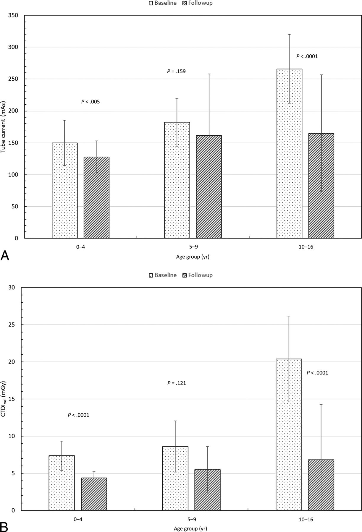
  • Fig 2.
    • Download figure
    • Open in new tab
    • Download powerpoint
    Fig 2.
    • Download figure
    • Open in new tab
    • Download powerpoint
    Fig 2.

    Comparison of the median values of tube current (milliampere-seconds) (A), radiation output of scanner (CTDIvol) (B), and scan length between the 2 cohorts (C). Also shown are P values from an unpaired t test.

  • Fig 3.
    • Download figure
    • Open in new tab
    • Download powerpoint
    Fig 3.

    Comparison of the median values of the dose-length product (A) and estimated effective dose (B) between the 2 cohorts. Also shown are the P values from an unpaired t test.
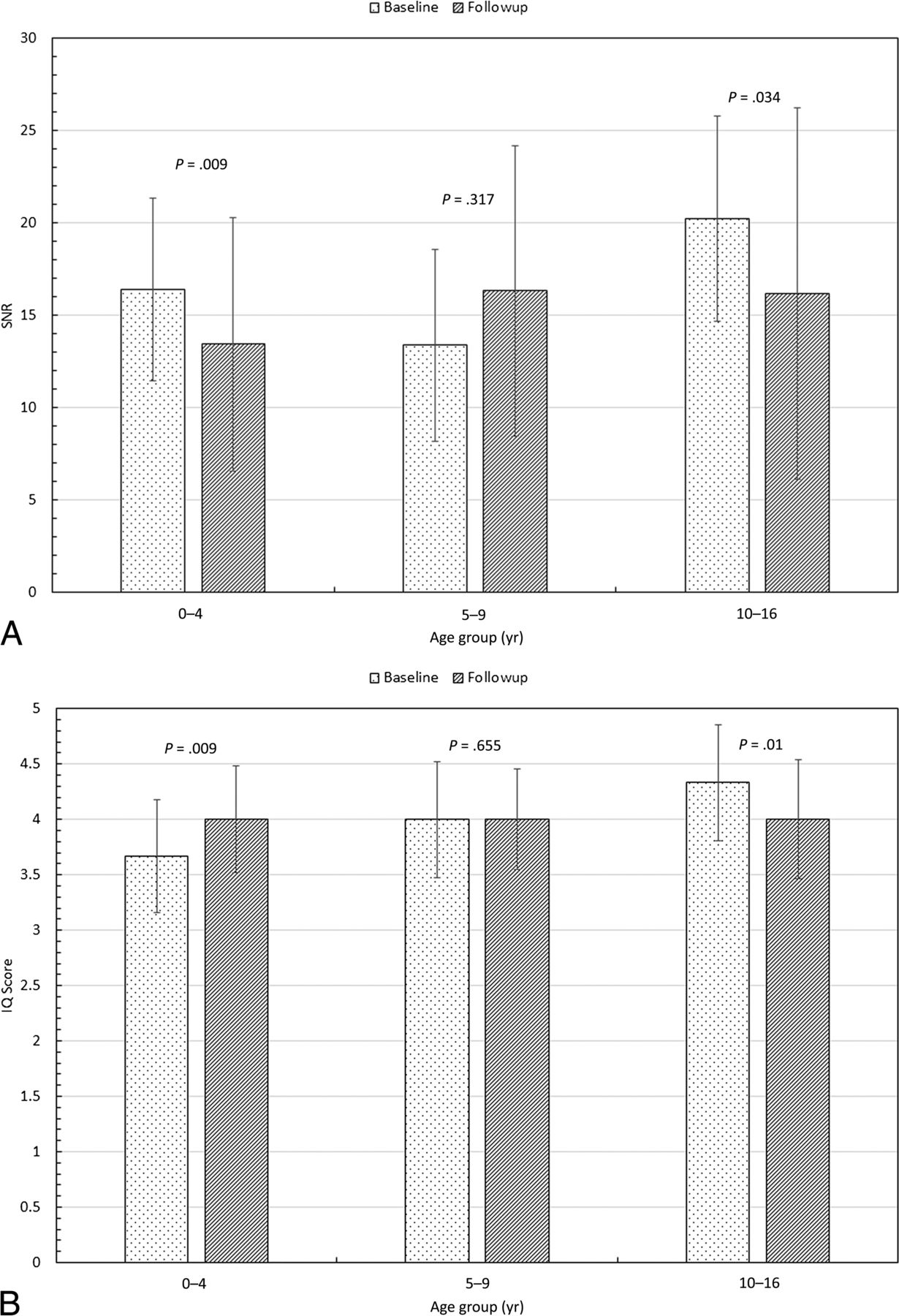
  • Fig 4.
    • Download figure
    • Open in new tab
    • Download powerpoint
    Fig 4.

    Comparison of the median values of the signal-to-noise ratio (A) and image-quality score (B) between the 2 cohorts. Also shown are the P values from an unpaired t test.

Tables

  • Figures
    • View popup
    Table 1:

    Total number of patients and distribution of patient age and sex in the 3 age groups of the study

    Group0–4 yr5–9 yr10–16 yr0–16 yr
    Baseline cohort
        ♂10112041
        ♀772034
        Total (No.)17184075
    Follow-up cohort
        ♂17172155
        ♀893249
        Total (No.)252653104
    • View popup
    Table 2:

    Interpretation of the subjective image-quality scores for the individual, prelabeled CT image slices at the level of the vocal cords

    ScoreComment
    1Image quality very poor (significant noise and/or artifacts, study uninterpretable)
    2Image quality poor (noise and/or artifacts, can only answer broad clinical questions)
    3Image quality adequate (some noise and/or artifacts, but study interpretable)
    4Above-average image quality (minimal noise and/or artifacts)
    5Excellent image quality (imperceptible noise and free of artifacts)
    • View popup
    Table 3:

    Median values of the scan parameters, signal-to-noise ratio, image-quality scores, estimated effective dose, and the percentage difference for the 2 cohorts

    Parameter0–4 yr5–9 yr10–16 yr
    III% DiffIII% DiffIII% Diff
    Tube current (mAs)150128−15182162−11266165−38
    CTDIvol (mGy)7.44.4−418.65.5−36207−67
    Scan length (cm)17.617.8120.321526271
    DLP (mGy-cm)12577−38175132−24522240−54
    ED (mSv)2.11.2−402.21.7−219.74.4−55
    SNR16.413.4−1813.416.32220.216.2−20
    IQ3.674.0094.004.0004.334.00−8
    • Note:—IQ indicates image quality; ED, effective dose; Diff, difference; I, baseline; II, follow-up.

PreviousNext
Back to top
Advertisement
Print
Download PDF
Email Article

Thank you for your interest in spreading the word on American Journal of Neuroradiology.

NOTE: We only request your email address so that the person you are recommending the page to knows that you wanted them to see it, and that it is not junk mail. We do not capture any email address.

Enter multiple addresses on separate lines or separate them with commas.
Radiation Dose and Image Quality in Pediatric Neck CT
(Your Name) has sent you a message from American Journal of Neuroradiology
(Your Name) thought you would like to see the American Journal of Neuroradiology web site.
CAPTCHA
This question is for testing whether or not you are a human visitor and to prevent automated spam submissions.
Cite this article
S.V. Tipnis, W.J. Rieter, D. Patel, S.T. Stalcup, M.G. Matheus, M.V. Spampinato
Radiation Dose and Image Quality in Pediatric Neck CT
American Journal of Neuroradiology May 2019, DOI: 10.3174/ajnr.A6073

Citation Manager Formats

  • BibTeX
  • Bookends
  • EasyBib
  • EndNote (tagged)
  • EndNote 8 (xml)
  • Medlars
  • Mendeley
  • Papers
  • RefWorks Tagged
  • Ref Manager
  • RIS
  • Zotero
0 Responses
Respond to this article
Share
Bookmark this article
Radiation Dose and Image Quality in Pediatric Neck CT
S.V. Tipnis, W.J. Rieter, D. Patel, S.T. Stalcup, M.G. Matheus, M.V. Spampinato
American Journal of Neuroradiology May 2019, DOI: 10.3174/ajnr.A6073
del.icio.us logo Twitter logo Facebook logo Mendeley logo
  • Tweet Widget
  • Facebook Like
  • Google Plus One
Purchase

Jump to section

  • Article
    • Abstract
    • ABBREVIATIONS:
    • Materials and Methods
    • Results
    • Discussion
    • Conclusions
    • Footnotes
    • REFERENCES
  • Figures & Data
  • Info & Metrics
  • Responses
  • References
  • PDF

Related Articles

  • No related articles found.
  • PubMed
  • Google Scholar

Cited By...

  • No citing articles found.
  • Crossref (5)
  • Google Scholar

This article has been cited by the following articles in journals that are participating in Crossref Cited-by Linking.

  • Variation in tube voltage for pediatric neck 64VCT: Effect on radiation dose and image quality
    Li-Guo Chen, Ping-An Wu, Hsing-Yang Tu, Ming-Huei Sheu, Li-Chuan Huang, Marco Giannelli
    PLOS ONE 2021 16 11
  • Congenital and Infantile Masses of the Head and Neck
    Alexandra M. Foust, Rekha Krishnasarma, Caroline D. Robson, Elizabeth Snyder, Dann Martin, Katharyn D. Cassella, Lindsey Johnstone, Joanne Rispoli, Sumit Pruthi, Asha Sarma
    RadioGraphics 2024 44 12
  • Evaluation of Radiation Exposure in Pediatric Cranial Trauma Patients
    Kamil DOĞAN, Adil DOĞAN, Seda Nida KARAKÜÇÜK, Hakan HAKKOYMAZ, Şükrü GÜNGÖR
    Kahramanmaraş Sütçü İmam Üniversitesi Tıp Fakültesi Dergisi 2023 18 3
  • Nontraumatic Pediatric Head and Neck Emergencies: Resource for On-Call Radiologists
    Alexandra M. Foust, Lindsey Johnstone, Rekha Krishnasarma, Dann C. Martin, Jennifer Vaughn, Karuna Shekdar, Elizabeth Snyder, Ty Todd, Sumit Pruthi, Asha Sarma
    RadioGraphics 2024 44 10
  • Radiopaedia.org
    Joachim Feger, Arlene Campos, Andrew Murphy
    2021

More in this TOC Section

  • Neuroimaging Delineation and Progression of SLSMD
  • fetal brain development of 10 weeks gestation
  • CHARGE fetal MRI clival cleft
Show more Pediatric Neuroimaging

Similar Articles

Advertisement

Indexed Content

  • Current Issue
  • Accepted Manuscripts
  • Article Preview
  • Past Issues
  • Editorials
  • Editor's Choice
  • Fellows' Journal Club
  • Letters to the Editor
  • Video Articles

Cases

  • Case Collection
  • Archive - Case of the Week
  • Archive - Case of the Month
  • Archive - Classic Case

More from AJNR

  • Trainee Corner
  • Imaging Protocols
  • MRI Safety Corner
  • Book Reviews

Multimedia

  • AJNR Podcasts
  • AJNR Scantastics

Resources

  • Turnaround Time
  • Submit a Manuscript
  • Submit a Video Article
  • Submit an eLetter to the Editor/Response
  • Manuscript Submission Guidelines
  • Statistical Tips
  • Fast Publishing of Accepted Manuscripts
  • Graphical Abstract Preparation
  • Imaging Protocol Submission
  • Evidence-Based Medicine Level Guide
  • Publishing Checklists
  • Author Policies
  • Become a Reviewer/Academy of Reviewers
  • News and Updates

About Us

  • About AJNR
  • Editorial Board
  • Editorial Board Alumni
  • Alerts
  • Permissions
  • Not an AJNR Subscriber? Join Now
  • Advertise with Us
  • Librarian Resources
  • Feedback
  • Terms and Conditions
  • AJNR Editorial Board Alumni

American Society of Neuroradiology

  • Not an ASNR Member? Join Now

© 2025 by the American Society of Neuroradiology All rights, including for text and data mining, AI training, and similar technologies, are reserved.
Print ISSN: 0195-6108 Online ISSN: 1936-959X

Powered by HighWire