
Article Figures & Data
Figures
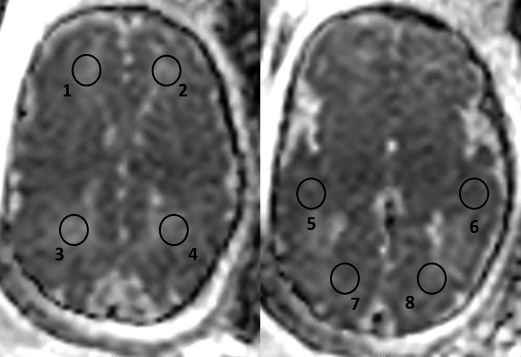
- Fig 1.
ADC calculation was performed on 8 circular ROIs: 2 on the white matter of both frontal (1, 2), parietal (3, 4), temporal (5, 6), and occipital lobes (7, 8). A circular ROI was placed over the desired anatomic area, ranging from 75 to 98 mm2.
- Fig 2.
T2 MR images (single-shot fast spin-echo T2-weighted sequences in 3 orthogonal planes using a half-Fourier technique, NEX = 0.53) of 2 fetuses at 33 weeks of gestational age. The CMV-positive fetus has diffuse WMHS (white arrowheads), unilateral ventriculomegaly, and intraventricular adhesions (black asterisks), suggesting ventriculitis (A–C). The CMV-negative fetus has WMHS located in the white matter in the temporal lobes (white arrowhead) (D–F).


Tables
- Table 1:
Demographic and clinical characteristics of fetuses with white matter T2 hyperintense signala
Characteristic CMV-Positive (n = 22) CMV-Negative (n = 22) P Value Maternal age (yr) 30.5 (26.7–34.2) 31 (28.7–34.2) .74 Gestational age at MRI (wk) 33 (32–34) 33.5 (32–35) .69 Previous pregnancies 2 (2–3) 2 (1–4) .94 Previous labors 1 (1–1.75) 1 (0–2.25) .95 Abnormal outcome in previous labors 6 (27%) 7 (32%) .74 Abnormal maternal medical background 0 6 (27%) .02 Mode of conception 19 (86%) 22 (100%) .35 Spontaneous IVF 2 (9%) 0 (0%) Induced pregnancy 1 (5%) 0 (0%) Sex (male) 12 (54%) 8 (38%) .36 Normal nuchal translucency scan results 21 (95%) 22 (100%) .99 Normal first trimester biochemical test results 21 (94%) 21 (94%) >.99 Normal second trimester biochemical test results 22 (100%) 22 (100%) >.99 Normal early anatomic scan findings 22 (100%) 16 (72%) .02 Normal late anatomic scan findings 21 (94%) 11 (60%) .009 Note:—IVF indicates in vitro fertilization.
↵a Data are presented as median (interquartile range) for continuous variables or number (percentage) for categoric variables.
CMV-Positive CMV-Negative P Value Gestational age at MRI (wk) 33 (32–34) 33.5 (32–35) .6 Radiologic finding Frontal T2 hyperintense signal 13/22 (59%) 6/22 (27.3%) .067 Parietal T2 hyperintense signal 14/22 (63.6%) 3/22 (13.6%) .002 Temporal T2 hyperintense signal 22/22 (100%) 21/22 (95.5%) >.999 Occipital T2 hyperintense signal 0/22 (0%) 0/22 (0%) >.999 T2 hyperintense signal at 3 lobes (frontal, parietal, and temporal) 13/22 (59%) 3/22 (13/6%) .004 Additional findings 1/22 (4.5%) 7/22 (31.8%) .046 ↵a Data are presented as median (interquartile range) for continuous variables or number (percentage) for categoric variables.
Lobe, Side Study Group ADC ADC Control P Value Frontal Right CMV− 1793 (191) 1809 (165) .754 Right CMV+ 1872 (160) .160 Left CMV− 1741 (141) .115 Left CMV+ 1858 (193) .302 Parietal Right CMV− 1659 (200) 1748 (192) .099 Right CMV+ 1840 (140) .057 Left CMV− 1690 (188) .268 Left CMV+ 1852 (138) .03 Temporal Right CMV− 1702 (169) 1692 (164) .843 Right CMV+ 1835 (161) .002 Left CMV− 1689 (193) .933 Left CMV+ 1846 (122) <.001 Occipital Right CMV− 1659 (224) 1731 (167) .171 Right CMV+ 1723 (129) .830 Left CMV− 1675 (169) .224 Left CMV+ 1725 (111) .882 Note:—CMV+ indicates CMV-positive; CMV−, CMV-negative.
↵a Data are presented as mean (SD). ADC units are 106 mm2/s.
- Table 4:
Comparison of ADC values between fetuses with CMV-positive infection and fetuses with isolated white matter hyperintense signala
Lobe, Side CMV-Positive CMV-Negative P Value Frontal Right 1872 (160) 1793 (192) .147 Left 1858 (193) 1741 (141) .026 Parietal Right 1840 (140) 1659 (200) .001 Left 1852 (138) 1690 (188) .002 Temporal Right 1835 (161) 1702 (169) .011 Left 1846 (122) 1689 (193) .002 Occipital Right 1723 (129) 1659 (224) .259 Left 1725 (111) 1675 (169) .254 ↵a Data are presented as mean (SD). ADC units are 106 mm2/s.
Characteristic CMV-Positive (n = 20) CMV-Negative (n = 18) P Value Birth week 39.2 (38.5–39.9) 37.9 (39.7–40.6) .55 Birth weight (percentile) 49 (31–73) 49 (13–68) .53 Apgar score at 5 min ≥9 20 (100%) 18 (100%) >.99 Hearing deficitb 1 (5%) 0 (0%) >.99 Age (mo) at VABS test 30 (17–49) 27.5 (17.2–28.5) .75 VABS motor skills 92.5 (81.75–104) 103 (95.25–108) .07 VABS daily living skills 103 (99.25–117) 109 (99.25–117) .28 VABS socialization 101 (98.5–105.75) 100 (96.75–115.75) .89 VABS communication 107.5 (100.25–113) 108 (99.25–117) .84 VABS adaptive score composite 102 (90–18) 107 (98–115) .16 Child with any VABS score <70 4 (20%) 1 (5.5%) .34


