Advertisement

Main menu

  • Home
  • Content
    • Current Issue
    • Accepted Manuscripts
    • Article Preview
    • Past Issue Archive
    • Video Articles
    • AJNR Case Collection
    • Case of the Week Archive
    • Case of the Month Archive
    • Classic Case Archive
  • Special Collections
    • AJNR Awards
    • Low-Field MRI
    • Alzheimer Disease
    • ASNR Foundation Special Collection
    • Photon-Counting CT
    • View All
  • Multimedia
    • AJNR Podcasts
    • AJNR SCANtastic
    • Trainee Corner
    • MRI Safety Corner
    • Imaging Protocols
  • For Authors
    • Submit a Manuscript
    • Submit a Video Article
    • Submit an eLetter to the Editor/Response
    • Manuscript Submission Guidelines
    • Statistical Tips
    • Fast Publishing of Accepted Manuscripts
    • Graphical Abstract Preparation
    • Imaging Protocol Submission
    • Author Policies
  • About Us
    • About AJNR
    • Editorial Board
    • Editorial Board Alumni
  • More
    • Become a Reviewer/Academy of Reviewers
    • Subscribers
    • Permissions
    • Alerts
    • Feedback
    • Advertisers
    • ASNR Home

User menu

  • Alerts
  • Log in

Search

  • Advanced search
American Journal of Neuroradiology

American Journal of Neuroradiology

ASHNR American Society of Functional Neuroradiology ASHNR American Society of Pediatric Neuroradiology ASSR
  • Alerts
  • Log in

Advanced Search

  • Home
  • Content
    • Current Issue
    • Accepted Manuscripts
    • Article Preview
    • Past Issue Archive
    • Video Articles
    • AJNR Case Collection
    • Case of the Week Archive
    • Case of the Month Archive
    • Classic Case Archive
  • Special Collections
    • AJNR Awards
    • Low-Field MRI
    • Alzheimer Disease
    • ASNR Foundation Special Collection
    • Photon-Counting CT
    • View All
  • Multimedia
    • AJNR Podcasts
    • AJNR SCANtastic
    • Trainee Corner
    • MRI Safety Corner
    • Imaging Protocols
  • For Authors
    • Submit a Manuscript
    • Submit a Video Article
    • Submit an eLetter to the Editor/Response
    • Manuscript Submission Guidelines
    • Statistical Tips
    • Fast Publishing of Accepted Manuscripts
    • Graphical Abstract Preparation
    • Imaging Protocol Submission
    • Author Policies
  • About Us
    • About AJNR
    • Editorial Board
    • Editorial Board Alumni
  • More
    • Become a Reviewer/Academy of Reviewers
    • Subscribers
    • Permissions
    • Alerts
    • Feedback
    • Advertisers
    • ASNR Home
  • Follow AJNR on Twitter
  • Visit AJNR on Facebook
  • Follow AJNR on Instagram
  • Join AJNR on LinkedIn
  • RSS Feeds

AJNR Awards, New Junior Editors, and more. Read the latest AJNR updates

Research ArticlePediatric Neuroimaging

Apparent Diffusion Coefficient Levels and Neurodevelopmental Outcome in Fetuses with Brain MR Imaging White Matter Hyperintense Signal

E. Katorza, G. Strauss, R. Cohen, M. Berkenstadt, C. Hoffmann, R. Achiron, E. Barzilay and O. Bar-Yosef
American Journal of Neuroradiology September 2018, DOI: https://doi.org/10.3174/ajnr.A5802
E. Katorza
aFrom the Antenatal Diagnostic Unit (E.K., G.S., R.C., R.A., E.B.)
eSackler School of Medicine (E.K., G.S., R.C., M.B., C.H., R.A., E.B., O.B.-Y.), Tel Aviv University, Tel Aviv, Israel.
  • Find this author on Google Scholar
  • Find this author on PubMed
  • Search for this author on this site
  • ORCID record for E. Katorza
G. Strauss
aFrom the Antenatal Diagnostic Unit (E.K., G.S., R.C., R.A., E.B.)
eSackler School of Medicine (E.K., G.S., R.C., M.B., C.H., R.A., E.B., O.B.-Y.), Tel Aviv University, Tel Aviv, Israel.
  • Find this author on Google Scholar
  • Find this author on PubMed
  • Search for this author on this site
  • ORCID record for G. Strauss
R. Cohen
aFrom the Antenatal Diagnostic Unit (E.K., G.S., R.C., R.A., E.B.)
eSackler School of Medicine (E.K., G.S., R.C., M.B., C.H., R.A., E.B., O.B.-Y.), Tel Aviv University, Tel Aviv, Israel.
  • Find this author on Google Scholar
  • Find this author on PubMed
  • Search for this author on this site
  • ORCID record for R. Cohen
M. Berkenstadt
cThe Danek Gertner Institute of Human Genetics (M.B.)
eSackler School of Medicine (E.K., G.S., R.C., M.B., C.H., R.A., E.B., O.B.-Y.), Tel Aviv University, Tel Aviv, Israel.
  • Find this author on Google Scholar
  • Find this author on PubMed
  • Search for this author on this site
  • ORCID record for M. Berkenstadt
C. Hoffmann
dNeuroradiology Unit (C.H.), Department of Diagnostic Radiology, Chaim Sheba Medical Center, Tel-Hashomer, Israel
eSackler School of Medicine (E.K., G.S., R.C., M.B., C.H., R.A., E.B., O.B.-Y.), Tel Aviv University, Tel Aviv, Israel.
  • Find this author on Google Scholar
  • Find this author on PubMed
  • Search for this author on this site
  • ORCID record for C. Hoffmann
R. Achiron
aFrom the Antenatal Diagnostic Unit (E.K., G.S., R.C., R.A., E.B.)
eSackler School of Medicine (E.K., G.S., R.C., M.B., C.H., R.A., E.B., O.B.-Y.), Tel Aviv University, Tel Aviv, Israel.
  • Find this author on Google Scholar
  • Find this author on PubMed
  • Search for this author on this site
  • ORCID record for R. Achiron
E. Barzilay
aFrom the Antenatal Diagnostic Unit (E.K., G.S., R.C., R.A., E.B.)
eSackler School of Medicine (E.K., G.S., R.C., M.B., C.H., R.A., E.B., O.B.-Y.), Tel Aviv University, Tel Aviv, Israel.
  • Find this author on Google Scholar
  • Find this author on PubMed
  • Search for this author on this site
  • ORCID record for E. Barzilay
O. Bar-Yosef
bDepartment of Obstetrics and Gynecology, Pediatric Neurology Unit (O.B.-Y.)
eSackler School of Medicine (E.K., G.S., R.C., M.B., C.H., R.A., E.B., O.B.-Y.), Tel Aviv University, Tel Aviv, Israel.
  • Find this author on Google Scholar
  • Find this author on PubMed
  • Search for this author on this site
  • ORCID record for O. Bar-Yosef
  • Article
  • Figures & Data
  • Info & Metrics
  • Responses
  • References
  • PDF
Loading

Abstract

BACKGROUND AND PURPOSE: One of the perplexing findings of fetal brain MR imaging is white matter T2 hyperintense signal. The aims of our study were initially to determine the main etiologies associated with white matter T2 hyperintense signal, then to examine whether the different etiologies have different ADC values, and, last, to assess the association of white matter T2 hyperintense signal with developmental outcome.

MATERIALS AND METHODS: This was a prospective cohort study of 44 MR imaging scans of fetal brains obtained for suspected brain pathologies at a tertiary medical center during 2011–2015. Clinical data were collected from electronic medical charts. ADC values were measured and averaged in the frontal, parietal, occipital, and temporal lobes. Neurodevelopmental assessments were performed with the Vineland Adaptive Behavior Scales II.

RESULTS: Half of the cases of MRI hyperintense T2 signal of the fetal brain were associated with congenital cytomegalovirus infection. The other half were mainly idiopathic. Thus, the study group was divided to subgroups positive and negative for cytomegalovirus. Both groups had hyperintense signal in the temporal lobe. The group positive for cytomegalovirus had involvement of the parietal lobe. Only this group had increased ADC values in the temporal and parietal lobes. There was no association between the neurodevelopment outcome and the etiologies or ADC values.

CONCLUSIONS: T2 hyperintense signal in fetal brain MRI associated with positive cytomegalovirus infection has increased ADC values in the temporal and parietal lobes, suggestive of brain edema in these areas. However, the association between this finding and neurodevelopment outcome requires further evaluation.

ABBREVIATIONS:

CMV
cytomegalovirus
fbMRI
fetal brain MRI
ICC
interclass correlation coefficient
VABS
Vineland Adaptive Behavior Scales II
WMHS
white matter hyperintense signal

Fetal brain MR imaging (fbMRI) has been increasingly used in recent years as a means of tracking normal and pathologic fetal brain maturation.1 One of the perplexing findings of fbMRI is white matter T2 hyperintense signal (WMHS). On the one hand, it has been associated with in utero brain pathologies, such as ischemia and cytomegalovirus (CMV) infection.2⇓–4 On the other hand, the validity and relevance of this finding have been questioned.5

In recent years, DWI and its ADC metric have become a quantitative method for evaluation of fetal brain maturation.6 Previous studies showed that ADC values of the developing fetal brain correlate with fetal brain maturation.7⇓⇓⇓–11 Deviation from normal ADC values has been shown to be associated with brain pathologies such as ischemia, CMV infection, and ventriculomegaly.2,12,13 Postmortem studies of animal and human fetuses with hypoxic-ischemic brain injury have demonstrated a transition from low ADC values after the injury to increased values 7 days after the injury. This transition was associated with histologic findings changing from initial cytotoxic edema and swollen astrocytes to vasogenic edema, astrogliosis, and abundance of macrophages.2,14 Thus, ADC and its association with histopathology could be used to test the validity and meaning of T2 hyperintensity.

The aims of our study were initially to determine the main etiologies associated with WMHS, then to examine whether the different etiologies have different ADC values, and, last, to assess the association of WMHS with developmental outcome.

Materials and Methods

Subjects

This was a prospective cohort study of women who were referred for fetal brain MR imaging to our tertiary medical center, Sheba Medical Center, Ramat Gan, Israel, between 2011 and 2015. The cohorts for this study were chosen on the basis of identification of a hyperintense signal on the T2-weighted sequences. Demographic and clinical data were collected from the electronic records of each patient.

Data obtained from the records included maternal history, prenatal screening tests, imaging results from anatomic sonography and MR imaging, maternal CMV status, and perinatal history. Fetal cytomegalovirus infection was confirmed by either amniocentesis performed during pregnancy or by the presence of CMV DNA in neonatal urine or saliva.

The study was approved by Sheba Medical Center, Ramat Gan, Israel, institutional ethics board. Informed consent was obtained from each participant in the study prospectively.

MR Imaging Scans

Fetal brain MR imaging was performed using a 1.5T system (Optima MR450w with GEM Suite; GE Healthcare, Milwaukee, Wisconsin). Single-shot fast spin-echo T2-weighted sequences in 3 orthogonal planes were performed using a half-Fourier technique (NEX = 0.53) with the following parameters: section thickness, 3 or 4 mm; no gap; flexible coil (8-channel cardiac coil). FOV was determined by the size of the fetal head with a range of 24 × 24 to 30 × 30 cm; acquisition time was between 40 and 45 seconds (matrix, 320 × 224; TE, 90 ms; TR, 1298 ms; pixel bandwidth, 122 Hz/pixel; specific absorption rate values, 1.1–1.7 W/kg). A DWI sequence in 1, 2, or 3 orthogonal planes was then performed, with a 40-cm FOV, b-values of 0 and 700 ms, and a slice thickness of 4 mm with no gap. All MR images were obtained by the same protocol at our institution and assessed by a specialist in fetal sonography and a neuroradiologist expert in MR imaging as previously published.15

Target Variables

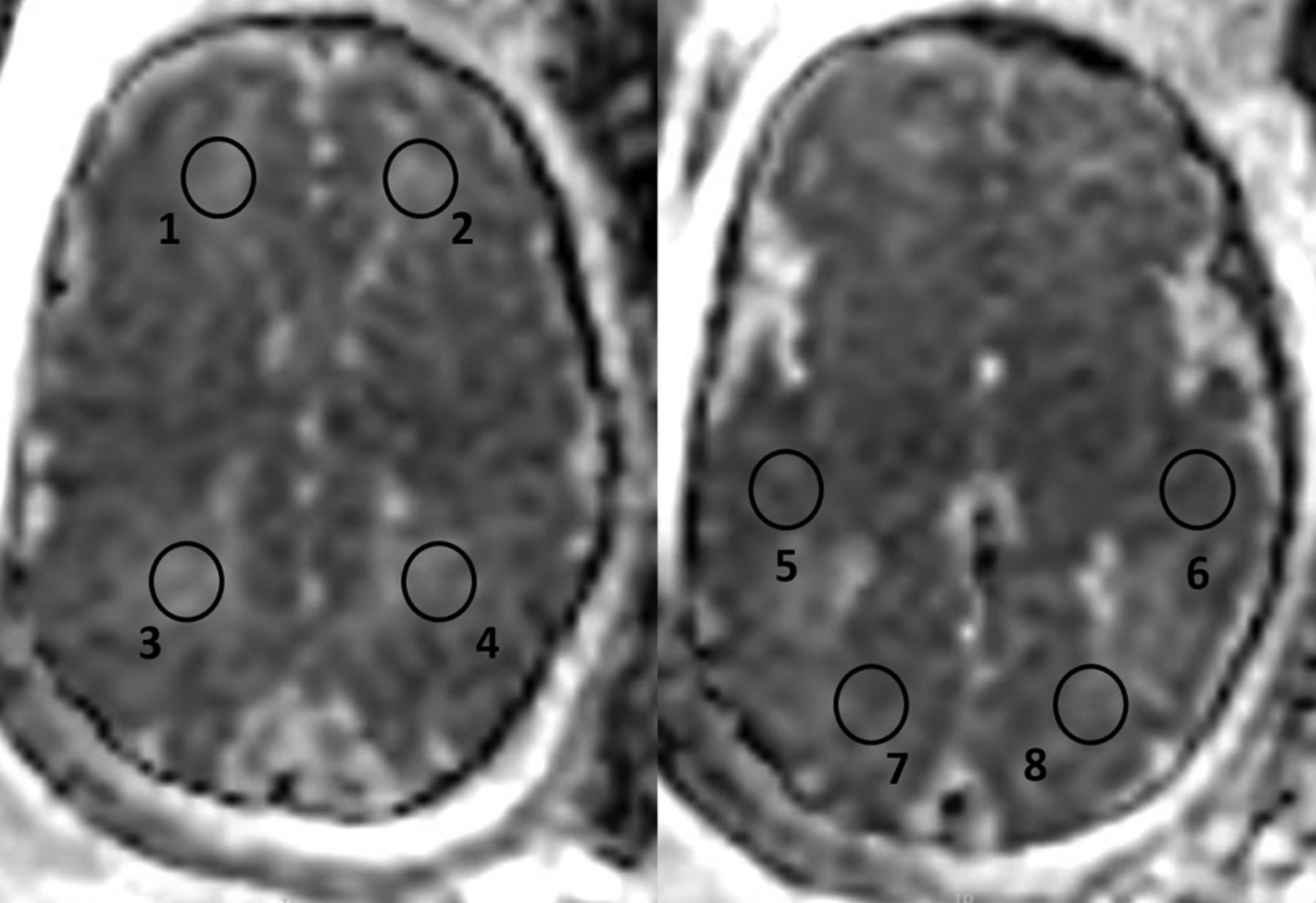
ADC calculation was performed on 8 circular ROIs: 2 on the white matter of both frontal, parietal, temporal, and occipital lobes. A circular ROI was used, placed over the desired anatomic area, ranging from 75 to 98 mm2, similar to that accepted in the recent literature.11 Examples of ROI placements over the various anatomic areas can be seen in Fig 1. The ROIs were placed with the following anatomic considerations: Frontal lobe ROIs were placed in the anterior part of the frontal lobes above the lateral ventricles, parietal lobe ROIs were placed in the posterior part of the parietal lobes above the lateral ventricles, temporal lobe ROIs were placed anterior to the temporal horns, and occipital lobe ROIs were placed posterior to the lateral ventricles. The ADC was calculated using IntelliSpace workstation software (Phillips Healthcare, Best, the Netherlands; https://www.usa.philips.com/healthcare/solutions/clinical-informatics/enterprise-imaging-pacs).

Fig 1.
  • Download figure
  • Open in new tab
  • Download powerpoint
Fig 1.

ADC calculation was performed on 8 circular ROIs: 2 on the white matter of both frontal (1, 2), parietal (3, 4), temporal (5, 6), and occipital lobes (7, 8). A circular ROI was placed over the desired anatomic area, ranging from 75 to 98 mm2.

White Matter T2 Hyperintense Signal

WMHS is a subjective interpretation of the signal from the fetal brain white matter on T2 sequences, made by the radiologist. This diagnosis is established when specific areas of the brain white matter appear hyperintense in comparison with other areas or with that expected according to the gestational age (Fig 2).16

Fig 2.
  • Download figure
  • Open in new tab
  • Download powerpoint
Fig 2.

T2 MR images (single-shot fast spin-echo T2-weighted sequences in 3 orthogonal planes using a half-Fourier technique, NEX = 0.53) of 2 fetuses at 33 weeks of gestational age. The CMV-positive fetus has diffuse WMHS (white arrowheads), unilateral ventriculomegaly, and intraventricular adhesions (black asterisks), suggesting ventriculitis (A–C). The CMV-negative fetus has WMHS located in the white matter in the temporal lobes (white arrowhead) (D–F).

Interobserver Validity of ADC Measurements

To validate the consistency of measurements and reliability of results, 2 observers evaluated 10 fetuses. Interobserver variability was assessed by the interclass correlation coefficient (ICC). We considered an ICC value of ≥0.8 as excellent agreement.

Neurodevelopmental and Hearing Outcome

Children were assessed by the Vineland Adaptive Behavior Scales, 2nd edition (VABS), which is a structured parent interview assessing 4 different domains of behavior: communication, daily living skills, socialization, and motor skills. All 4 domains are included in an adaptive composite score.17,18 VABS assessment was conducted by a phone interview by 2 medical students trained and supervised by a pediatric neurologist and child development expert experienced in conducting VABS (O.B.-Y.). Validation of the phone interviews were assessed by correlation of the ICC to the VABS scores of 15 children (divided into 7 and 8 children between the students) evaluated by the pediatric neurologist (O.B.-Y.). The ICC was >0.83 for the 4 VABS domains and the composite score. Scores of children were considered abnormal if the standard score was <70.

Hearing outcome was assessed 2–3 days after delivery by Transient Evoked Otoacoustic Emissions. Neonates positive for CMV were tested routinely before 1 month of age by brain stem auditory evoked potential, then at 3, 6, and 12 months by Transient Evoked Otoacoustic Emissions and behavioral assessment.

Statistical Analysis

Categoric variables were expressed as number and percentage. Distribution of continuous variables was assessed using a histogram and Q-Q plot. Continuous variables were described using median and interquartile range or mean and SD as appropriate. Categoric variables were compared using the χ2 test, Fisher exact test, or McNemar test as appropriate. Continuous variables were compared using the Student t test or Mann-Whitney test as appropriate. A 2-tailed P < .05 was considered statistically significant. Analyses were performed with SPSS (Version 24.0, 2016; IBM, Armonk, New York).

Results

Demographic and Clinical Characteristics of the Study Population

The study subjects comprised 43 singleton pregnant women and 1 single twins pregnant woman who underwent fbMRI scans. All 44 fetuses included in this study were in their third trimester of pregnancy at the time of the fetal MR imaging, with a median gestational age of 32 weeks (interquartile range, 28.25–35 weeks). The average maternal age was 33 years (interquartile range, 25–32 years). The indications for MR imaging included the following: maternal cytomegalovirus infection (n = 31), family history of central nervous system pathology (n = 3), and abnormal findings during fetal sonography (n = 10). The abnormal sonographic findings included the following: lateral ventricular asymmetry (n = 4), mega cisterna magna (n = 2), small head circumference (n = 2), fisted hands (n = 1), and hyperechogenic bowel (n = 1). Twenty-two (50%) of the fetuses were found to be positive for CMV by amniocentesis or by saliva or urinary CMV DNA testing after birth. Thus, to examine the association between ADC values and etiology, we divided the main study group into a CMV-positive group consisting of 22 fetuses with confirmed CMV infection and a CMV-negative group consisting of 22 fetuses with WMHS from an unknown etiology.

These 2 distinct patient populations are described separately and compared by their demographic, clinical, imaging, and neurodevelopment characteristics.

The median gestational age, pregnancy history, and sex did not differ statistically between the groups. All mothers in the CMV-positive group had normal medical background, compared with 6 (27%) mothers in the CMV-negative group who had significant abnormal medical background including thrombophilia, Raynaud disease, and a history of cerebellar infarction (P = .02).

In the CMV-positive group, there were significantly less early and late anatomic scan findings (P = .02, P = .009, respectively) (Table 1).

View this table:
  • View inline
  • View popup
Table 1:

Demographic and clinical characteristics of fetuses with white matter T2 hyperintense signala

Distribution of Hyperintense Signal and Additional MR Imaging Findings

Both groups had similar rates of WMHS in the temporal lobe (P > .999). WMHS was not depicted in the occipital lobe in both groups. However, the CMV-positive group had statistically higher rates of parietal hyperintense signal (P = .002) and a trend toward higher rates in the frontal lobe (P = .067). The CMV-positive group had more extended WMHS involving all 3 lobes (P = .004). The CMV-negative fetuses had statistically significantly higher rates (P = .046) of minor additional findings on imaging, including subarachnoid cyst (n = 2), slightly enlarged subarachnoid space (n = 1), and lateral ventricle asymmetry (n = 4) (Table 2 and Fig 2).

View this table:
  • View inline
  • View popup
Table 2:

Radiologic MR imaging findingsa

Interobserver Validity of ADC Measurements

ADC measurements showed excellent interobserver agreement for all regions, with the ICC ranging between 0.81 and 0.97.

ADC Value Measurements

ADC values for each group were compared with the normal ADC values as published by Hoffmann et al.11 Fetuses in the CMV-positive group were found to have statistically higher ADC values in the temporal lobe bilaterally (P = .002 and P < .001) and the left parietal lobe (P = .033) and a trend in the right parietal lobe (P = .057) (Table 3).

View this table:
  • View inline
  • View popup
Table 3:

Comparison of ADC values of the study group with the control groupa

ADC values were compared for each region between the 2 study groups. Fetuses in the CMV-positive group were found to have higher ADC values in the left frontal lobe (P = .026), the parietal lobe bilaterally (right, P = .001; left, P = .002), and the temporal lobe bilaterally (right, P = .011; left, P = .002) (Table 4).

View this table:
  • View inline
  • View popup
Table 4:

Comparison of ADC values between fetuses with CMV-positive infection and fetuses with isolated white matter hyperintense signala

Delivery Data, Hearing, and Neurodevelopmental Assessment

Neurodevelopmental assessment (VABS) and delivery data were analyzed for 20 children in the CMV-positive group and 18 children in the CMV-negative group (Table 5). Six patients (2 from the CMV-positive group and 4 from the CMV-negative group) refused to participate in the neurodevelopmental assessment. Delivery data and age at developmental assessment were not statistically different between the groups. Comparison of the VABS score for the 4 domains and the adaptive composite score showed only a trend for motor skills (P = .07). Four children in the CMV-positive group and 1 child in the CMV-negative group had at least 1 VABS score <70; however, this was not statistically significant. Only 1 child had a hearing deficit; this child was CMV-positive with a low VABS score.

View this table:
  • View inline
  • View popup
Table 5:

Delivery data, hearing, and VABS assessmenta

Discussion

WMHS is a puzzling finding in fetal MR imaging, and its significance is not completely understood. The aims of our study were initially to explore the etiologies associated with this finding, then to examine whether the different etiologies are characterized by different ADC values, and, last, to investigate the association of WMHS with neurodevelopmental outcome.

Diverse etiologies were found to be associated with WMHS in this study. The major one (half of the study group), in accordance with fetal MR imaging literature, is congenital CMV infection.13,19,20 The precise etiologies of the second half of the group are not completely clear. Ten fetuses had further imaging findings; thus, the WMHS might be a different aspect of the overall brain pathology.

Comparison between the 2 groups demonstrated a difference in the distribution of the WMHS. Both groups had the same prevalence of WMHS in the temporal lobes, but the CMV-positive group had a higher rate in the parietal lobes and a trend in the frontal lobes. In general, the CMV-positive group had more diffuse WMHS.

The median ADC values were higher in the CMV-positive group than in the CMV-negative group in the temporal, parietal, and left frontal lobes. The ADC values of the CMV-negative group did not differ from those of the control group. Thus, although the incidences of WMHS were similar in the temporal lobes, the median ADC values were different. Because ADC is a quantitative measure and not a subjective interpretation, it is possible that some WMHS of the CMV-negative group is actually overinterpretation.

As for the CMV-positive group, the specific involvement of the temporal lobes was previously described in fbMRI. WMHS is part of a spectrum of findings localized to this area, including cysts, dilation of the temporal horns, and reduced temporal lobe volume.4,11,21,22 The reason for the specific vulnerability of the temporal lobe is unclear. Most interesting, in addition to the presence of WMHS in the temporal lobe, most of the fbMRIs in our study depicted a similar signal in the parietal lobe. This finding was previously described in children with congenital CMV.23⇓–25 Thus, the combination of temporal and parietal hyperintense T2 signal is potentially a sign of CMV infection involving the brain. Further involvement of WMHS including the frontal lobes may serve as an additional sign of congenital CMV infection.

The combination of WMHS and high ADC values in the CMV-positive group suggests a possible etiology for this finding. Hyperintense signal was associated at postmortem examination with astrogliosis and extracellular brain edema in the subacute stage of hypoxic-ischemic injury.2 Although hypoxic-ischemic injury pathogenesis is different from CMV infection, the simplest explanation of the T2 finding is an increase in extracellular fluid content in the white matter. Kotovich et al26 found low ADC values in fetuses with congenital CMV infection without T2 hyperintense signal. In a previous study of the same group, Yaniv et al4 described postmortem histology consisting of edema and cellular infiltration of plasma cells, lymphocytes, and microglia. The difference in T2 hyperintense signal and ADC values between the 2 latter studies and ours might reflect a different ratio between the edema and cellular component in the brain tissue. When the microgliosis and edema component are more prominent, the ADC values are larger than those of controls; whereas when the component of cellular degeneration and immune cell infiltration is more prominent, the ADC values are lower than those of controls.2,14,27⇓–29

The association between WMHS and abnormal neurodevelopmental outcome was not found in this study. In the CMV-positive group, 4 children (20%) had abnormal neurodevelopmental findings or hearing loss. Based on neurodevelopmental data from previous studies, the expected range of these abnormal findings in children with asymptomatic congenital CMV (normal prenatal imaging findings) is 10%–15%.30,31 Thus, it is difficult to assess whether the WMHS and the increased ADC values predict worse prognosis.

Combining the results from our study and others2,11⇓⇓–14,26 investigating the association of ADC and brain pathologies suggests the following practical advice for clinicians: The combination of WMHS and normal ADC suggests that the source of WMHS is most likely without clinical significance. As for cases of abnormal ADC either below or above the expected value for gestational age, they raise the suspicion of brain pathology such as CMV infection, ventriculomegaly, or ischemia. However, the association of abnormal ADC with neurodevelopmental outcome is unclear.

Our study is limited by the size of the study group. A larger number of patients could be used to correlate the distribution of T2 hyperintense signal or ADC values with neurodevelopment. On the other hand, the small number of children in each group strengthens the significance of the difference in T2 hyperintense signal and ADC values between the CMV-positive and -negative groups.

Conclusions

ADC measurements supported the validity of T2 hyperintense signal in fbMRI only in fetuses with CMV infection. The association of this finding with neurodevelopmental outcome requires further investigation.

Footnotes

  • E. Katorza and G. Strauss contributed equally to this work.

References

  1. 1.↵
    1. Griffiths PD,
    2. Bradburn M,
    3. Campbell MJ, et al.
    MERIDIAN collaborative group. Use of MRI in the diagnosis of fetal brain abnormalities in utero (MERIDIAN): a multicentre, prospective cohort study. Lancet 2017;389:538–46 doi:10.1016/S0140-6736(16)31723-8 pmid:27988140
    CrossRefPubMed
  2. 2.↵
    1. Guimiot F,
    2. Garel C,
    3. Fallet-Bianco C, et al
    . Contribution of diffusion-weighted imaging in the evaluation of diffuse white matter ischemic lesions in fetuses: correlations with fetopathologic findings. AJNR Am J Neuroradiol 2008;29:110–115 doi:10.3174/ajnr.A0754 pmid:17947368
    Abstract/FREE Full Text
  3. 3.↵
    1. Malinger G,
    2. Lev D,
    3. Lerman-Sagie T
    . Imaging of fetal cytomegalovirus infection. Fetal Diagn Ther 2011;29:117–26 doi:10.1159/000321346 pmid:21088375
    CrossRefPubMed
  4. 4.↵
    1. Yaniv G,
    2. Hoffmann C,
    3. Weisz B, et al
    . Region-specific reductions in brain apparent diffusion coefficient in cytomegalovirus-infected fetuses. Ultrasound Obstet Gynecol 2016;47:600–07 doi:10.1002/uog.14737 pmid:25412951
    CrossRefPubMed
  5. 5.↵
    1. Fazekas F,
    2. Kleinert R,
    3. Offenbacher H, et al
    . Pathologic correlates of incidental MRI white matter signal hyperintensities. Neurology 1993;43:1683–89 doi:10.1212/WNL.43.9.1683 pmid:8414012
    CrossRefPubMed
  6. 6.↵
    1. Schneider JF,
    2. Confort-Gouny S,
    3. Le Fur Y, et al
    . Diffusion-weighted imaging in normal fetal brain maturation. Eur Radiol 2007;17:2422–29 doi:10.1007/s00330-007-0634-x pmid:17404738
    CrossRefPubMed
  7. 7.↵
    1. Sartor A,
    2. Arthurs O,
    3. Alberti C, et al
    . Apparent diffusion coefficient measurements of the fetal brain during the third trimester of pregnancy: how reliable are they in clinical practice? Prenat Diagn 2014;34:357–66 doi:10.1002/pd.4309 pmid:24375489
    CrossRefPubMed
  8. 8.↵
    1. Neil JJ,
    2. Shiran SI,
    3. McKinstry RC, et al
    . Normal brain in human newborns: apparent diffusion coefficient and diffusion anisotropy measured by using diffusion tensor MR imaging. Radiology 1998;209:57–66 doi:10.1148/radiology.209.1.9769812 pmid:9769812
    CrossRefPubMed
  9. 9.↵
    1. Girard N,
    2. Confort-Gouny S,
    3. Schneider J, et al
    . MR imaging of brain maturation. J Neuroradiol 2007;34:290–310 doi:10.1016/j.neurad.2007.07.007 pmid:17822767
    CrossRefPubMed
  10. 10.↵
    1. Toft PB,
    2. Leth H,
    3. Peitersen B, et al
    . The apparent diffusion coefficient of water in gray and white matter of the infant brain. J Comput Assist Tomograph 1996;20:1006–11 doi:10.1097/00004728-199611000-00029 pmid:8933811
    CrossRefPubMed
  11. 11.↵
    1. Hoffmann C,
    2. Weisz B,
    3. Lipitz S, et al
    . Regional apparent diffusion coefficient values in 3rd trimester fetal brain. Neuroradiology 2014;56:561–67 doi:10.1007/s00234-014-1359-6 pmid:24748534
    CrossRefPubMed
  12. 12.↵
    1. Erdem G1,
    2. Celik O,
    3. Hascalik, et al
    . Diffusion-weighted imaging evaluation of subtle cerebral microstructural changes in intrauterine fetal hydrocephalus. Magn Reson Imaging 2001;25:417–22 pmid:17513078
    PubMed
  13. 13.↵
    1. Yaniv G,
    2. Katorza E,
    3. Bercovitz R, et al
    . Region-specific changes in brain diffusivity in fetal isolated mild ventriculomegaly. Eur Radiol 2016;26:840–48 doi:10.1007/s00330-015-3893-y pmid:26135001
    CrossRefPubMed
  14. 14.↵
    1. Baud O,
    2. Daire JL,
    3. Dalmaz Y, et al
    . Gestational hypoxia induces white matter damage in neonatal rats: a new model of periventricular leukomalacia. Brain Pathol 2004;14:1–10 doi:10.1111/j.1750-3639.2004.tb00492.x pmid:14997932
    CrossRefPubMed
  15. 15.↵
    1. Gat I,
    2. Hoffmann C,
    3. Shashar D, et al
    . Fetal brain MRI: novel classification and contribution to sonography. Ultraschall Med 2016;37:176–84 doi:10.1055/s-0034-1384935 pmid:25535813
    CrossRefPubMed
  16. 16.↵
    1. Lipitz S,
    2. Hoffmann C,
    3. Feldman B, et al
    . Value of prenatal ultrasound and magnetic resonance imaging in assessment of congenital primary cytomegalovirus infection. Ultrasound Obstet Gynecol 2010;36:709–17 doi:10.1002/uog.7657 pmid:20503234
    CrossRefPubMed
  17. 17.↵
    1. Limperopoulos C,
    2. Majnemer A,
    3. Steinbach CL, et al
    . Equivalence reliability of the Vineland Adaptive Behavior Scale between in-person and telephone administration. Phys Occup Ther Pediatr 2006;26:115–27 pmid:16938828
    CrossRefPubMed
  18. 18.↵
    1. Sparrow SS.
    2. Cicchetti D,
    3. Balla DA
    . Vineland Adaptive Behavior Scales. 2nd ed. San Antonio; Pearson: 2005
  19. 19.↵
    1. Oosterom N,
    2. Nijman J,
    3. Gunkel J, et al
    . Neuro-imaging findings in infants with congenital cytomegalovirus infection: relation to trimester of infection. Neonatology 2015;107:289–96 doi:10.1159/000375439 pmid:25790782
    CrossRefPubMed
  20. 20.↵
    1. de Vries LS,
    2. Gunardi H,
    3. Barth PG, et al
    . The spectrum of cranial ultrasound and magnetic resonance imaging abnormalities in congenital cytomegalovirus infection. Neuropediatrics 2004;35:113–19 doi:10.1055/s-2004-815833 pmid:15127310
    CrossRefPubMed
  21. 21.↵
    1. Cannie MM,
    2. Devlieger R,
    3. Leyder M, et al
    . Congenital cytomegalovirus infection: contribution and best timing of prenatal MR imaging. Eur Radiol 2016;26:3760–69 doi:10.1007/s00330-015-4187-0 pmid:26984434
    CrossRefPubMed
  22. 22.↵
    1. Doneda C,
    2. Parazzini C,
    3. Righini A, et al
    . Early cerebral lesions in cytomegalovirus infection: prenatal MR imaging. Radiology 2010;255:613–21 doi:10.1148/radiol.10090749 pmid:20413771
    CrossRefPubMed
  23. 23.↵
    1. van der Knaap MS,
    2. Vermeulen G,
    3. Barkhof F, et al
    . Pattern of white matter abnormalities at MR imaging: use of polymerase chain reaction testing of Guthrie cards to link pattern with congenital cytomegalovirus infection. Radiology 2004;230:529–36 doi:10.1148/radiol.2302021459 pmid:14752192
    CrossRefPubMed
  24. 24.↵
    1. Manara R,
    2. Balao L,
    3. Baracchini C, et al
    . Brain magnetic resonance findings in symptomatic congenital cytomegalovirus infection. Pediatr Radiol 2011;41:962–70 doi:10.1007/s00247-011-2120-5 pmid:21597906
    CrossRefPubMed
  25. 25.↵
    1. Hart CK,
    2. Wiley S,
    3. Choo DI, et al
    . Developmental disabilities and intracranial abnormalities in children with symptomatic cytomegalovirus and cochlear implants. ISRN Otolaryngol 2012;2012:502746 doi:10.5402/2012/502746 pmid:23762614
    CrossRefPubMed
  26. 26.↵
    1. Kotovich D,
    2. Guedalia JS,
    3. Hoffmann C, et al
    . Apparent diffusion coefficient value changes and clinical correlation in 90 cases of cytomegalovirus-infected fetuses with unremarkable fetal MRI results. AJNR Am J Neuroradiol 2017;38:1443–48 doi:10.3174/ajnr.A5222 pmid:28522662
    Abstract/FREE Full Text
  27. 27.↵
    1. Vannucci RC,
    2. Christensen MA,
    3. Yager JY
    . Nature, time-course, and extent of cerebral edema in perinatal hypoxic-ischemic brain damage. Pediatr Neurol 1993;9:29–34 doi:10.1016/0887-8994(93)90006-X pmid:8452596
    CrossRefPubMed
  28. 28.↵
    1. Lodygensky GA,
    2. West T,
    3. Moravec MD, et al
    . Diffusion characteristics associated with neuronal injury and glial activation following hypoxia-ischemia in the immature brain. Magn Reson Med 2011;66:839–45 doi:10.1002/mrm.22869 pmid:21394776
    CrossRefPubMed
  29. 29.↵
    1. Miller SP,
    2. Vigneron DB,
    3. Henry RG, et al
    . Serial quantitative diffusion tensor MRI of the premature brain: development in newborns with and without injury. J Magn Reson Imaging 2002;16:621–62 doi:10.1002/jmri.10205 pmid:12451575
    CrossRefPubMed
  30. 30.↵
    1. Naing ZW,
    2. Scott GM,
    3. Shand A, et al
    . Congenital cytomegalovirus infection in pregnancy: a review of prevalence, clinical features, diagnosis and prevention. Aust N Z J Obstet Gynaecol 2016;56:9–18 doi:10.1111/ajo.12408 pmid:26391432
    CrossRefPubMed
  31. 31.↵
    1. Lipitz S,
    2. Yinon Y,
    3. Malinger G, et al
    . Risk of cytomegalovirus-associated sequelae in relation to time of infection and findings on prenatal imaging. Ultrasound Obstet Gynecol 2013;508–14 doi:10.1002/uog.12377pmid:23288698
    CrossRefPubMed
  • Received February 26, 2018.
  • Accepted after revision July 19, 2018.
  • © 2018 by American Journal of Neuroradiology
View Abstract
Next
Back to top
Advertisement
Print
Download PDF
Email Article

Thank you for your interest in spreading the word on American Journal of Neuroradiology.

NOTE: We only request your email address so that the person you are recommending the page to knows that you wanted them to see it, and that it is not junk mail. We do not capture any email address.

Enter multiple addresses on separate lines or separate them with commas.
Apparent Diffusion Coefficient Levels and Neurodevelopmental Outcome in Fetuses with Brain MR Imaging White Matter Hyperintense Signal
(Your Name) has sent you a message from American Journal of Neuroradiology
(Your Name) thought you would like to see the American Journal of Neuroradiology web site.
CAPTCHA
This question is for testing whether or not you are a human visitor and to prevent automated spam submissions.
Cite this article
E. Katorza, G. Strauss, R. Cohen, M. Berkenstadt, C. Hoffmann, R. Achiron, E. Barzilay, O. Bar-Yosef
Apparent Diffusion Coefficient Levels and Neurodevelopmental Outcome in Fetuses with Brain MR Imaging White Matter Hyperintense Signal
American Journal of Neuroradiology Sep 2018, DOI: 10.3174/ajnr.A5802

Citation Manager Formats

  • BibTeX
  • Bookends
  • EasyBib
  • EndNote (tagged)
  • EndNote 8 (xml)
  • Medlars
  • Mendeley
  • Papers
  • RefWorks Tagged
  • Ref Manager
  • RIS
  • Zotero
0 Responses
Respond to this article
Share
Bookmark this article
Apparent Diffusion Coefficient Levels and Neurodevelopmental Outcome in Fetuses with Brain MR Imaging White Matter Hyperintense Signal
E. Katorza, G. Strauss, R. Cohen, M. Berkenstadt, C. Hoffmann, R. Achiron, E. Barzilay, O. Bar-Yosef
American Journal of Neuroradiology Sep 2018, DOI: 10.3174/ajnr.A5802
del.icio.us logo Twitter logo Facebook logo Mendeley logo
  • Tweet Widget
  • Facebook Like
  • Google Plus One
Purchase

Jump to section

  • Article
    • Abstract
    • ABBREVIATIONS:
    • Materials and Methods
    • Results
    • Discussion
    • Conclusions
    • Footnotes
    • References
  • Figures & Data
  • Info & Metrics
  • Responses
  • References
  • PDF

Related Articles

  • No related articles found.
  • PubMed
  • Google Scholar

Cited By...

  • Volumetric Brain MRI Study in Fetuses with Intrauterine Growth Restriction Using a Semiautomated Method
  • Crossref (14)
  • Google Scholar

This article has been cited by the following articles in journals that are participating in Crossref Cited-by Linking.

  • Cytomegalovirus infection during pregnancy: state of the science
    Marianne Leruez-Ville, Ina Foulon, Robert Pass, Yves Ville
    American Journal of Obstetrics and Gynecology 2020 223 3
  • Subtle findings on fetal brain imaging in CMV infected pregnancies: What is the clinical significance? A retrospective analysis with outcome correlation
    Birnbaum Roee, Winsteen Adi, Brusilov Michael, Wolman Igal, Krajden Haratz Karina, Ben‐Sira Liat, Malinger Gustavo
    Prenatal Diagnosis 2020 40 4
  • The Influence of Various Cerebral and Extracerebral Pathologies on Apparent Diffusion Coefficient Values in the Fetal Brain
    Nadja Schönberg, Christian Weisstanner, Roland Wiest, Harald M. Bonél, Eike I. Piechowiak, Jennifer L. Cullmann, Luigi Raio, Manuela Pastore‐Wapp, Nedelina Slavova
    Journal of Neuroimaging 2020 30 4
  • Fetal periventricular pseudocysts: is MRI evaluation needed? What is the long-term neurodevelopmental outcome? Systematic review and meta-analysis
    Li Qiu, Ni Chen, Hong Luo
    Archives of Gynecology and Obstetrics 2022 307 6
  • The association between fetal intracranial hemorrhages detected on MRI and neurodevelopment
    Katorza Eldad, Gutman Ya'ara, Lassman Simon, Bar-Yosef Omer
    European Journal of Radiology 2024 173
  • Brain fetal magnetic resonance imaging to evaluate maturation of normal white matter during the third trimester of pregnancy
    Camille Letissier, Amandine Crombé, Lydie Chérier, Jean Delmas, Jean-François Chateil
    Pediatric Radiology 2021 51 10
  • Volumetric Brain MRI Study in Fetuses with Intrauterine Growth Restriction Using a Semiautomated Method
    R. Peretz, T. Halevy, M. Gafner, S. Fried, Y. Revesz, A. Mayer, E. Katorza
    American Journal of Neuroradiology 2022 43 11
  • Brain cortical maturation assessed by magnetic resonance imaging in unaffected or mildly affected fetuses with cytomegalovirus infection
    A. Hawkins‐Villarreal, A. L. Moreno‐Espinosa, K. Castillo, N. Hahner, O. Picone, L. Mandelbrot, I. Simon, E. Gratacós, A. Goncé, E. Eixarch
    Ultrasound in Obstetrics & Gynecology 2023 61 5
  • Diffusion‐Weighted MRI of the Fetal Brain in Fetal Growth Restriction With Maternal Preeclampsia or Gestational Hypertension
    Weizeng Zheng, Guohui Yan, Ying Jiang, Zhongkun Bao, Kui Li, Meixiang Deng, Baohua Li, Yu Zou
    Journal of Magnetic Resonance Imaging 2024 59 4
  • Fetal brain volumes and neurodevelopmental outcome of intrauterine growth restricted fetuses
    Jorden Halevy, Reut Peretz, Tomer Ziv-Baran, Eldad Katorza
    European Journal of Radiology 2023 168

More in this TOC Section

  • Neuroimaging Delineation and Progression of SLSMD
  • fetal brain development of 10 weeks gestation
  • CHARGE fetal MRI clival cleft
Show more Pediatric Neuroimaging

Similar Articles

Advertisement

Indexed Content

  • Current Issue
  • Accepted Manuscripts
  • Article Preview
  • Past Issues
  • Editorials
  • Editor's Choice
  • Fellows' Journal Club
  • Letters to the Editor
  • Video Articles

Cases

  • Case Collection
  • Archive - Case of the Week
  • Archive - Case of the Month
  • Archive - Classic Case

More from AJNR

  • Trainee Corner
  • Imaging Protocols
  • MRI Safety Corner
  • Book Reviews

Multimedia

  • AJNR Podcasts
  • AJNR Scantastics

Resources

  • Turnaround Time
  • Submit a Manuscript
  • Submit a Video Article
  • Submit an eLetter to the Editor/Response
  • Manuscript Submission Guidelines
  • Statistical Tips
  • Fast Publishing of Accepted Manuscripts
  • Graphical Abstract Preparation
  • Imaging Protocol Submission
  • Evidence-Based Medicine Level Guide
  • Publishing Checklists
  • Author Policies
  • Become a Reviewer/Academy of Reviewers
  • News and Updates

About Us

  • About AJNR
  • Editorial Board
  • Editorial Board Alumni
  • Alerts
  • Permissions
  • Not an AJNR Subscriber? Join Now
  • Advertise with Us
  • Librarian Resources
  • Feedback
  • Terms and Conditions
  • AJNR Editorial Board Alumni

American Society of Neuroradiology

  • Not an ASNR Member? Join Now

© 2025 by the American Society of Neuroradiology All rights, including for text and data mining, AI training, and similar technologies, are reserved.
Print ISSN: 0195-6108 Online ISSN: 1936-959X

Powered by HighWire