Advertisement

Main menu

  • Home
  • Content
    • Current Issue
    • Accepted Manuscripts
    • Article Preview
    • Past Issue Archive
    • Video Articles
    • AJNR Case Collection
    • Case of the Week Archive
    • Case of the Month Archive
    • Classic Case Archive
  • Special Collections
    • AJNR Awards
    • Low-Field MRI
    • Alzheimer Disease
    • ASNR Foundation Special Collection
    • Photon-Counting CT
    • View All
  • Multimedia
    • AJNR Podcasts
    • AJNR SCANtastic
    • Trainee Corner
    • MRI Safety Corner
    • Imaging Protocols
  • For Authors
    • Submit a Manuscript
    • Submit a Video Article
    • Submit an eLetter to the Editor/Response
    • Manuscript Submission Guidelines
    • Statistical Tips
    • Fast Publishing of Accepted Manuscripts
    • Graphical Abstract Preparation
    • Imaging Protocol Submission
    • Author Policies
  • About Us
    • About AJNR
    • Editorial Board
    • Editorial Board Alumni
  • More
    • Become a Reviewer/Academy of Reviewers
    • Subscribers
    • Permissions
    • Alerts
    • Feedback
    • Advertisers
    • ASNR Home

User menu

  • Alerts
  • Log in

Search

  • Advanced search
American Journal of Neuroradiology

American Journal of Neuroradiology

ASHNR American Society of Functional Neuroradiology ASHNR American Society of Pediatric Neuroradiology ASSR
  • Alerts
  • Log in

Advanced Search

  • Home
  • Content
    • Current Issue
    • Accepted Manuscripts
    • Article Preview
    • Past Issue Archive
    • Video Articles
    • AJNR Case Collection
    • Case of the Week Archive
    • Case of the Month Archive
    • Classic Case Archive
  • Special Collections
    • AJNR Awards
    • Low-Field MRI
    • Alzheimer Disease
    • ASNR Foundation Special Collection
    • Photon-Counting CT
    • View All
  • Multimedia
    • AJNR Podcasts
    • AJNR SCANtastic
    • Trainee Corner
    • MRI Safety Corner
    • Imaging Protocols
  • For Authors
    • Submit a Manuscript
    • Submit a Video Article
    • Submit an eLetter to the Editor/Response
    • Manuscript Submission Guidelines
    • Statistical Tips
    • Fast Publishing of Accepted Manuscripts
    • Graphical Abstract Preparation
    • Imaging Protocol Submission
    • Author Policies
  • About Us
    • About AJNR
    • Editorial Board
    • Editorial Board Alumni
  • More
    • Become a Reviewer/Academy of Reviewers
    • Subscribers
    • Permissions
    • Alerts
    • Feedback
    • Advertisers
    • ASNR Home
  • Follow AJNR on Twitter
  • Visit AJNR on Facebook
  • Follow AJNR on Instagram
  • Join AJNR on LinkedIn
  • RSS Feeds

AJNR Awards, New Junior Editors, and more. Read the latest AJNR updates

Research ArticleNeurointervention

Risk of Branch Occlusion and Ischemic Complications with the Pipeline Embolization Device in the Treatment of Posterior Circulation Aneurysms

N. Adeeb, C.J. Griessenauer, A.A. Dmytriw, H. Shallwani, R. Gupta, P.M. Foreman, H. Shakir, J. Moore, N. Limbucci, S. Mangiafico, A. Kumar, C. Michelozzi, Y. Zhang, V.M. Pereira, C.C. Matouk, M.R. Harrigan, A.H. Siddiqui, E.I. Levy, L. Renieri, T.R. Marotta, C. Cognard, C.S. Ogilvy and A.J. Thomas
American Journal of Neuroradiology June 2018, DOI: https://doi.org/10.3174/ajnr.A5696
N. Adeeb
aFrom the Beth Israel Deaconess Medical Center (N.A., C.J.G., A.A.D., R.G., J.M., C.S.O., A.J.T.), Harvard Medical School, Boston, Massachusetts
  • Find this author on Google Scholar
  • Find this author on PubMed
  • Search for this author on this site
  • ORCID record for N. Adeeb
C.J. Griessenauer
aFrom the Beth Israel Deaconess Medical Center (N.A., C.J.G., A.A.D., R.G., J.M., C.S.O., A.J.T.), Harvard Medical School, Boston, Massachusetts
bDepartment of Neurosurgery (C.J.G.), Geisinger Medical Center, Geisinger Commonwealth School of Medicine, Danville, Pennsylvania
  • Find this author on Google Scholar
  • Find this author on PubMed
  • Search for this author on this site
  • ORCID record for C.J. Griessenauer
A.A. Dmytriw
aFrom the Beth Israel Deaconess Medical Center (N.A., C.J.G., A.A.D., R.G., J.M., C.S.O., A.J.T.), Harvard Medical School, Boston, Massachusetts
cDepartment of Medical Imaging (A.A.D., Y.Z., V.M.P.), Toronto Western Hospital, University Health Network, Toronto, Ontario, Canada
dDivision of Diagnostic and Therapeutic Neuroradiology (A.A.D., A.K., T.R.M.), St. Michael's Hospital, Toronto, Ontario, Canada
  • Find this author on Google Scholar
  • Find this author on PubMed
  • Search for this author on this site
  • ORCID record for A.A. Dmytriw
H. Shallwani
eDepartment of Neurosurgery (H. Shallwani, H. Shakir, A.H.S., E.I.L.), State University of New York at Buffalo, Buffalo, New York
  • Find this author on Google Scholar
  • Find this author on PubMed
  • Search for this author on this site
  • ORCID record for H. Shallwani
R. Gupta
aFrom the Beth Israel Deaconess Medical Center (N.A., C.J.G., A.A.D., R.G., J.M., C.S.O., A.J.T.), Harvard Medical School, Boston, Massachusetts
  • Find this author on Google Scholar
  • Find this author on PubMed
  • Search for this author on this site
  • ORCID record for R. Gupta
P.M. Foreman
fDepartment of Neurosurgery (P.M.F., M.R.H.), University of Alabama at Birmingham, Birmingham, Alabama
  • Find this author on Google Scholar
  • Find this author on PubMed
  • Search for this author on this site
  • ORCID record for P.M. Foreman
H. Shakir
eDepartment of Neurosurgery (H. Shallwani, H. Shakir, A.H.S., E.I.L.), State University of New York at Buffalo, Buffalo, New York
  • Find this author on Google Scholar
  • Find this author on PubMed
  • Search for this author on this site
  • ORCID record for H. Shakir
J. Moore
aFrom the Beth Israel Deaconess Medical Center (N.A., C.J.G., A.A.D., R.G., J.M., C.S.O., A.J.T.), Harvard Medical School, Boston, Massachusetts
  • Find this author on Google Scholar
  • Find this author on PubMed
  • Search for this author on this site
  • ORCID record for J. Moore
N. Limbucci
gDepartment of Interventional Neuroradiology (N.L., S.M., L.R.), University of Florence, Florence, Italy
  • Find this author on Google Scholar
  • Find this author on PubMed
  • Search for this author on this site
  • ORCID record for N. Limbucci
S. Mangiafico
gDepartment of Interventional Neuroradiology (N.L., S.M., L.R.), University of Florence, Florence, Italy
  • Find this author on Google Scholar
  • Find this author on PubMed
  • Search for this author on this site
  • ORCID record for S. Mangiafico
A. Kumar
dDivision of Diagnostic and Therapeutic Neuroradiology (A.A.D., A.K., T.R.M.), St. Michael's Hospital, Toronto, Ontario, Canada
  • Find this author on Google Scholar
  • Find this author on PubMed
  • Search for this author on this site
  • ORCID record for A. Kumar
C. Michelozzi
hDepartment of Diagnostic and Therapeutic Neuroradiology (C.M., C.C.), Toulouse University Hospital, Toulouse, France
  • Find this author on Google Scholar
  • Find this author on PubMed
  • Search for this author on this site
  • ORCID record for C. Michelozzi
Y. Zhang
cDepartment of Medical Imaging (A.A.D., Y.Z., V.M.P.), Toronto Western Hospital, University Health Network, Toronto, Ontario, Canada
  • Find this author on Google Scholar
  • Find this author on PubMed
  • Search for this author on this site
  • ORCID record for Y. Zhang
V.M. Pereira
cDepartment of Medical Imaging (A.A.D., Y.Z., V.M.P.), Toronto Western Hospital, University Health Network, Toronto, Ontario, Canada
  • Find this author on Google Scholar
  • Find this author on PubMed
  • Search for this author on this site
  • ORCID record for V.M. Pereira
C.C. Matouk
iDepartment of Neurosurgery (C.C.M.), Yale School of Medicine, New Haven, Connecticut.
  • Find this author on Google Scholar
  • Find this author on PubMed
  • Search for this author on this site
  • ORCID record for C.C. Matouk
M.R. Harrigan
fDepartment of Neurosurgery (P.M.F., M.R.H.), University of Alabama at Birmingham, Birmingham, Alabama
  • Find this author on Google Scholar
  • Find this author on PubMed
  • Search for this author on this site
  • ORCID record for M.R. Harrigan
A.H. Siddiqui
eDepartment of Neurosurgery (H. Shallwani, H. Shakir, A.H.S., E.I.L.), State University of New York at Buffalo, Buffalo, New York
  • Find this author on Google Scholar
  • Find this author on PubMed
  • Search for this author on this site
  • ORCID record for A.H. Siddiqui
E.I. Levy
eDepartment of Neurosurgery (H. Shallwani, H. Shakir, A.H.S., E.I.L.), State University of New York at Buffalo, Buffalo, New York
  • Find this author on Google Scholar
  • Find this author on PubMed
  • Search for this author on this site
  • ORCID record for E.I. Levy
L. Renieri
gDepartment of Interventional Neuroradiology (N.L., S.M., L.R.), University of Florence, Florence, Italy
  • Find this author on Google Scholar
  • Find this author on PubMed
  • Search for this author on this site
  • ORCID record for L. Renieri
T.R. Marotta
dDivision of Diagnostic and Therapeutic Neuroradiology (A.A.D., A.K., T.R.M.), St. Michael's Hospital, Toronto, Ontario, Canada
  • Find this author on Google Scholar
  • Find this author on PubMed
  • Search for this author on this site
  • ORCID record for T.R. Marotta
C. Cognard
hDepartment of Diagnostic and Therapeutic Neuroradiology (C.M., C.C.), Toulouse University Hospital, Toulouse, France
  • Find this author on Google Scholar
  • Find this author on PubMed
  • Search for this author on this site
  • ORCID record for C. Cognard
C.S. Ogilvy
aFrom the Beth Israel Deaconess Medical Center (N.A., C.J.G., A.A.D., R.G., J.M., C.S.O., A.J.T.), Harvard Medical School, Boston, Massachusetts
  • Find this author on Google Scholar
  • Find this author on PubMed
  • Search for this author on this site
  • ORCID record for C.S. Ogilvy
A.J. Thomas
aFrom the Beth Israel Deaconess Medical Center (N.A., C.J.G., A.A.D., R.G., J.M., C.S.O., A.J.T.), Harvard Medical School, Boston, Massachusetts
  • Find this author on Google Scholar
  • Find this author on PubMed
  • Search for this author on this site
  • ORCID record for A.J. Thomas
  • Article
  • Figures & Data
  • Supplemental
  • Info & Metrics
  • Responses
  • References
  • PDF
Loading

Article Figures & Data

Figures

  • Tables
  • Fig 1.
    • Download figure
    • Open in new tab
    • Download powerpoint
    Fig 1.

    Digital subtraction angiography shows a basilar tip saccular aneurysm before treatment with the Pipeline Embolization Device (A). The PED was placed spanning the lower part of the basilar truck into the left posterior cerebral artery. On 4-month follow-up (B), DSA shows complete aneurysm occlusion, along with complete occlusion of the right PCA. The anterior inferior cerebral arteries and superior cerebellar arteries remained patent. The patient remained neurologically intact.
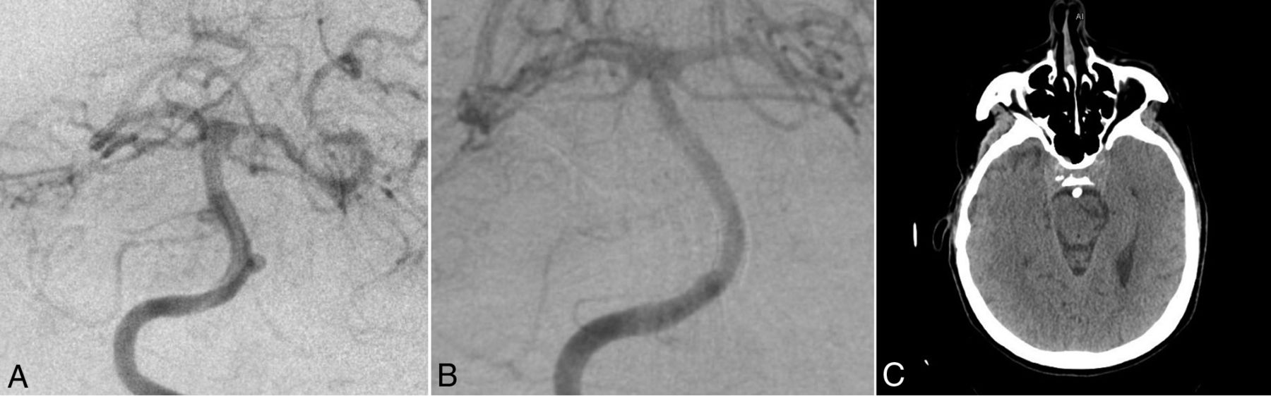
  • Fig 2.
    • Download figure
    • Open in new tab
    • Download powerpoint
    Fig 2.

    Digital subtraction angiography shows 2 basilar trunk saccular aneurysms before treatment with the Pipeline Embolization Device (A). The PED was placed spanning the AICA bilaterally. On follow-up DSA (B), there was complete occlusion of the left AICA. The patient had a symptomatic left-sided pontine stroke that remained symptomatic at 10-month follow-up (C).

Tables

  • Figures
    • View popup
    Table 1:

    Baseline characteristics

    ParameterNo.
    No. of procedures129
    No. of aneurysms131
    No. of branches covered228
    Sex
        Female82 (63.6%)
        Male47 (36.4%)
    Median age (range) (yr)58 (29–82)
    Smokinga40 (33.3%)
    Multiple aneurysms32 (24.8%)
    Presenting symptoms
        Asymptomatic28 (21.7%)
        Headache/dizziness30 (23.3%)
        Neurologic deficit71 (55%)
    Subarachnoid hemorrhage
        Immediate (<24 hr)18 (14.0%)
        Acute (>24 hr and <2 wk)7 (5.4%)
        Remote (>2 wk)14 (10.9%)
    Pretreatment mRS
        0–2101 (78.3%)
        3–528 (21.7%)
    Aneurysm shape
        Saccular49 (37.4%)
        Fusiform53 (40.5%)
        Dissecting29 (22.1%)
    Aneurysm location
        Vertebral artery46 (35.1%)
        PICA10 (7.6%)
        Vertebrobasilar artery18 (13.6%)
        Basilar artery45 (34.5%)
        SCA4 (3.1%)
        PCA8 (6.1%)
    Aneurysm measurements (median) (range) (mm)
        Maximal diameter12 (2–73)
        Neck size (for saccular aneurysms)5.35 (2–15)
    Daughter sac26 (19.8%)
    Prior treatment
        Endovascular14 (10.7%)
        Surgery2 (1.5%)
        Both1 (0.8%)
    Platelet function test77 (59.7%)
    Clopidogrel nonresponders14 (18.2%)
    Treatment of nonresponders
        Continue clopidogrel8 (57.1%)
        Switch to ticagrelor5 (35.8%)
        Other1 (7.1%)
    • Note:—SCA indicates superior cerebellar artery.

    • ↵a Data are missing for 9 procedures.

    • View popup
    Table 2:

    Outcome measures

    ParameterNo.
    No. of Pipelines deployed (median) (range)1 (1–14)
    Adjunctive coiling40 (31%)
    Procedures with covered branches103 (79.8%)
    No. of covered branches (median) (range)2 (0–6)
    Last angiographic follow-up (median) (range) (mo)a11 (1–72)
    Follow-up aneurysm occlusion ratea
        Complete (100%)85 (66.4%)
        Near-complete (90%–99%)15 (11.7%)
        Partial (<90%)28 (21.9%)
    Retreatment
        Endovascular11 (8.4%)
    Outcome of covered branchesb228
        Occluded (overall)25 (11%)
        VA23
            Occluded8 (34.8%)
        PICA49
            Occluded4 (8.2%)
        AICA78
            Occluded6 (7.7%)
        SCA61
            Occluded3 (4.9%)
        PCA17
            Occluded4 (23.5%)
    Last clinical follow-up (median) (range) (mo)c8 (0.3–72)
    Posttreatment mRSc
        0–299 (79.2%)
        3–512 (9.6%)
        6 (Death)14 (11.2%)
    Follow-up mRSc
        Improved43 (34.4%)
        No change54 (43.2%)
        Worsened28 (22.4%)
    Ischemic complications29 (22.5%)
        Timing
            Intraprocedural2 (1.6%)
            Postprocedural27 (20.9%)
        Location
            Brain stem13 (10.1%)
            Cerebellum13 (10.1%)
            PCA territory10 (7.8%)
        Symptomatic17 (13.2%)
            Temporary6 (4.7%)
            Permanent11 (8.5%)
    Territory of ischemic complications
        Same as covered branch21 (72.4%)
        Different8 (27.6%)
    • Note:—SCA indicates superior cerebellar artery.

    • ↵a Data were missing on 3 aneurysms.

    • ↵b Each covered branch was considered separately.

    • ↵c Data were missing on 4 procedures.

    • View popup
    Table 3:

    Predictors of branch occlusion following coverage

    ParameterBranch Occlusion (No.) (%)P Value
    Not Occluded (n = 203)Occluded (n = 25)
    Sex
        Female108 (87.1%)16 (12.9%).3
        Male95 (91.3%)9 (8.7%)
    Median age (range) (yr)59.5 (29–82)61 (42–78).56
    Smokinga
        Yes53 (82.8%)11 (17.2%).1
        No137 (90.7%)14 (9.3%)
    Presenting symptoms
        Asymptomatic32 (72.7%)12 (27.3%)<.001
        Headache/dizziness38 (88.4%)5 (11.5%)
        Neurologic deficit133 (94.3%)8 (5.7%)
    Subarachnoid hemorrhage
        No141 (87.6%)20 (12.4%).36
        Acute (<24 hr)25 (96.2%)1 (3.8%)
        Immediate (<2 wk)11 (100%)0%
        Remote (>2 wk)26 (86.7%)4 (13.3%)
    Pretreatment mRS
        0–2160 (87.4%)23 (12.6%).12
        3–543 (95.6%)2 (4.4%)
    Aneurysm shape
        Saccular74 (84.1%)14 (15.9%).18
        Fusiform89 (91.8%)8 (8.2%)
        Dissecting38 (92.7%)3 (7.3%)
    Aneurysm measurements
        Maximal diameter
            <7 mm32 (84.2%)6 (15.8%).23
            7–12 mm50 (84.7%)9 (15.3%)
            13–24 mm52 (89.7%)6 (10.3%)
            >24 mm69 (94.5%)4 (5.5%)
    Daughter sac
        Yes45 (90%)5 (10%).8
        No158 (88.8%)20 (11.2%)
    Intra-aneurysmal thrombus
        Yes68 (94.4%)4 (5.6%).08
        No135 (86.5%)21 (13.5%)
    Prior treatment
        No174 (88.3%)23 (11.7%).8
        Endovascular25 (92.6%)2 (7.4%)
        Surgery3 (100%)0%
        Both1 (100%)0%
    Platelet function testing
        Yes128 (85.3%)22 (14.7%).01
        No75 (96.2%)3 (3.8%)
    Clopidogrel respondersb
        Yes110 (86.6%)17 (13.4%).3
        No18 (78.3%)5 (21.7%)
    Adjunctive coiling
        Yes75 (88.2%)10 (11.8%).77
        No128 (89.5%)15 (10.5%)
    No. of Pipelines deployed (median) (range)1 (1–14)1 (1–4).33
    Length of procedure (median) (range) (min)110 (22–410)114 (32–271).8
    Branch
        VA15 (65.2%)8 (34.8%).001
        PICA45 (91.8%)4 (8.2%)
        AICA72 (92.3%)6 (7.7%)
        SCA58 (95.1%)3 (4.9%)
        PCA13 (76.5%)4 (23.5%)
    No. of covered branches in same procedure
        137 (97.4%)1 (2.6%).07
        ≥2166 (87.4%)24 (12.6%)
    • Note:—SCA indicates superior cerebellar artery.

    • ↵a Data are missing for 13 branches.

    • ↵b Among patients with platelet function testing.

Next
Back to top
Advertisement
Print
Download PDF
Email Article

Thank you for your interest in spreading the word on American Journal of Neuroradiology.

NOTE: We only request your email address so that the person you are recommending the page to knows that you wanted them to see it, and that it is not junk mail. We do not capture any email address.

Enter multiple addresses on separate lines or separate them with commas.
Risk of Branch Occlusion and Ischemic Complications with the Pipeline Embolization Device in the Treatment of Posterior Circulation Aneurysms
(Your Name) has sent you a message from American Journal of Neuroradiology
(Your Name) thought you would like to see the American Journal of Neuroradiology web site.
CAPTCHA
This question is for testing whether or not you are a human visitor and to prevent automated spam submissions.
Cite this article
N. Adeeb, C.J. Griessenauer, A.A. Dmytriw, H. Shallwani, R. Gupta, P.M. Foreman, H. Shakir, J. Moore, N. Limbucci, S. Mangiafico, A. Kumar, C. Michelozzi, Y. Zhang, V.M. Pereira, C.C. Matouk, M.R. Harrigan, A.H. Siddiqui, E.I. Levy, L. Renieri, T.R. Marotta, C. Cognard, C.S. Ogilvy, A.J. Thomas
Risk of Branch Occlusion and Ischemic Complications with the Pipeline Embolization Device in the Treatment of Posterior Circulation Aneurysms
American Journal of Neuroradiology Jun 2018, DOI: 10.3174/ajnr.A5696

Citation Manager Formats

  • BibTeX
  • Bookends
  • EasyBib
  • EndNote (tagged)
  • EndNote 8 (xml)
  • Medlars
  • Mendeley
  • Papers
  • RefWorks Tagged
  • Ref Manager
  • RIS
  • Zotero
0 Responses
Respond to this article
Share
Bookmark this article
Risk of Branch Occlusion and Ischemic Complications with the Pipeline Embolization Device in the Treatment of Posterior Circulation Aneurysms
N. Adeeb, C.J. Griessenauer, A.A. Dmytriw, H. Shallwani, R. Gupta, P.M. Foreman, H. Shakir, J. Moore, N. Limbucci, S. Mangiafico, A. Kumar, C. Michelozzi, Y. Zhang, V.M. Pereira, C.C. Matouk, M.R. Harrigan, A.H. Siddiqui, E.I. Levy, L. Renieri, T.R. Marotta, C. Cognard, C.S. Ogilvy, A.J. Thomas
American Journal of Neuroradiology Jun 2018, DOI: 10.3174/ajnr.A5696
del.icio.us logo Twitter logo Facebook logo Mendeley logo
  • Tweet Widget
  • Facebook Like
  • Google Plus One
Purchase

Jump to section

  • Article
    • Abstract
    • ABBREVIATIONS:
    • Materials and Methods
    • Results
    • Discussion
    • Conclusions
    • Footnotes
    • References
  • Figures & Data
  • Supplemental
  • Info & Metrics
  • Responses
  • References
  • PDF

Related Articles

  • PubMed
  • Google Scholar

Cited By...

  • Endovascular structures of the basilar artery: forms of the basilar nonfusion spectrum
  • Learning Curve for Flow Diversion of Posterior Circulation Aneurysms: A Long-Term International Multicenter Cohort Study
  • Treatment of posterior circulation non-saccular aneurysms with flow diversion versus stent-assisted coiling: a systematic review and meta-analysis
  • Periprocedural to 1-year safety and efficacy outcomes with the Pipeline Embolization Device with Shield technology for intracranial aneurysms: a prospective, post-market, multi-center study
  • The FRED for Cerebral Aneurysms of the Posterior Circulation: A Subgroup Analysis of the EuFRED Registry
  • Crossref
  • Google Scholar

This article has not yet been cited by articles in journals that are participating in Crossref Cited-by Linking.

More in this TOC Section

  • Cavernous dural AVF treated by transfacial route
  • A Retrospective Study in Tentorial DAVFs
  • Proximal Protection Devices for Carotid Stenting
Show more NEUROINTERVENTION

Similar Articles

Advertisement

Indexed Content

  • Current Issue
  • Accepted Manuscripts
  • Article Preview
  • Past Issues
  • Editorials
  • Editor's Choice
  • Fellows' Journal Club
  • Letters to the Editor
  • Video Articles

Cases

  • Case Collection
  • Archive - Case of the Week
  • Archive - Case of the Month
  • Archive - Classic Case

More from AJNR

  • Trainee Corner
  • Imaging Protocols
  • MRI Safety Corner
  • Book Reviews

Multimedia

  • AJNR Podcasts
  • AJNR Scantastics

Resources

  • Turnaround Time
  • Submit a Manuscript
  • Submit a Video Article
  • Submit an eLetter to the Editor/Response
  • Manuscript Submission Guidelines
  • Statistical Tips
  • Fast Publishing of Accepted Manuscripts
  • Graphical Abstract Preparation
  • Imaging Protocol Submission
  • Evidence-Based Medicine Level Guide
  • Publishing Checklists
  • Author Policies
  • Become a Reviewer/Academy of Reviewers
  • News and Updates

About Us

  • About AJNR
  • Editorial Board
  • Editorial Board Alumni
  • Alerts
  • Permissions
  • Not an AJNR Subscriber? Join Now
  • Advertise with Us
  • Librarian Resources
  • Feedback
  • Terms and Conditions
  • AJNR Editorial Board Alumni

American Society of Neuroradiology

  • Not an ASNR Member? Join Now

© 2025 by the American Society of Neuroradiology All rights, including for text and data mining, AI training, and similar technologies, are reserved.
Print ISSN: 0195-6108 Online ISSN: 1936-959X

Powered by HighWire