Advertisement

Main menu

  • Home
  • Content
    • Current Issue
    • Accepted Manuscripts
    • Article Preview
    • Past Issue Archive
    • Video Articles
    • AJNR Case Collection
    • Case of the Week Archive
    • Case of the Month Archive
    • Classic Case Archive
  • Special Collections
    • AJNR Awards
    • Low-Field MRI
    • Alzheimer Disease
    • ASNR Foundation Special Collection
    • Photon-Counting CT
    • View All
  • Multimedia
    • AJNR Podcasts
    • AJNR SCANtastic
    • Trainee Corner
    • MRI Safety Corner
    • Imaging Protocols
  • For Authors
    • Submit a Manuscript
    • Submit a Video Article
    • Submit an eLetter to the Editor/Response
    • Manuscript Submission Guidelines
    • Statistical Tips
    • Fast Publishing of Accepted Manuscripts
    • Graphical Abstract Preparation
    • Imaging Protocol Submission
    • Author Policies
  • About Us
    • About AJNR
    • Editorial Board
    • Editorial Board Alumni
  • More
    • Become a Reviewer/Academy of Reviewers
    • Subscribers
    • Permissions
    • Alerts
    • Feedback
    • Advertisers
    • ASNR Home

User menu

  • Alerts
  • Log in

Search

  • Advanced search
American Journal of Neuroradiology

American Journal of Neuroradiology

ASHNR American Society of Functional Neuroradiology ASHNR American Society of Pediatric Neuroradiology ASSR
  • Alerts
  • Log in

Advanced Search

  • Home
  • Content
    • Current Issue
    • Accepted Manuscripts
    • Article Preview
    • Past Issue Archive
    • Video Articles
    • AJNR Case Collection
    • Case of the Week Archive
    • Case of the Month Archive
    • Classic Case Archive
  • Special Collections
    • AJNR Awards
    • Low-Field MRI
    • Alzheimer Disease
    • ASNR Foundation Special Collection
    • Photon-Counting CT
    • View All
  • Multimedia
    • AJNR Podcasts
    • AJNR SCANtastic
    • Trainee Corner
    • MRI Safety Corner
    • Imaging Protocols
  • For Authors
    • Submit a Manuscript
    • Submit a Video Article
    • Submit an eLetter to the Editor/Response
    • Manuscript Submission Guidelines
    • Statistical Tips
    • Fast Publishing of Accepted Manuscripts
    • Graphical Abstract Preparation
    • Imaging Protocol Submission
    • Author Policies
  • About Us
    • About AJNR
    • Editorial Board
    • Editorial Board Alumni
  • More
    • Become a Reviewer/Academy of Reviewers
    • Subscribers
    • Permissions
    • Alerts
    • Feedback
    • Advertisers
    • ASNR Home
  • Follow AJNR on Twitter
  • Visit AJNR on Facebook
  • Follow AJNR on Instagram
  • Join AJNR on LinkedIn
  • RSS Feeds

AJNR Awards, New Junior Editors, and more. Read the latest AJNR updates

Research ArticleAdult Brain
Open Access

Arterial Spin-Labeling Improves Detection of Intracranial Dural Arteriovenous Fistulas with MRI

S.A. Amukotuwa, M.P. Marks, G. Zaharchuk, F. Calamante, R. Bammer and N. Fischbein
American Journal of Neuroradiology March 2018, DOI: https://doi.org/10.3174/ajnr.A5570
S.A. Amukotuwa
aFrom the Department of Radiology (S.A.A., M.P.M., G.Z., R.B., N.F.), Stanford University, Stanford, California
bFlorey Department of Neuroscience and Mental Health (S.A.A., F.C.), University of Melbourne, Melbourne, Victoria, Australia.
  • Find this author on Google Scholar
  • Find this author on PubMed
  • Search for this author on this site
  • ORCID record for S.A. Amukotuwa
M.P. Marks
aFrom the Department of Radiology (S.A.A., M.P.M., G.Z., R.B., N.F.), Stanford University, Stanford, California
  • Find this author on Google Scholar
  • Find this author on PubMed
  • Search for this author on this site
  • ORCID record for M.P. Marks
G. Zaharchuk
aFrom the Department of Radiology (S.A.A., M.P.M., G.Z., R.B., N.F.), Stanford University, Stanford, California
  • Find this author on Google Scholar
  • Find this author on PubMed
  • Search for this author on this site
  • ORCID record for G. Zaharchuk
F. Calamante
bFlorey Department of Neuroscience and Mental Health (S.A.A., F.C.), University of Melbourne, Melbourne, Victoria, Australia.
  • Find this author on Google Scholar
  • Find this author on PubMed
  • Search for this author on this site
  • ORCID record for F. Calamante
R. Bammer
aFrom the Department of Radiology (S.A.A., M.P.M., G.Z., R.B., N.F.), Stanford University, Stanford, California
  • Find this author on Google Scholar
  • Find this author on PubMed
  • Search for this author on this site
  • ORCID record for R. Bammer
N. Fischbein
aFrom the Department of Radiology (S.A.A., M.P.M., G.Z., R.B., N.F.), Stanford University, Stanford, California
  • Find this author on Google Scholar
  • Find this author on PubMed
  • Search for this author on this site
  • ORCID record for N. Fischbein
  • Article
  • Figures & Data
  • Supplemental
  • Info & Metrics
  • Responses
  • References
  • PDF
Loading

Article Figures & Data

Figures

  • Tables
  • Fig 1.
    • Download figure
    • Open in new tab
    • Download powerpoint
    Fig 1.

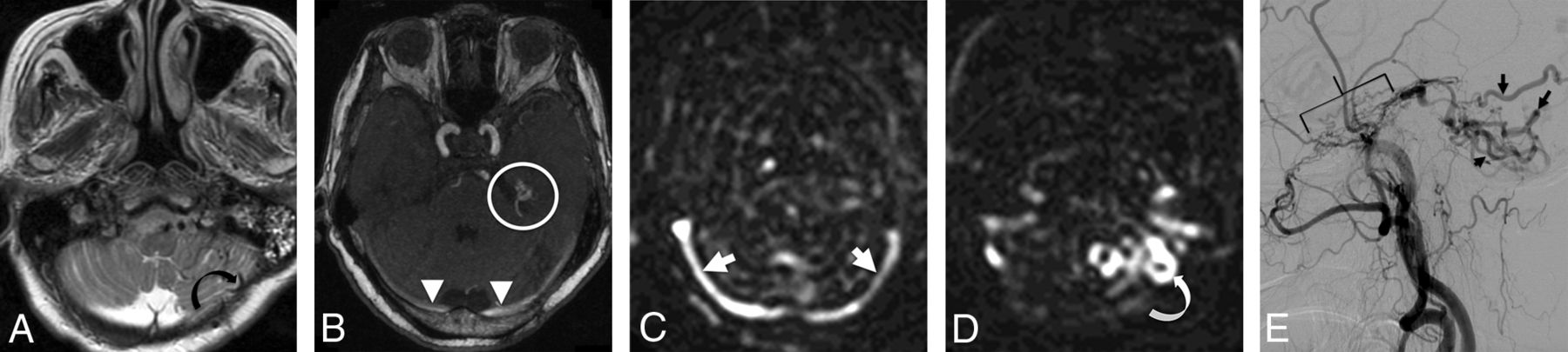
    A 79-year-old woman presenting with ataxia. A, T2-weighted imaging demonstrates tortuous and ectatic pial veins (curved black arrow) along the inferior surface of the left cerebellar hemisphere. B, MIP TOF-MRA image demonstrates nodular and curvilinear hyperintensities immediately superior to the left petrous apex, representing a left tentorial fistula (white circle) and high signal in the transverse sinuses (white arrowheads). C, pCASL image shows venous ASL signal in the transverse sinuses (white arrows) due to shunting. D, More inferiorly, venous ASL signal is seen in draining pial veins (white curved arrow). E, DSA image following left external carotid artery injection confirms a Cognard type IV left tentorial DAVF (bracket) with a middle meningeal artery supply and drainage directly into ectatic cerebellar cortical veins (black arrows).

  • Fig 2.
    • Download figure
    • Open in new tab
    • Download powerpoint
    Fig 2.

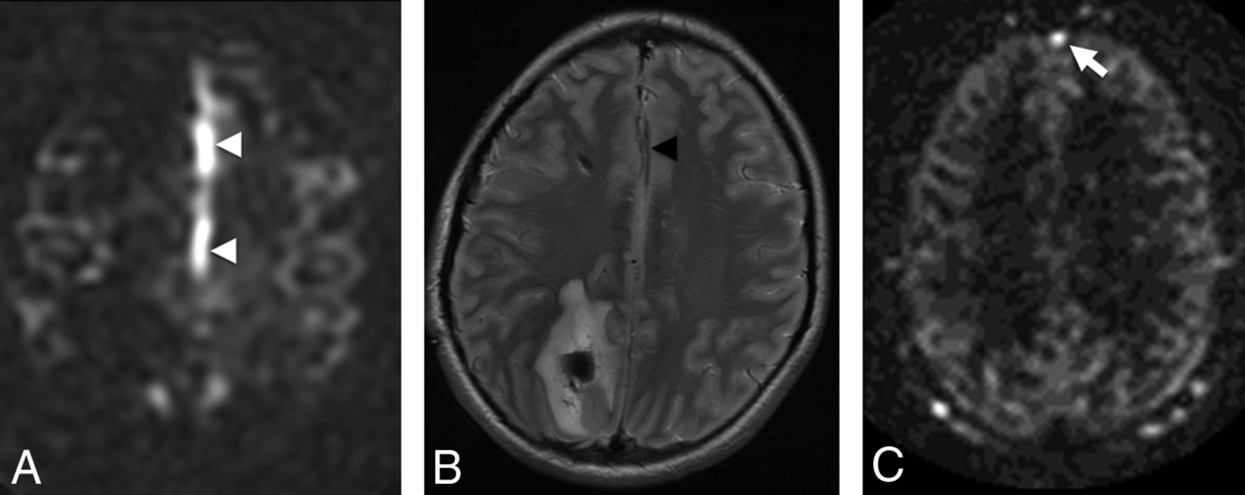
    False-positive venous ASL signal in 2 patients. A, ASL signal in the A3 branches of the anterior cerebral arteries (white arrowheads) was mistaken for venous ASL signal in a 15-year-old male patient who presented with a right parietal parenchymal hematoma. B, T2-weighted images show localization of this signal to the anterior cerebral arteries (black arrowhead). C, ASL signal in the anterior aspect of the superior sagittal sinus in a 70-year-old man with subarachnoid hemorrhage (white arrow). This patient had no evidence of a DAVF or shunting on DSA.

  • Fig 3.
    • Download figure
    • Open in new tab
    • Download powerpoint
    Fig 3.

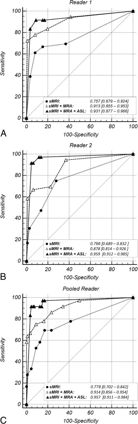
    ROC curves for each reader's individual diagnostic performance (A and B) and their pooled diagnostic performance (C) with each of the following: structural MR imaging (dotted line), structural MR imaging and TOF-MRA (sMRI + MRA, dashed line), and structural MR imaging with TOF-MRA and pCASL (sMRI + MRA + pCASL, solid line). The light gray diagonal is the line of no discrimination. The triangle and dot symbols on the curve indicate true-positive rate/false-positive rate pairs computed at different discrimination thresholds. With the addition of TOF-MRA and then pCASL to structural MR imaging, the ROC curve becomes more well-rounded with an incrementally higher AUC. This indicates increased diagnostic sensitivity for detection of a DAVF at a set specificity.

  • Fig 4.
    • Download figure
    • Open in new tab
    • Download powerpoint
    Fig 4.

    Distribution of Likert scale scores for patients with (A) and without (B) a DAVF on structural MR imaging alone, structural MR imaging with TOF-MRA (sMRI/MRA), and structural MR imaging with both TOF-MRA and pCASL (sMRI/MRA/pCASL). A, In the DAVF group, there is a marked incremental increase in reader accuracy and confidence in the presence of a DAVF—with a higher percentage considered “very likely” to have a DAVF—with the addition of pCASL. B, In the control group, reader accuracy and confidence in the absence of a DAVF decrease with the addition of TOF-MRA to sMRI due to a high number of false-positives for venous hyperintensity on TOF-MRA. Reader certainty as to the absence of a fistula increased (and was highest) following review of pCASL.

Tables

  • Figures
    • View popup
    Table 1:

    Individual imaging features—univariate binary logistic regression analysis on DSA and interreader agreement

    Imaging FeatureORSEWald TestPSensitivity (95% CI) (%)Specificity (95% CI) (%)PPV (95% CI) (%)NPV (95% CI) (%)Interobserver Agreement (κ) (95% CI)
    Structural MRI
        ICH0.181.3237.84.00137.2 (26.5–47.9)23.1 (17.7–28.5)13.9 (9.2–18.6)52.4 (43.0–62.1)0.99 (0.97–1.00)
        Vasogenic edema or gliosis0.361.3710.59.00120.8 (11.7–58.1)58.1 (51.8–64.4)14.0 (7.7–20.4)69.0 (62.6–75.5)1.00 (1.00–1.00)
        Abnormal vessels5.571.3237.24.00159.0 (48.1–69.9)79.5 (74.3–84.7)48.9 (38.8–59.0)85.3 (80.6–90.0)0.73 (0.67–0.79)
        Enlarged sinus2.581.369.50.00231.2 (20.8–41.5)79.9 (73.9–85.8)40.7 (28.1–53.2)72.4 (66.1–78.7)0.69 (0.61–0.76)
        Enlarged SOV3.681.627.36.00712.7 (5.3–20.0)96.2 (93.7–98.6)52.6 (30.2–75.1)76.5 (71.7–81.4)0.83 (0.74–0.93)
        Cavernous sinus enlargement34.802.1421.85.00123.1 (13.7–32.4)99.2 (98.0–100.3)90.0 (76.9–103.2)79.5 (74.8–84.1)0.89 (0.82–0.97)
        Orbital edema/proptosis12.262.2310.38.00110.3 (3.5–17.0)99.2 (98.0–100.3)80.0 (55.2–104.8)76.8 (72.1–81.6)1.00 (1.00–1.00)
    Time-of-flight MRA
        NCH114.701.8856.75.00161.1 (49.9–72.4)98.7 (97.1–100.2)93.6 (86.6–100.6)88.7 (84.7–92.6)0.87 (0.82–0.93)
        Abnormal vessels6.001.3437.28.00159.7 (48.4–71.1)80.2 (74.9–85.4)49.4 (38.9–59.9)86.0 (81.3–90.7)0.38 (0.30–0.46)
        Venous signal20.751.4760.96.00187.5 (79.9–95.1)74.8 (69.1–80.5)52.9 (44.0–61.9)94.9 (91.6–98.1)0.87 (0.83–0.91)
        Enlarged extracranial arteries17.811.4170.98.00162.5 (51.3–73.7)91.4 (87.8–95.1)70.3 (59.1–81.5)88.3 (84.1–92.4)0.76 (0.69–0.83)
    ASL
        Venous ASL signal103.201.6584.95.00193.6 (88.2–99.0)87.6 (83.4–91.8)71.5 (62.8–80.3)97.6 (95.6–99.7)0.94 (0.9–0.97)
    • Note:—SOV indicates superior ophthalmic vein; SE, standard error; ICH, intracerebral hemorrhage.

    • View popup
    Table 2:

    Pair-wise comparison of ROC curves

    Reader, Diagnostic InstrumentΔAUCSE95% CIzP
    Reader 1
        sMRI vs sMRI + MRA0.1560.0460.066–0.2473.381<.01
        sMRI vs sMRI + MRA + pCASL0.1740.0470.082–0.2663.701<.01
        sMRI + MRA vs sMRI+MRA + pCASL0.0180.028−0.036–0.0720.644.52
    Reader 2
        sMRI vs sMRI + MRA0.1120.0440.027–0.1972.581<.01
        sMRI vs sMRI + MRA + pCASL0.1930.0430.108–0.2774.475<.01
        sMRI + MRA vs sMRI + MRA + pCASL0.0810.0300.022–0.1402.675<.01
    Pooled Readers 1 + 2
        sMRI vs sMRI + MRA0.1360.0420.053–0.2193.208<.01
        sMRI vs sMRI + MRA + pCASL0.1790.0450.091–0.2673.965<.01
        sMRI + MRA vs sMRI + MRA + pCASL0.0430.027−0.009–0.1001.615.11
    • View popup
    Table 3:

    Pair-wise likelihood ratio tests for the multivariate models

    Reader, Diagnostic Instrument−2LLχ2PΔdf
    sMRI260.18
    sMRI + MRA117.09
    sMRI + MRA + ASL84.23
    (sMRI) vs (sMRI + MRA)143.09<.001a4
    (sMRI + MRA) vs (sMRI + MRA + ASL)32.86<.001a1
    (sMRI) vs (sMRI + MRA + ASL)175.95<.001a8
    • Note:—LL indicates log likelihood.

    • ↵a Significance (P < .001)—that is, that the null hypothesis (difference in −2LLs = 0) is rejected and that the −2LLs are different.

    • View popup
    Table 4:

    Classification tables for the multivariate binary logistic regression modelsa

    MRI SequencesSensitivity (95% CI) (%)Specificity (95% CI) (%)PPV (95% CI) (%)NPV (95% CI) (%)
    Structural MRI alone32.9 (22.3–43.5)97.0 (94.8–99.2)78.1 (63.8–92.5)81.7 (77.1–86.2)
    Structural MRI and TOF-MRA75.7 (65.7–85.8)98.7 (97.1–100.2)94.6 (88.8–100.5)92.8 (89.5–96.1)
    Structural MRI, TOF-MRA and pCASL88.6 (81.1–96.0)96.4 (93.9–98.9)88.5 (81.1–96.0)96.4 (93.9–98.9)
    • ↵a Classification table generated for a probability value of P = .05 for each of the 3 multivariate models.

Next
Back to top
Advertisement
Print
Download PDF
Email Article

Thank you for your interest in spreading the word on American Journal of Neuroradiology.

NOTE: We only request your email address so that the person you are recommending the page to knows that you wanted them to see it, and that it is not junk mail. We do not capture any email address.

Enter multiple addresses on separate lines or separate them with commas.
Arterial Spin-Labeling Improves Detection of Intracranial Dural Arteriovenous Fistulas with MRI
(Your Name) has sent you a message from American Journal of Neuroradiology
(Your Name) thought you would like to see the American Journal of Neuroradiology web site.
CAPTCHA
This question is for testing whether or not you are a human visitor and to prevent automated spam submissions.
Cite this article
S.A. Amukotuwa, M.P. Marks, G. Zaharchuk, F. Calamante, R. Bammer, N. Fischbein
Arterial Spin-Labeling Improves Detection of Intracranial Dural Arteriovenous Fistulas with MRI
American Journal of Neuroradiology Mar 2018, DOI: 10.3174/ajnr.A5570

Citation Manager Formats

  • BibTeX
  • Bookends
  • EasyBib
  • EndNote (tagged)
  • EndNote 8 (xml)
  • Medlars
  • Mendeley
  • Papers
  • RefWorks Tagged
  • Ref Manager
  • RIS
  • Zotero
0 Responses
Respond to this article
Share
Bookmark this article
Arterial Spin-Labeling Improves Detection of Intracranial Dural Arteriovenous Fistulas with MRI
S.A. Amukotuwa, M.P. Marks, G. Zaharchuk, F. Calamante, R. Bammer, N. Fischbein
American Journal of Neuroradiology Mar 2018, DOI: 10.3174/ajnr.A5570
del.icio.us logo Twitter logo Facebook logo Mendeley logo
  • Tweet Widget
  • Facebook Like
  • Google Plus One
Purchase

Jump to section

  • Article
    • Abstract
    • ABBREVIATIONS:
    • Materials and Methods
    • Results
    • Discussion
    • Conclusions
    • Acknowledgments
    • Footnotes
    • References
  • Figures & Data
  • Supplemental
  • Info & Metrics
  • Responses
  • References
  • PDF

Related Articles

  • PubMed
  • Google Scholar

Cited By...

  • Time-Saving 3D MR Imaging Protocols with Millimeter and Submillimeter Isotropic Spatial Resolution for Face and Neck Imaging as Implemented at a Single-Site Major Referral Center
  • Four dimensional-flow magnetic resonance imaging analysis of carotid-cavernous fistula, dural arteriovenous fistula and spinal arteriovenous fistula: Detecting shunt point and diagnosing based on flow dynamics analysis
  • Arterial Spin-Labeling MR Imaging for the Differential Diagnosis of Venous-Predominant AVMs and Developmental Venous Anomalies
  • Assessment of 4D MR Angiography at 3T Compared with DSA for the Follow-up of Embolized Brain Dural Arteriovenous Fistula: A Dual-Center Study
  • Gadolinium Deposition Safety: Seeking the Patients Perspective
  • Follow-Up MRI for Small Brain AVMs Treated by Radiosurgery: Is Gadolinium Really Necessary?
  • Crossref (40)
  • Google Scholar

This article has been cited by the following articles in journals that are participating in Crossref Cited-by Linking.

  • Recent advances in MRI of the head and neck, skull base and cranial nerves: new and evolving sequences, analyses and clinical applications
    Philip Touska, Steve E. J. Connor
    The British Journal of Radiology 2019 92 1104
  • Gadolinium Deposition Safety: Seeking the Patient’s Perspective
    C.A. Mallio, C.C. Quattrocchi, À. Rovira, P.M. Parizel
    American Journal of Neuroradiology 2020 41 6
  • A visual quality control scale for clinical arterial spin labeling images
    S. M. Fallatah, F. B. Pizzini, B. Gomez-Anson, J. Magerkurth, E. De Vita, S. Bisdas, H. R. Jäger, H. J. M. M. Mutsaerts, X. Golay
    European Radiology Experimental 2018 2 1
  • Follow-Up MRI for Small Brain AVMs Treated by Radiosurgery: Is Gadolinium Really Necessary?
    X. Leclerc, O. Guillaud, N. Reyns, J. Hodel, O. Outteryck, F. Bala, N. Bricout, M. Bretzner, N. Ramdane, J.-P. Pruvo, L. Hacein-Bey, G. Kuchcinski
    American Journal of Neuroradiology 2020 41 3
  • Assessment of 4D MR Angiography at 3T Compared with DSA for the Follow-up of Embolized Brain Dural Arteriovenous Fistula: A Dual-Center Study
    B. Dissaux, F. Eugène, J. Ognard, J.-Y. Gauvrit, J.-C. Gentric, J.-C. Ferré
    American Journal of Neuroradiology 2021 42 2
  • MRI and MR angiography evaluation of pulsatile tinnitus: A focused, physiology‐based protocol
    Daniel D. Cummins, Michael T. Caton, Vinil Shah, Karl Meisel, Christine Glastonbury, Matthew R. Amans
    Journal of Neuroimaging 2022 32 2
  • Venous cerebral blood flow quantification and cognition in patients with sickle cell anemia
    Hanne Stotesbury, Patrick W Hales, Melanie Koelbel, Anna M Hood, Jamie M Kawadler, Dawn E Saunders, Sati Sahota, David C Rees, Olu Wilkey, Mark Layton, Maria Pelidis, Baba PD Inusa, Jo Howard, Subarna Chakravorty, Chris A Clark, Fenella J Kirkham
    Journal of Cerebral Blood Flow & Metabolism 2022 42 6
  • Arterial spin-labeling perfusion MR images with dual postlabeling delay reveals hemodynamic changes in dural arteriovenous fistulas following endovascular surgery
    So Tokunaga, Takato Morioka, Noritoshi Shirozu, Yuichiro Tsurusaki, Shoji Arihiro, Takafumi Shimogawa, Masahiro Mizoguchi, Sei Haga
    Interdisciplinary Neurosurgery 2020 21
  • Utility of silent magnetic resonance angiography in the evaluation and characterisation of intracranial dural arteriovenous fistula
    A. Prasad Balasubramanian, S. Kumar Kannath, J. Enakshy Rajan, G. Singh, C. Kesavadas, B. Thomas
    Clinical Radiology 2021 76 9
  • Contrecoup Injury-Induced Middle Meningeal Arteriovenous Fistula Detected by Time-of-Flight Magnetic Resonance Angiography and Magnetic Resonance Arterial Spin Labeling: Case Report and Review of the Literature
    Kikutaro Tokairin, Toshiya Osanai, Ken Kazumata, Ryosuke Sawaya, Kiyohiro Houkin
    World Neurosurgery 2019 127

More in this TOC Section

  • Diagnostic Neuroradiology of Monoclonal Antibodies
  • Clinical Outcomes After Chiari I Decompression
  • Segmentation of Brain Metastases with BLAST
Show more Adult Brain

Similar Articles

Advertisement

Indexed Content

  • Current Issue
  • Accepted Manuscripts
  • Article Preview
  • Past Issues
  • Editorials
  • Editor's Choice
  • Fellows' Journal Club
  • Letters to the Editor
  • Video Articles

Cases

  • Case Collection
  • Archive - Case of the Week
  • Archive - Case of the Month
  • Archive - Classic Case

More from AJNR

  • Trainee Corner
  • Imaging Protocols
  • MRI Safety Corner
  • Book Reviews

Multimedia

  • AJNR Podcasts
  • AJNR Scantastics

Resources

  • Turnaround Time
  • Submit a Manuscript
  • Submit a Video Article
  • Submit an eLetter to the Editor/Response
  • Manuscript Submission Guidelines
  • Statistical Tips
  • Fast Publishing of Accepted Manuscripts
  • Graphical Abstract Preparation
  • Imaging Protocol Submission
  • Evidence-Based Medicine Level Guide
  • Publishing Checklists
  • Author Policies
  • Become a Reviewer/Academy of Reviewers
  • News and Updates

About Us

  • About AJNR
  • Editorial Board
  • Editorial Board Alumni
  • Alerts
  • Permissions
  • Not an AJNR Subscriber? Join Now
  • Advertise with Us
  • Librarian Resources
  • Feedback
  • Terms and Conditions
  • AJNR Editorial Board Alumni

American Society of Neuroradiology

  • Not an ASNR Member? Join Now

© 2025 by the American Society of Neuroradiology All rights, including for text and data mining, AI training, and similar technologies, are reserved.
Print ISSN: 0195-6108 Online ISSN: 1936-959X

Powered by HighWire