Advertisement

Main menu

  • Home
  • Content
    • Current Issue
    • Accepted Manuscripts
    • Article Preview
    • Past Issue Archive
    • Video Articles
    • AJNR Case Collection
    • Case of the Week Archive
    • Case of the Month Archive
    • Classic Case Archive
  • Special Collections
    • AJNR Awards
    • Low-Field MRI
    • Alzheimer Disease
    • ASNR Foundation Special Collection
    • Photon-Counting CT
    • View All
  • Multimedia
    • AJNR Podcasts
    • AJNR SCANtastic
    • Trainee Corner
    • MRI Safety Corner
    • Imaging Protocols
  • For Authors
    • Submit a Manuscript
    • Submit a Video Article
    • Submit an eLetter to the Editor/Response
    • Manuscript Submission Guidelines
    • Statistical Tips
    • Fast Publishing of Accepted Manuscripts
    • Graphical Abstract Preparation
    • Imaging Protocol Submission
    • Author Policies
  • About Us
    • About AJNR
    • Editorial Board
    • Editorial Board Alumni
  • More
    • Become a Reviewer/Academy of Reviewers
    • Subscribers
    • Permissions
    • Alerts
    • Feedback
    • Advertisers
    • ASNR Home

User menu

  • Alerts
  • Log in

Search

  • Advanced search
American Journal of Neuroradiology

American Journal of Neuroradiology

ASHNR American Society of Functional Neuroradiology ASHNR American Society of Pediatric Neuroradiology ASSR
  • Alerts
  • Log in

Advanced Search

  • Home
  • Content
    • Current Issue
    • Accepted Manuscripts
    • Article Preview
    • Past Issue Archive
    • Video Articles
    • AJNR Case Collection
    • Case of the Week Archive
    • Case of the Month Archive
    • Classic Case Archive
  • Special Collections
    • AJNR Awards
    • Low-Field MRI
    • Alzheimer Disease
    • ASNR Foundation Special Collection
    • Photon-Counting CT
    • View All
  • Multimedia
    • AJNR Podcasts
    • AJNR SCANtastic
    • Trainee Corner
    • MRI Safety Corner
    • Imaging Protocols
  • For Authors
    • Submit a Manuscript
    • Submit a Video Article
    • Submit an eLetter to the Editor/Response
    • Manuscript Submission Guidelines
    • Statistical Tips
    • Fast Publishing of Accepted Manuscripts
    • Graphical Abstract Preparation
    • Imaging Protocol Submission
    • Author Policies
  • About Us
    • About AJNR
    • Editorial Board
    • Editorial Board Alumni
  • More
    • Become a Reviewer/Academy of Reviewers
    • Subscribers
    • Permissions
    • Alerts
    • Feedback
    • Advertisers
    • ASNR Home
  • Follow AJNR on Twitter
  • Visit AJNR on Facebook
  • Follow AJNR on Instagram
  • Join AJNR on LinkedIn
  • RSS Feeds

AJNR Awards, New Junior Editors, and more. Read the latest AJNR updates

Research ArticleNeurointervention

Endovascular Thrombectomy in Wake-Up Stroke and Stroke with Unknown Symptom Onset

P. Bücke, M. Aguilar Pérez, V. Hellstern, M. AlMatter, H. Bäzner and H. Henkes
American Journal of Neuroradiology February 2018, DOI: https://doi.org/10.3174/ajnr.A5540
P. Bücke
aFrom the Psychiatric University Hospital Zurich (P.B.), University of Zurich, Zurich, Switzerland
bNeurological Clinic (P.B., H.B.)
  • Find this author on Google Scholar
  • Find this author on PubMed
  • Search for this author on this site
  • ORCID record for P. Bücke
M. Aguilar Pérez
cNeuroradiological Clinic (M.A.P., V.H., M.A., H.H.), Klinikum Stuttgart, Stuttgart, Germany
  • Find this author on Google Scholar
  • Find this author on PubMed
  • Search for this author on this site
  • ORCID record for M. Aguilar Pérez
V. Hellstern
cNeuroradiological Clinic (M.A.P., V.H., M.A., H.H.), Klinikum Stuttgart, Stuttgart, Germany
  • Find this author on Google Scholar
  • Find this author on PubMed
  • Search for this author on this site
  • ORCID record for V. Hellstern
M. AlMatter
cNeuroradiological Clinic (M.A.P., V.H., M.A., H.H.), Klinikum Stuttgart, Stuttgart, Germany
  • Find this author on Google Scholar
  • Find this author on PubMed
  • Search for this author on this site
  • ORCID record for M. AlMatter
H. Bäzner
bNeurological Clinic (P.B., H.B.)
  • Find this author on Google Scholar
  • Find this author on PubMed
  • Search for this author on this site
  • ORCID record for H. Bäzner
H. Henkes
cNeuroradiological Clinic (M.A.P., V.H., M.A., H.H.), Klinikum Stuttgart, Stuttgart, Germany
dMedical Faculty (H.H.), University Duisburg-Essen, Essen, Germany.
  • Find this author on Google Scholar
  • Find this author on PubMed
  • Search for this author on this site
  • ORCID record for H. Henkes
  • Article
  • Figures & Data
  • Supplemental
  • Info & Metrics
  • Responses
  • References
  • PDF
Loading

Abstract

BACKGROUND AND PURPOSE: Mechanical thrombectomy in acute ischemic stroke within 6 hours of symptom onset is effective and safe. However, in many patients, information on the beginning of symptoms is not available. Patients can be divided into those with wake-up stroke and daytime-unwitnessed stroke. Evidence on outcome and complications after mechanical thrombectomy in wake-up stroke and daytime-unwitnessed stroke is rare. A potential beneficial effect of mechanical thrombectomy in selected patients with wake-up stroke or daytime-unwitnessed stroke is suspected.

MATERIALS AND METHODS: We analyzed 1073 patients with anterior circulation stroke undergoing mechanical thrombectomy between 2010 and 2016. Patients with wake-up stroke and daytime-unwitnessed stroke were compared with controls receiving mechanical thrombectomy as the standard of care. We assessed good functional outcome (mRS ≤ 2 at 3 months), mortality rates, and frequencies of symptomatic intracranial hemorrhage. Subgroup analyses tried to detect influences of patient selection via further imaging modalities (MR imaging, CTP; wake-up stroke [advanced], daytime-unwitnessed stroke [advanced]) on outcome and safety.

RESULTS: There was no significant difference in good functional outcome between patients with wake-up stroke and controls (35.9% versus 38.3%, P = .625). Outcome in patients with daytime-unwitnessed stroke was inferior compared with controls (27.3%, P = .007). Groups did not differ in all-cause mortality at day 90 (P = .224) and the rate of symptomatic intracranial hemorrhage (P = .292). Advanced imaging improved the frequency of good functional outcome (non-wake-up stroke [advanced] versus wake-up stroke [advanced]: OR, 2.92; 95% CI, 1.32–6.45; non-daytime-unwitnessed stroke [advanced] versus daytime-unwitnessed stroke [advanced]: OR, 2.09; 95% CI, 1.03–4.25) with an additional reduction in all-cause mortality (non-daytime-unwitnessed stroke [advanced] versus daytime-unwitnessed stroke [advanced]: OR, 0.42; 95% CI, 0.20–0.88).

CONCLUSIONS: Mechanical thrombectomy in selected patients with wake-up stroke allows a good functional outcome comparable with that of controls. Outcome after mechanical thrombectomy in daytime-unwitnessed stroke seems to be inferior compared with that in controls. Advanced imaging modalities may increase the frequency of good functional outcome in both patients with wake-up stroke and daytime-unwitnessed stroke.

ABBREVIATIONS:

aTE
aspiration thrombectomy
DUS
daytime-unwitnessed stroke
mTE
mechanical thrombectomy
WUS
wake-up stroke

Mechanical thrombectomy (mTE) in acute ischemic stroke due to embolic large-vessel occlusion has been shown to be effective and safe. Several randomized controlled trials have demonstrated the superiority of mTE in combination with intravenous thrombolysis compared with intravenous thrombolysis alone.1⇓⇓⇓–5 Subsequently, specific recommendations for patient selection and execution of mTE and/or aspiration thrombectomy (aTE) were implemented (eg, initiation within 6 hours after symptom onset).6 However, in many patients, information on the beginning of stroke symptoms is not available. They might therefore be excluded from beneficial endovascular therapy. Evidence on mTE in a prolonged time window is inconsistent, and data on the efficacy and safety of mTE in patients with unknown symptom onset are rare.7⇓–9

Materials and Methods

From our ongoing prospective single-center stroke registry, consecutive patients treated with mTE/aTE between January 2010 and December 2016 were considered for this retrospective noninterventional analysis. Patients were either primarily treated in our center (Neurozentrum, Klinikum Stuttgart) or secondarily transferred from hospitals in surrounding cities.10 Nonetheless, enrollment was based on the initial intention to treat patients via mTE/aTE. There was no secondary triage, selection procedure, or additional imaging before the intervention. The study was approved by the local institutional review board.

Study Population

Patients with an anterior circulation stroke due to an occlusion of the ICA, the intracranial carotid bifurcation, or an M1 or M2 branch of the MCA were included. Patients with an occlusion of an MCA M3 branch, the anterior cerebral artery, or the posterior circulation were excluded. In case of an initial proximal vessel occlusion that was later found recanalized during angiography (spontaneously or after intravenous thrombolysis), datasets were removed from further analysis. We did exclude patients who were not treated according to current recommendations (eg, because of a delayed treatment onset) unless they were classified as having wake-up stroke (WUS) or daytime-unwitnessed stroke (DUS).6 Further exclusion criteria were the following: stent angioplasty without mTE/aTE due to high-grade intra- or extracranial stenosis or dissection because of anticipated differences in clinical outcome; the application of older generation stent retrievers and aspiration systems not used in recent randomized controlled trials (the following stent retrievers and aspiration systems were included: Solitaire FR, Medtronic, Minneapolis, Minnesota; pREset, phenox, Bochum, Germany; ACE aspiration catheters, Penumbra, Alameda, California; and Sofia, MicroVention, Tustin, California); and the lack of a 3-month follow-up. Datasets with inconsistent information that could not be verified were excluded.

Patients treated within 6 hours of symptom onset (according to current guidelines) were included in the control group (C). Because of expected differences in pathophysiology and outcome, patients with an unknown symptom onset were divided in the following manner: 1) WUS, occurring out of sleep in the early morning hours; 2) DUS.11⇓–13 WUS was defined as stroke symptoms being present during awakening. Patients had to be asymptomatic when going to sleep and during the night. DUS included patients who were asymptomatic while waking up or had recognized symptoms at some point during day or night. Patients without information on “last seen well” who could not be specified as having WUS by the assessing neurologist were subsumed in DUS.

Subgroup analyses were conducted using consecutive steps in patient selection: 1) further imaging modalities with CTP or MR imaging allowing patient selection due to a mismatch concept (CBV versus TTP or MTT in CTP, FLAIR-DWI mismatch in MR imaging; WUS[advanced], DUS[advanced]); and 2) advanced imaging with MR imaging only (WUS[mri], DUS[mri]).14⇓⇓⇓⇓–19 Examples of mismatch in both MR imaging and CTP are shown in On-line Figs 1–3. The decision to perform advanced imaging was made by the respective stroke specialist. In patients without CTP or MR imaging, we routinely opted for mTE when there was a proved vessel occlusion (eg, in CTA, hyperdense vessel sign), no major demarcation of infarcted tissue on plain CT, or an ASPECTS score of ≥4. There was no fixed CTP protocol (eg, quantitative measurement or thresholds defining mismatch). Information on a possible mismatch was reported by the referring hospital or our neuroradiology department. The results were validated by the neurointerventionalist before endovascular therapy. WUS[mri] and DUS[mri] were subsequently compared with a selected control group (C[mri]) with MR imaging as the initial imaging technique. To detect a possible effect of imaging selection on outcome, we compared patients with WUS and DUS in subgroups 1 and 2 with the remaining patients with WUS or DUS not part of the respective group (eg, WUS[mri] versus non-WUS[mri]).

Outcome Measures

Primary outcome measure was mRS at day 90, with mRS 0–2 indicating good functional outcome. Secondary outcome measures were the following: 1) development of a symptomatic intracranial hemorrhage according to the Solitaire with the Intention for Thrombectomy as Primary Endovascular Treatment for Acute Ischemic Stroke (SWIFT PRIME) criteria (parenchymal hemorrhage type 1 or 2, SAH, or intraventricular hemorrhage within 24 hours after mTE with a deterioration in the NIHSS score of ≥4 points or leading to death)20; 2) in-hospital mortality; and 3) all-cause mortality at day 90.

Data Collection

Information on age, sex, medical history, stroke onset, NIHSS, imaging technique, mRS score, and stroke etiology was drawn from referral letters, admission notes, or discharge papers. Imaging times were stored in our PACS. Periprocedural information (eg, TICI scores) was documented by the neuroradiology department. Follow-up data were collected by our study nurse via telephone calls.

Statistical Analysis

Numeric baseline characteristics were described in medians (quartiles) or means (SDs). Categoric baseline parameters were described in frequencies. For comparing groups, the Fisher exact test or the χ2 test was used for categoric parameters. Numeric parameters were analyzed with the Kruskal-Wallis-test or the Mann-Whitney U test as appropriate. Dichotomized outcome (head-to-head comparison of groups) was evaluated in a univariate logistic regression model adjusting for possible confounders (based on literature research; baseline-NIHSS, age, ICA occlusion, stroke etiology, imaging-to-groin time, diabetes mellitus, and hypertension). A P value < .05 was considered statistically significant. STATA/IC 13.1 for Windows (StataCorp, College Station, Texas) was used for statistical analysis.

Results

Between January 2010 and December 2016, one thousand nine hundred sixty-one patients were treated with mTE/aTE, 888 patients (45.2%) did not meet the predefined inclusion criteria (Fig 1), and 1073 patients (54.8%) would eventually be analyzed. Most (n = 780, 72.7%) formed the control group. In 293 patients (27.3%), symptom onset was unclear. One hundred twenty-eight patients (11.9%) were categorized as having WUS; and 165 (15.4%), DUS.

Fig 1.
  • Download figure
  • Open in new tab
  • Download powerpoint
Fig 1.

Flowchart depicting patient selection according to predefined inclusion and exclusion criteria. IVT indicates intravenous thrombolysis.

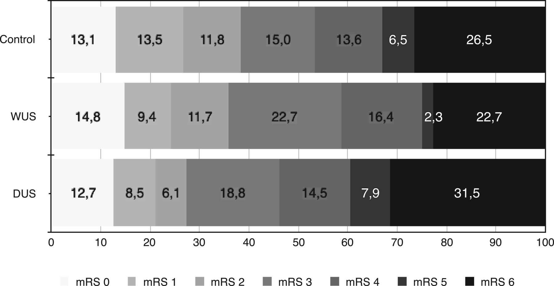
The baseline characteristics are shown in On-line Table 1. There was no difference in the frequency of good functional outcome (mRS ≤ 2) between patients with WUS and controls (35.9% versus 38.3%, P = .625; Table 1). Good functional outcome was reduced in those with DUS compared with controls (27.3%, P = .007). WUS and DUS did not differ significantly (P = .127). Figure 2 illustrates the distribution of mRS scores at day 90. In-hospital mortality (C: 18.9%; WUS: 12.6%; DUS: 23.3%; P = .067) and all-cause mortality at day 90 (C: 26.5%; WUS: 22.7%; DUS: 31.5%; P = .224) did not differ significantly. The rate of symptomatic intracranial hemorrhage was similar in all groups (P = .292, Table 1). Results did not change in the head-to-head comparison of groups adjusted for the abovementioned confounders (good functional outcome; WUS versus C: OR, 0.74; 95% CI, 0.48–1.16; P = .193; DUS versus C: OR, 0.49; 95% CI, 0.31–0.79; P = .003; data not shown).

View this table:
  • View inline
  • View popup
Table 1:

Primary and secondary outcome parameters

Fig 2.
  • Download figure
  • Open in new tab
  • Download powerpoint
Fig 2.

Distribution of mRS scores at 90 days.

Sixty-eight of the 128 patients with WUS (53.1%) underwent CTP or MR imaging (DUS: n = 63; 38.2%). MR imaging was performed in 36.7% of patients with WUS (n = 47) and in 31.5% of those with DUS (n = 52). In WUS[advanced], mismatch was present in 52 patients (76.5%; DUS[advanced]: n = 46, 73.0%; WUS[mri]: n = 36, 76.6%; DUS[mri]: n = 37, 71.2%). Subgroup analyses based on imaging selection are summarized in Table 2. When we compared WUS[advanced] and DUS[advanced] with unselected controls, there was no statistically significant difference in good functional outcome (C: 38.3%; WUS[advanced]: 47.1%; DUS[advanced]: 36.5%; P = .344). In-house mortality was reduced significantly in WUS[advanced] compared with controls (7.5% versus 18.9%, P = .019). Similar results were seen in patients with WUS[mri] and DUS[mri] (Table 2).

View this table:
  • View inline
  • View popup
Table 2:

Subgroup analysis—patient selection via imaging modalities

When we compared controls with patients in the WUS and DUS subgroups presenting with a verified mismatch only, the results did not change. Mortality rates remained stable with a nonsignificant increase in the percentage of good functional outcome (WUS[advanced]: 51.9%; DUS[advanced]: 39.1%; WUS[mri]: 52.8%; DUS[mri]: 43.2%; On-line Table 2). There was no significant difference in good functional outcome among WUS[mri] (51.1%), DUS[mri] (38.5%), and C[mri] (48.8%; P = .357; Table 2). The same was true for all-cause mortality at 3 months (C[mri]: 20.0%; WUS[mri]: 12.8%; DUS[mri]: 23.1%; P = .385) and the rate of symptomatic intracranial hemorrhage (P = .875).

We did compare WUS[advanced] and WUS[mri] with WUS not part of the respective groups (non-WUS[advanced], non-WUS[mri]; Table 3). Non-WUS[advanced] versus WUS[advanced] showed an increase in the rate of good functional outcome (23.3% versus 47.1%; OR, 2.92; 95% CI, 1.32–6.45; P = .006). In non-WUS[mri] versus WUS[mri], good functional outcome increased similarly (27.2% versus 51.1%; OR, 2.80; 95% CI, 1.28–6.10; P = .008) with an additional decrease in in-hospital mortality (17.3% versus 4.3%; OR, 0.22; 95% CI, 0.05–1.04; P = .049) and all-cause mortality at day 90 (28.4% versus 12.8%; OR, 0.37; 95% CI, 0.14–1.01; P = .05). For DUS, advanced imaging provided a significant improvement in the frequency of good functional outcome (non-DUS[advanced] versus DUS[advanced]: 21.6% versus 36.5%; OR, 2.09; 95% CI, 1.03–4.24; P = .048; non-DUS[mri] versus DUS[mri]: 22.1% versus 38.5%; OR, 2.20; 95% CI, 1.06–4.55; P = .038). A significant reduction of in-hospital mortality (30.0% versus 12.7%; OR, 0.34; 95% CI, 0.14–0.82; P = .013) and all-cause mortality at day 90 (38.2% versus 20.6%; OR, 0.42; 95% CI, 0.20–0.88; P = .025) was seen in non-DUS[advanced] versus DUS[advanced].

View this table:
  • View inline
  • View popup
Table 3:

Effect of advanced imaging on outcome and safety

Discussion

Stroke with an unknown symptom onset is frequent and accounts for up to 30% of ischemic stroke cases.11⇓–13 WUS (in which symptoms are realized during awakening) and DUS (unrealized symptom onset during daytime) can be distinguished. Endovascular therapy is currently not approved in those patients.6 On the basis of a considerable real-world dataset, we tried to detect differences in outcome among patients with WUS, DUS, and controls (excluding patients with known symptom onset presenting >6 hours after notification of symptoms). In our cohort, outcome in WUS did not differ from that in controls (mRS 0–2: 35.9% versus 38.3%), suggesting a beneficial effect of mTE in WUS. No increase in mortality rates or the frequency of symptomatic intracranial hemorrhage was observed. Functional outcome in DUS overall was significantly inferior compared with that of controls (mRS 0–2: 27.3%).

The first evidence indicating a possible beneficial effect of intravenous thrombolysis in carefully selected patients presenting in a prolonged time-window was published several years ago.21,22 Similar results were obtained for endovascular therapy in late-presenting stroke cases.23,24 From a present-day perspective, the use of older generation thrombectomy devices and small sample sizes limited generalizability. Subsequent retrospective and observational data suggested a potential beneficial effect of mTE in WUS with unknown symptom onset.7,9 However, DUS was not addressed. Recently, preliminary results of the diffusion-weighted imaging or CT perfusion assessment with Clinical Mismatch in the Triage of Wake Up and Late Presenting Strokes Undergoing Neurointervention with Trevo (DAWN) and Endovascular Therapy Following Imaging Evaluation for Ischemic Stroke 3 (DEFUSE 3) trials were presented.25⇓⇓–28 DAWN included WUS and patients “last seen well” up to 24 hours prior to endovascular therapy; DEFUSE 3 recruited patients treated between 6 and 16 hours after notification of symptoms. Both randomized controlled trials were terminated after interim analysis demonstrated superiority of the interventional group. It was proved that a strict imaging selection can identify patients eligible for mTE well after the currently approved time window. So far there is still little information on possible differences between WUS and DUS.

In our data, patient selection due to administration of CTP or MR imaging likewise led to a significant increase in the frequency of good functional outcome in patients with WUS and DUS. Consecutive steps in patient selection (1, all patients; 2, patients with CTP or MR imaging; 3, patients with MR imaging; 4, patients with documented mismatch in CTP or MR imaging; 5, patients with documented mismatch in MR imaging) increased the frequency of good functional outcome in both WUS and DUS (WUS 1: 35.9%; WUS 2: 47.1%; WUS 3: 51.1%; WUS 4: 51.9%; WUS 5: 52.8%; DUS 1: 27.3%; DUS 2: 36.5%; DUS 3: 38.5%; DUS 4: 39.1%; DUS 5: 43.2%). Outcome in DUS still was reduced. However, with the application of advanced imaging techniques, the statistically significant difference disappeared.

Besides outcome, WUS and DUS also seem to differ in pathophysiologic characteristics. An increase in platelet aggregation as well as a blood pressure surge during awakening are said to be associated with WUS but not DUS.29,30 WUS might occur right before awakening and therefore be comparable with a stroke population with documented symptom onset.31 Indeed, it was shown that clinical and imaging characteristics in WUS and stroke eligible for intravenous thrombolysis seem to be comparable, whereas DUS—as in our cohort—tends to have a worse prognosis.13,32,33 We did not observe significant differences in baseline characteristics. Longer imaging-to-groin times in WUS and DUS can be explained by a higher percentage of MR imaging scans and might indicate a careful patient selection. An average 138 minutes in controls is attributed to many patients being secondarily transferred for mTE/aTE.10

This study has several limitations. First, due to the retrospective design, selection bias can be suspected because we do not know in which cases mTE or aTE was not considered. Also, a certain inconsistency in decision-making can be assumed. Second, the definition of WUS and DUS could lead to some overlap of groups. Some of the patients categorized as having DUS by the assessing neurologist (eg, patients arriving late in the evening) could be WUS instead. There was no common CTP protocol or quantitative mismatch measurement, which may reduce comparability. An imbalance in group size might introduce a power problem (especially in subgroup analysis).

Conclusions

Our data suggest that mTE/aTE in selected patients with WUS allows a good functional outcome comparable with that in patients treated within 6 hours of symptom onset. Patients with DUS seem to be inferior to controls regarding outcome and mortality rates. The application of advanced imaging modalities (MR imaging, CTP) significantly increases the frequency of good functional outcome in both WUS and DUS and seems to reduce mortality.

Acknowledgments

We were supported by Hiltrud Niggemann in performing the statistical analysis. Casjupea Knispel, stroke and study nurse of the Neuroradiological Clinic, participated in data collection and conducted the follow-up interviews.

Footnotes

  • Disclosures: Marta Aguilar Pérez—UNRELATED: Consultancy: phenox, Comments: proctor and consultant. Hansjörg Bäzner—UNRELATED: Payment for Lectures Including Service on Speakers Bureaus: Bayer AG, Vital Pharma GmbH, Boehringer Ingelheim, Bristol-Myers Squibb, Daiichi Sankyo. Hans Henkes—UNRELATED: Board Membership: phenox, Comments: I am Speaker of the Board without payment; phenox compensates travel expenses; Consultancy: phenox, Comments: I am a consultant phenox and receive financial compensation for this effort; Patents (Planned, Pending or Issued): phenox, Medtronic, Comments: I am a coinventor of several intellectual property claims held by Medtronic and phenox; Stock/Stock Options: phenox, Comments: I am cofounder and shareholder of phenox; Travel/Accommodations/Meeting Expenses Unrelated to Activities Listed: phenox, Comments: I receive compensation for travel expenses for activities on behalf of phenox.

References

  1. 1.↵
    1. Berkhemer OA,
    2. Fransen PS,
    3. Beumer D, et al
    . A randomized trial of intraarterial treatment for acute ischemic stroke. N Engl J Med 2015;372:11–20 doi:10.1056/NEJMoa1411587 pmid:25517348
    CrossRefPubMed
  2. 2.↵
    1. Goyal M,
    2. Demchuk AM,
    3. Menon BK, et al
    ; ESCAPE Trial Investigators. Randomized assessment of rapid endovascular treatment of ischemic stroke. N Engl J Med 2015;372:1019–30 doi:10.1056/NEJMoa1414905 pmid:25671798
    CrossRefPubMed
  3. 3.↵
    1. Saver JL,
    2. Goyal M,
    3. Bonafe A, et al
    ; SWIFT PRIME Investigators. Stent-retriever thrombectomy after intravenous t-PA vs. t-PA alone in stroke. N Engl J Med 2015;372:2285–95 doi:10.1056/NEJMoa1415061 pmid:25882376
    CrossRefPubMed
  4. 4.↵
    1. Campbell BC,
    2. Mitchell PJ,
    3. Kleinig TJ, et al
    ; EXTEND-IA Investigators. Endovascular therapy for ischemic stroke with perfusion-imaging selection. N Engl J Med 2015;372:1009–18 doi:10.1056/NEJMoa1414792 pmid:25671797
    CrossRefPubMed
  5. 5.↵
    1. Jovin TG,
    2. Chamorro A,
    3. Cobo E, et al
    ; REVASCAT Trial Investigators. Thrombectomy within 8 hours after symptom onset in ischemic stroke. N Engl J Med 2015;372:2296–306 doi:10.1056/NEJMoa1503780 pmid:25882510
    CrossRefPubMed
  6. 6.↵
    1. Wahlgren N,
    2. Moreira T,
    3. Michel P, et al
    ; ESO-KSU, ESO, ESMINT, ESNR and EAN. Mechanical thrombectomy in acute ischemic stroke: consensus statement by ESO-Karolinska Stroke Update 2014/2015, supported by ESO, ESMINT, ESNR and EAN. Int J Stroke 2016;11:134–47 doi:10.1177/1747493015609778 pmid:26763029
    CrossRefPubMed
  7. 7.↵
    1. Mokin M,
    2. Kan P,
    3. Sivakanthan S, et al
    . Endovascular therapy of wake-up strokes in the modern era of stent retriever thrombectomy. J Neurointerv Surg 2016;8:240–43 doi:10.1136/neurintsurg-2014-011586 pmid:25634902
    Abstract/FREE Full Text
  8. 8.↵
    1. Qureshi AI,
    2. Miley JT,
    3. Chaudhry SA, et al
    . Safety and effectiveness of endovascular treatment after 6 hours of symptom onset in patients with anterior circulation ischemic stroke: a matched case control study. J Stroke Cerebrovasc Dis 2013;22:1076–81 doi:10.1016/j.jstrokecerebrovasdis.2012.07.015 pmid:23099041
    CrossRefPubMed
  9. 9.↵
    1. Kuntze Söderqvist Å,
    2. Andersson T,
    3. Wahlgren N, et al
    . Mechanical thrombectomy in acute ischemic stroke: patients with wake-up stroke and the elderly may benefit as well. J Stroke Cerebrovasc Dis 2016;25:2276–83 doi:10.1016/j.jstrokecerebrovasdis.2016.05.019 pmid:27282301
    CrossRefPubMed
  10. 10.↵
    1. Bücke P,
    2. Pérez MA,
    3. Schmid E, et al
    . Endovascular thrombectomy in acute ischemic stroke: outcome in referred versus directly admitted patients. Clin Neuroradiol 2017 Jan 31. [Epub ahead of print] doi:10.1007/s00062-017-0558-z pmid:28144702
    CrossRefPubMed
  11. 11.↵
    1. Kim YJ,
    2. Kim BJ,
    3. Kwon SU, et al
    . Unclear-onset stroke: daytime-unwitnessed stroke vs. wake-up stroke. Int J Stroke 2016;11:212–20 doi:10.1177/1747493015616513 pmid:26783313
    CrossRefPubMed
  12. 12.↵
    1. Wouters A,
    2. Lemmens R,
    3. Dupont P, et al
    . Wake-up stroke and stroke of unknown onset: a critical review. Front Neurol 2014;5:153 doi:10.3389/fneur.2014.00153 pmid:25161646
    CrossRefPubMed
  13. 13.↵
    1. Reid JM,
    2. Dai D,
    3. Cheripelli B, et al
    . Differences in wake-up and unknown onset stroke examined in a stroke registry. Int J Stroke 2015;10:331–35 doi:10.1111/ijs.12388 pmid:25338933
    CrossRefPubMed
  14. 14.↵
    1. Bouslama M,
    2. Haussen DC,
    3. Grossberg JA, et al
    . Computed tomographic perfusion selection and clinical outcomes after endovascular therapy in large vessel occlusion stroke. Stroke 2017;48:1271–77 doi:10.1161/STROKEAHA.116.015636 pmid:28389614
    Abstract/FREE Full Text
  15. 15.↵
    1. Campbell BC,
    2. Yassi N,
    3. Ma H, et al
    . Imaging selection in ischemic stroke: feasibility of automated CT-perfusion analysis. Int J Stroke 2015;10:51–54 doi:10.1111/ijs.12381 pmid:25319251
    CrossRefPubMed
  16. 16.↵
    1. Thomalla G,
    2. Cheng B,
    3. Ebinger M, et al
    ; VISTA Imaging Investigators. DWI-FLAIR mismatch for the identification of patients with acute ischaemic stroke within 4·5 h of symptom onset (PRE-FLAIR): a multicentre observational study. Lancet Neurol 2011;10:978–86 doi:10.1016/S1474-4422(11)70192-2 pmid:21978972
    CrossRefPubMed
  17. 17.↵
    1. Thomalla G,
    2. Boutitie F,
    3. Fiebach JB, et al
    ; WAKE-UP Investigators. Stroke with unknown time of symptom onset: baseline clinical and magnetic resonance imaging data of the first thousand patients in WAKE-UP (efficacy and safety of MRI-based thrombolysis in wake-up stroke: a randomized, doubleblind, placebo-controlled trial). Stroke 2017;48:770–73 doi:10.1161/STROKEAHA.116.015233 pmid:28174327
    Abstract/FREE Full Text
  18. 18.↵
    1. Turk A,
    2. Magarik JA,
    3. Chaudry I, et al
    . CT perfusion-guided patient selection for endovascular treatment of acute ischemic stroke is safe and effective. J Neurointerv Surg 2012;4:261–65 doi:10.1136/neurintsurg-2011-010067 pmid:21990520
    CrossRefPubMed
  19. 19.↵
    1. Michel P,
    2. Ntaios G,
    3. Reichhart M, et al
    . Perfusion-CT guided intravenous thrombolysis in patients with unknown-onset stroke: a randomized, double-blind, placebo-controlled, pilot feasibility trial. Neuroradiology 2012;54:579–88 doi:10.1007/s00234-011-0944-1 pmid:21808985
    CrossRefPubMed
  20. 20.↵
    1. Saver JL,
    2. Goyal M,
    3. Bonafe A, et al
    ; SWIFT PRIME Investigators. Solitaire™ with the Intention for Thrombectomy as Primary Endovascular Treatment for Acute Ischemic Stroke (SWIFT PRIME) trial: protocol for a randomized, controlled, multicenter study comparing the Solitaire revascularization device with IV tPA with IV tPA alone in acute ischemic stroke. Int J Stroke 2015;10:439–48 doi:10.1111/ijs.12459 pmid:25777831
    CrossRefPubMed
  21. 21.↵
    1. Cho AH,
    2. Sohn SI,
    3. Han MK, et al
    . Safety and efficacy of MRI-based thrombolysis in unclear-onset stroke: a preliminary report. Cerebrovasc Dis 2008;25:572–79 doi:10.1159/000132204 pmid:18483457
    CrossRefPubMed
  22. 22.↵
    1. Schellinger PD,
    2. Thomalla G,
    3. Fiehler J, et al
    . MRI-based and CT-based thrombolytic therapy in acute stroke within and beyond established time windows: an analysis of 1210 patients. Stroke 2007;38:2640–45 doi:10.1161/STROKEAHA.107.483255 pmid:17702961
    Abstract/FREE Full Text
  23. 23.↵
    1. Abou-Chebl A
    . Endovascular treatment of acute ischemic stroke may be safely performed with no time window limit in appropriately selected patients. Stroke 2010;41:1996–2000 doi:10.1161/STROKEAHA.110.578997 pmid:20651271
    Abstract/FREE Full Text
  24. 24.↵
    1. Natarajan SK,
    2. Snyder KV,
    3. Siddiqui AH, et al
    . Safety and effectiveness of endovascular therapy after 8 hours of acute ischemic stroke onset and wake-up strokes. Stroke 2009;40:3269–74 doi:10.1161/STROKEAHA.109.555102 pmid:19628808
    Abstract/FREE Full Text
  25. 25.↵
    Third European Stroke Organisation Conference. Prague, Czech Republic. May 16–18, 2017; https://eso-stroke.org/kategorie-1/data-highlights-opening-plenary-esoc-2017/. Accessed October 18, 2017
  26. 26.↵
    NeuroNews. DEFUSE 3 terminated early with high likelihood of benefit in the endovascular group. https://neuronewsinternational.com/defuse-3-terminated-early-high-likelihood-benefit-endovascular-group/. Accessed October 18, 2017
  27. 27.↵
    1. Jovin TG,
    2. Saver JL,
    3. Ribo M, et al
    . Diffusion-weighted imaging or computerized tomography perfusion assessment with clinical mismatch in the triage of wake up and late presenting strokes undergoing neurointervention with Trevo (DAWN) trial methods. Int J Stroke 2017;12:641–52 doi:10.1177/1747493017710341 pmid:28569123
    CrossRefPubMed
  28. 28.↵
    1. Albers GW,
    2. Lansberg MG,
    3. Kemp S, et al
    . A multicenter randomized controlled trial of endovascular therapy following imaging evaluation for ischemic stroke (DEFUSE 3). Int J Stroke 2017;12:896–905 doi:10.1177/1747493017701147 pmid:28946832
    CrossRefPubMed
  29. 29.↵
    1. Lundholm MD,
    2. Rooney M,
    3. Maas MB, et al
    . Wake-up stroke is associated with greater nocturnal mean arterial pressure variability. Stroke 2017;48:1668–70 doi:10.1161/STROKEAHA.116.016202 pmid:28455315
    Abstract/FREE Full Text
  30. 30.↵
    1. Kario K,
    2. Yano Y,
    3. Matsuo T, et al
    . Morning blood pressure surge, morning platelet aggregation, and silent cerebral infarction in older Japanese hypertensive patients. J Hypertens 2011;29:2433–39 doi:10.1097/HJH.0b013e32834cf1c0 pmid:22025234
    CrossRefPubMed
  31. 31.↵
    1. Rubin MN,
    2. Barrett KM
    . What to do with wake-up stroke. Neurohospitalist 2015;5:161–72 doi:10.1177/1941874415576204 pmid:26288674
    CrossRefPubMed
  32. 32.↵
    1. Mackey J,
    2. Kleindorfer D,
    3. Sucharew H, et al
    . Population-based study of wake-up strokes. Neurology 2011;76:1662–67 doi:10.1212/WNL.0b013e318219fb30 pmid:21555734
    Abstract/FREE Full Text
  33. 33.↵
    1. Costa R,
    2. Pinho J,
    3. Alves JN, et al
    . Wake-up stroke and stroke within the therapeutic window for thrombolysis have similar clinical severity, imaging characteristics, and outcome. J Stroke Cerebrovasc Dis 2016;25:511–14 doi:10.1016/j.jstrokecerebrovasdis.2015.10.032 pmid:26639403
    CrossRefPubMed
  • Received August 21, 2017.
  • Accepted after revision November 13, 2017.
  • © 2018 by American Journal of Neuroradiology
View Abstract
Next
Back to top
Advertisement
Print
Download PDF
Email Article

Thank you for your interest in spreading the word on American Journal of Neuroradiology.

NOTE: We only request your email address so that the person you are recommending the page to knows that you wanted them to see it, and that it is not junk mail. We do not capture any email address.

Enter multiple addresses on separate lines or separate them with commas.
Endovascular Thrombectomy in Wake-Up Stroke and Stroke with Unknown Symptom Onset
(Your Name) has sent you a message from American Journal of Neuroradiology
(Your Name) thought you would like to see the American Journal of Neuroradiology web site.
CAPTCHA
This question is for testing whether or not you are a human visitor and to prevent automated spam submissions.
Cite this article
P. Bücke, M. Aguilar Pérez, V. Hellstern, M. AlMatter, H. Bäzner, H. Henkes
Endovascular Thrombectomy in Wake-Up Stroke and Stroke with Unknown Symptom Onset
American Journal of Neuroradiology Feb 2018, DOI: 10.3174/ajnr.A5540

Citation Manager Formats

  • BibTeX
  • Bookends
  • EasyBib
  • EndNote (tagged)
  • EndNote 8 (xml)
  • Medlars
  • Mendeley
  • Papers
  • RefWorks Tagged
  • Ref Manager
  • RIS
  • Zotero
0 Responses
Respond to this article
Share
Bookmark this article
Endovascular Thrombectomy in Wake-Up Stroke and Stroke with Unknown Symptom Onset
P. Bücke, M. Aguilar Pérez, V. Hellstern, M. AlMatter, H. Bäzner, H. Henkes
American Journal of Neuroradiology Feb 2018, DOI: 10.3174/ajnr.A5540
del.icio.us logo Twitter logo Facebook logo Mendeley logo
  • Tweet Widget
  • Facebook Like
  • Google Plus One
Purchase

Jump to section

  • Article
    • Abstract
    • ABBREVIATIONS:
    • Materials and Methods
    • Results
    • Discussion
    • Conclusions
    • Acknowledgments
    • Footnotes
    • References
  • Figures & Data
  • Supplemental
  • Info & Metrics
  • Responses
  • References
  • PDF

Related Articles

  • PubMed
  • Google Scholar

Cited By...

  • Functional Outcome, Recanalization, and Hemorrhage Rates After Large Vessel Occlusion Stroke Treated With Tenecteplase Before Thrombectomy
  • Crossref (16)
  • Google Scholar

This article has been cited by the following articles in journals that are participating in Crossref Cited-by Linking.

  • Risk factors of intracranial hemorrhage after mechanical thrombectomy of anterior circulation ischemic stroke
    Ulf Neuberger, Philipp Kickingereder, Silvia Schönenberger, Simon Schieber, Peter A. Ringleb, Martin Bendszus, Johannes Pfaff, Markus A. Möhlenbruch
    Neuroradiology 2019 61 4
  • Functional Outcome, Recanalization, and Hemorrhage Rates After Large Vessel Occlusion Stroke Treated With Tenecteplase Before Thrombectomy
    Gaspard Gerschenfeld, Didier Smadja, Guillaume Turc, Stephane Olindo, François-Xavier Laborne, Marion Yger, Jildaz Caroff, Bruno Gonçalves, Pierre Seners, Marie Cantier, Yann l'Hermitte, Manvel Aghasaryan, Cosmin Alecu, Gaultier Marnat, Wagih Ben Hassen, Erwah Kalsoum, Frédéric Clarençon, Michel Piotin, Laurent Spelle, Christian Denier, Igor Sibon, Sonia Alamowitch, Nicolas Chausson, Vincent Degos, Mariana Sarov, Nicolas Legris, Olivier Chassin, Djibril Soumah, Tony Altarcha, Carole Imbernon, Pauline Renou, Mathilde Poli, Sabrina Debruxelles, Sharmila Sagnier, François Rouanet, Jean-Sebastien Liegey, David Calvet, Jean-Claude Baron, Laure Bottin, Stephen Delorme, Jean Capron, Diana Doukhi, Sam Ghazanfari, David Weisenburger, Edwige Lescieux, Florent Gariel, Xavier Barreau, Patrice Menegon, Thomas Tourdias, Catherine Oppenheim, Olivier Naggara, Titien Tuilier, Nader Sourour, Nader Sourour, Eimad Shotar, Stéphanie Lenck, Kévin Premat, Raphaël Blanc, Simon Escalard, Robert Fahed, Stanislas Smajda, Mikael Mazighi
    Neurology 2021 97 22
  • Mechanical Thrombectomy in the Late Presentation of Anterior Circulation Large Vessel Occlusion Stroke: A Guideline From the Society of Vascular and Interventional Neurology Guidelines and Practice Standards Committee
    Thanh N. Nguyen, Alicia C. Castonguay, James E. Siegler, Simon Nagel, Maarten G. Lansberg, Adam de Havenon, Sunil A. Sheth, Mohamad Abdalkader, Jenny P. Tsai, Gregory W. Albers, Hesham E. Masoud, Tudor G. Jovin, Sheila O. Martins, Raul G. Nogueira, Osama O. Zaidat
    Stroke: Vascular and Interventional Neurology 2023 3 1
  • Severe Leukoaraiosis Is Associated with Poor Outcome after Successful Recanalization of M1 Middle Cerebral Artery Occlusion Strokes
    Johannes Sebastian Mutzenbach, Erasmia Müller-Thies-Broussalis, Monika Killer-Oberpfalzer, Christoph Johannes Griessenauer, Constantin Hecker, Luis Rafael Moscote-Salazar, Pia Paradaiser, Slaven Pikija
    Cerebrovascular Diseases 2020 49 3
  • Benefits of Endovascular Treatment in Late Window for Acute Ischemic Stroke Selected without CT Perfusion: A Real-World Study
    Yuan Yang, Ting Cui, Zuoxiao Li, Jinglun Li, Ting Duan, Zhengzhou Yuan, Changyi Wang, Jincheng Wan, Cao Li, Shujiang Zhang, Ling Li, Fayun Hu, Bo Wu
    Clinical Interventions in Aging 2022 Volume 17
  • Cerebral ischaemia with unknown onset: Outcome after recanalization procedure
    R. Tortuyaux, M. Ferrigno, N. Dequatre-Ponchelle, S. Djelad, C. Cordonnier, H. Hénon, D. Leys
    Revue Neurologique 2020 176 1-2
  • Early Versus Late Initiation of Endovascular Therapy in Patients with Severe Cerebral Venous Sinus Thrombosis
    Philipp Bücke, Hans Henkes, Johannes Kaesmacher, Mirjam R. Heldner, Adrian Scutelnic, Marcel Arnold, Thomas R. Meinel, Alexandru Cimpoca, Thomas Horvath, Elina Henkes, Hansjörg Bäzner, Victoria Hellstern
    Neurocritical Care 2024 41 3
  • Endovascular treatment for ischemic stroke beyond the time window: A meta‐analysis
    Xuefei Li, Lingshan Wu, Hongxian Xie, Yuxian Bao, Dan He, Xiang Luo
    Acta Neurologica Scandinavica 2020 141 1
  • Outcome of Stroke Patients with Unknown Onset and Unknown Time Last Known Well Undergoing Endovascular Therapy
    Sebastian Stösser, Felix J. Bode, Julius N. Meissner, Johannes M. Weller, Christine Kindler, Malte Sauer, Daniel Paech, Christoph Riegler, Christian H. Nolte, Amitis Pourian, Joachim Röther, Nadja Selo, Ulrike Ernemann, Sven Poli, Rosa Marie Eckert, Georg Bohner, Korbinian Scherling, Franziska Dorn, Gabor C. Petzold
    Clinical Neuroradiology 2023 33 1
  • Unknown Onset Stroke: Differences Between Patients with Wake-Up Stroke and Daytime-Unwitnessed Stroke
    Katharina Vedder, Dr. Anne Ebert, Prof. Dr. Kristina Szabo, Prof. Dr. Alex Förster, Prof. Dr. Angelika Alonso
    Journal of Stroke and Cerebrovascular Diseases 2021 30 7

More in this TOC Section

  • Rescue Reentry in Carotid Near-Occlusion
  • Contour Neurovascular System: Five Year Follow Up
  • Effect of SARS-CoV2 on Endovascular Thrombectomy
Show more NEUROINTERVENTION

Similar Articles

Advertisement

Indexed Content

  • Current Issue
  • Accepted Manuscripts
  • Article Preview
  • Past Issues
  • Editorials
  • Editor's Choice
  • Fellows' Journal Club
  • Letters to the Editor
  • Video Articles

Cases

  • Case Collection
  • Archive - Case of the Week
  • Archive - Case of the Month
  • Archive - Classic Case

More from AJNR

  • Trainee Corner
  • Imaging Protocols
  • MRI Safety Corner
  • Book Reviews

Multimedia

  • AJNR Podcasts
  • AJNR Scantastics

Resources

  • Turnaround Time
  • Submit a Manuscript
  • Submit a Video Article
  • Submit an eLetter to the Editor/Response
  • Manuscript Submission Guidelines
  • Statistical Tips
  • Fast Publishing of Accepted Manuscripts
  • Graphical Abstract Preparation
  • Imaging Protocol Submission
  • Evidence-Based Medicine Level Guide
  • Publishing Checklists
  • Author Policies
  • Become a Reviewer/Academy of Reviewers
  • News and Updates

About Us

  • About AJNR
  • Editorial Board
  • Editorial Board Alumni
  • Alerts
  • Permissions
  • Not an AJNR Subscriber? Join Now
  • Advertise with Us
  • Librarian Resources
  • Feedback
  • Terms and Conditions
  • AJNR Editorial Board Alumni

American Society of Neuroradiology

  • Not an ASNR Member? Join Now

© 2025 by the American Society of Neuroradiology All rights, including for text and data mining, AI training, and similar technologies, are reserved.
Print ISSN: 0195-6108 Online ISSN: 1936-959X

Powered by HighWire