Advertisement
  • Main menu
  • User menu
  • Search
  • English ▼
    • English
    • Afrikaans
    • Albanian
    • Amharic
    • Arabic
    • Armenian
    • Azerbaijani
    • Basque
    • Belarusian
    • Bengali
    • Bosnian
    • Bulgarian
    • Catalan
    • Cebuano
    • Chichewa
    • Chinese (Simplified)
    • Chinese (Traditional)
    • Corsican
    • Croatian
    • Czech
    • Danish
    • Dutch
    • Esperanto
    • Estonian
    • Filipino
    • Finnish
    • French
    • Frisian
    • Galician
    • Georgian
    • German
    • Greek
    • Gujarati
    • Haitian Creole
    • Hausa
    • Hawaiian
    • Hebrew
    • Hindi
    • Hmong
    • Hungarian
    • Icelandic
    • Igbo
    • Indonesian
    • Irish
    • Italian
    • Japanese
    • Javanese
    • Kannada
    • Kazakh
    • Khmer
    • Korean
    • Kurdish (Kurmanji)
    • Kyrgyz
    • Lao
    • Latin
    • Latvian
    • Lithuanian
    • Luxembourgish
    • Macedonian
    • Malagasy
    • Malay
    • Malayalam
    • Maltese
    • Maori
    • Marathi
    • Mongolian
    • Myanmar (Burmese)
    • Nepali
    • Norwegian
    • Pashto
    • Persian
    • Polish
    • Portuguese
    • Punjabi
    • Romanian
    • Russian
    • Samoan
    • Scottish Gaelic
    • Serbian
    • Sesotho
    • Shona
    • Sindhi
    • Sinhala
    • Slovak
    • Slovenian
    • Somali
    • Spanish
    • Sudanese
    • Swahili
    • Swedish
    • Tajik
    • Tamil
    • Telugu
    • Thai
    • Turkish
    • Ukrainian
    • Urdu
    • Uzbek
    • Vietnamese
    • Welsh
    • Xhosa
    • Yiddish
    • Yoruba
    • Zulu

Main menu

  • Home
  • Content
    • Current Issue
    • Accepted Manuscripts
    • Article Preview
    • Past Issue Archive
    • Video Articles
    • AJNR Case Collection
    • Case of the Week Archive
    • Case of the Month Archive
    • Classic Case Archive
  • Special Collections
    • AJNR Awards
    • Low-Field MRI
    • Alzheimer Disease
    • ASNR Foundation Special Collection
    • Photon-Counting CT
    • View All
  • Multimedia
    • AJNR Podcasts
    • AJNR SCANtastic
    • Trainee Corner
    • MRI Safety Corner
    • Imaging Protocols
  • For Authors
    • Submit a Manuscript
    • Submit a Video Article
    • Submit an eLetter to the Editor/Response
    • Manuscript Submission Guidelines
    • Statistical Tips
    • Fast Publishing of Accepted Manuscripts
    • Graphical Abstract Preparation
    • Imaging Protocol Submission
    • Author Policies
  • About Us
    • About AJNR
    • Editorial Board
    • Editorial Board Alumni
  • More
    • Become a Reviewer/Academy of Reviewers
    • Subscribers
    • Permissions
    • Alerts
    • Feedback
    • Advertisers
    • ASNR Home

User menu

  • Alerts
  • Log in

Search

  • Advanced search
American Journal of Neuroradiology

American Journal of Neuroradiology

ASHNR American Society of Functional Neuroradiology ASHNR American Society of Pediatric Neuroradiology ASSR
  • Alerts
  • Log in

Advanced Search

English ▼
  • English
  • Afrikaans
  • Albanian
  • Amharic
  • Arabic
  • Armenian
  • Azerbaijani
  • Basque
  • Belarusian
  • Bengali
  • Bosnian
  • Bulgarian
  • Catalan
  • Cebuano
  • Chichewa
  • Chinese (Simplified)
  • Chinese (Traditional)
  • Corsican
  • Croatian
  • Czech
  • Danish
  • Dutch
  • Esperanto
  • Estonian
  • Filipino
  • Finnish
  • French
  • Frisian
  • Galician
  • Georgian
  • German
  • Greek
  • Gujarati
  • Haitian Creole
  • Hausa
  • Hawaiian
  • Hebrew
  • Hindi
  • Hmong
  • Hungarian
  • Icelandic
  • Igbo
  • Indonesian
  • Irish
  • Italian
  • Japanese
  • Javanese
  • Kannada
  • Kazakh
  • Khmer
  • Korean
  • Kurdish (Kurmanji)
  • Kyrgyz
  • Lao
  • Latin
  • Latvian
  • Lithuanian
  • Luxembourgish
  • Macedonian
  • Malagasy
  • Malay
  • Malayalam
  • Maltese
  • Maori
  • Marathi
  • Mongolian
  • Myanmar (Burmese)
  • Nepali
  • Norwegian
  • Pashto
  • Persian
  • Polish
  • Portuguese
  • Punjabi
  • Romanian
  • Russian
  • Samoan
  • Scottish Gaelic
  • Serbian
  • Sesotho
  • Shona
  • Sindhi
  • Sinhala
  • Slovak
  • Slovenian
  • Somali
  • Spanish
  • Sudanese
  • Swahili
  • Swedish
  • Tajik
  • Tamil
  • Telugu
  • Thai
  • Turkish
  • Ukrainian
  • Urdu
  • Uzbek
  • Vietnamese
  • Welsh
  • Xhosa
  • Yiddish
  • Yoruba
  • Zulu
  • Home
  • Content
    • Current Issue
    • Accepted Manuscripts
    • Article Preview
    • Past Issue Archive
    • Video Articles
    • AJNR Case Collection
    • Case of the Week Archive
    • Case of the Month Archive
    • Classic Case Archive
  • Special Collections
    • AJNR Awards
    • Low-Field MRI
    • Alzheimer Disease
    • ASNR Foundation Special Collection
    • Photon-Counting CT
    • View All
  • Multimedia
    • AJNR Podcasts
    • AJNR SCANtastic
    • Trainee Corner
    • MRI Safety Corner
    • Imaging Protocols
  • For Authors
    • Submit a Manuscript
    • Submit a Video Article
    • Submit an eLetter to the Editor/Response
    • Manuscript Submission Guidelines
    • Statistical Tips
    • Fast Publishing of Accepted Manuscripts
    • Graphical Abstract Preparation
    • Imaging Protocol Submission
    • Author Policies
  • About Us
    • About AJNR
    • Editorial Board
    • Editorial Board Alumni
  • More
    • Become a Reviewer/Academy of Reviewers
    • Subscribers
    • Permissions
    • Alerts
    • Feedback
    • Advertisers
    • ASNR Home
  • Follow AJNR on Twitter
  • Visit AJNR on Facebook
  • Follow AJNR on Instagram
  • Join AJNR on LinkedIn
  • RSS Feeds

AJNR Awards, New Junior Editors, and more. Read the latest AJNR updates

Research ArticleAdult Brain

Iodine Extravasation Quantification on Dual-Energy CT of the Brain Performed after Mechanical Thrombectomy for Acute Ischemic Stroke Can Predict Hemorrhagic Complications

M. Bonatti, F. Lombardo, G.A. Zamboni, F. Vittadello, R. Currò Dossi, B. Bonetti, R. Pozzi Mucelli and G. Bonatti
American Journal of Neuroradiology January 2018, DOI: https://doi.org/10.3174/ajnr.A5513
M. Bonatti
aFrom the Departments of Radiology (M.B., F.L., G.B.)
  • Find this author on Google Scholar
  • Find this author on PubMed
  • Search for this author on this site
  • ORCID record for M. Bonatti
F. Lombardo
aFrom the Departments of Radiology (M.B., F.L., G.B.)
  • Find this author on Google Scholar
  • Find this author on PubMed
  • Search for this author on this site
  • ORCID record for F. Lombardo
G.A. Zamboni
cDepartment of Radiology (G.A.Z., R.P.M.), University of Verona, Verona, Italy
  • Find this author on Google Scholar
  • Find this author on PubMed
  • Search for this author on this site
  • ORCID record for G.A. Zamboni
F. Vittadello
dExplora-Research and Statistical Analysis (F.V.), Vigodarzere, Italy.
  • Find this author on Google Scholar
  • Find this author on PubMed
  • Search for this author on this site
  • ORCID record for F. Vittadello
R. Currò Dossi
bNeurology (R.C.D., B.B.), Bolzano Central Hospital, Bolzano, Italy
  • Find this author on Google Scholar
  • Find this author on PubMed
  • Search for this author on this site
  • ORCID record for R. Currò Dossi
B. Bonetti
bNeurology (R.C.D., B.B.), Bolzano Central Hospital, Bolzano, Italy
  • Find this author on Google Scholar
  • Find this author on PubMed
  • Search for this author on this site
  • ORCID record for B. Bonetti
R. Pozzi Mucelli
cDepartment of Radiology (G.A.Z., R.P.M.), University of Verona, Verona, Italy
  • Find this author on Google Scholar
  • Find this author on PubMed
  • Search for this author on this site
  • ORCID record for R. Pozzi Mucelli
G. Bonatti
aFrom the Departments of Radiology (M.B., F.L., G.B.)
  • Find this author on Google Scholar
  • Find this author on PubMed
  • Search for this author on this site
  • ORCID record for G. Bonatti
  • Article
  • Figures & Data
  • Info & Metrics
  • Responses
  • References
  • PDF
Loading

Article Figures & Data

Figures

  • Tables
  • Fig 1.
    • Download figure
    • Open in new tab
    • Download powerpoint
    Fig 1.

    A, Postoperative DECT with an iodine map overlay shows a cortical-subcortical hyperdensity secondary to iodine extravasation with a maximum iodine concentration of 3.0 mg/mL; no hyperdensity was visible on virtual unenhanced images (not included). B, A follow-up CT performed 48 hours later because of sudden clinical worsening shows a large parenchymal hematoma with a contralateral shift of the midline structures.
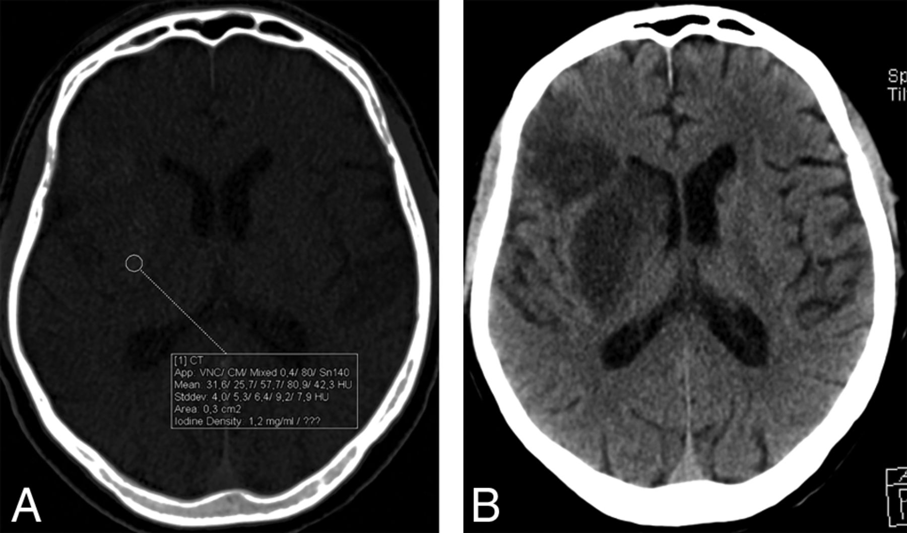
  • Fig 2.
    • Download figure
    • Open in new tab
    • Download powerpoint
    Fig 2.

    A, Postoperative DECT with an iodine map overlay shows a cortical-subcortical and deep white matter hyperdensity secondary to iodine extravasation with a maximum iodine concentration of 1.2 mg/mL; no hyperdensity was visible on virtual unenhanced images (not included). B, The follow-up CT performed before discharge, 4 days later, shows ischemic lesions at the sites of iodine extravasation but no hemorrhage.
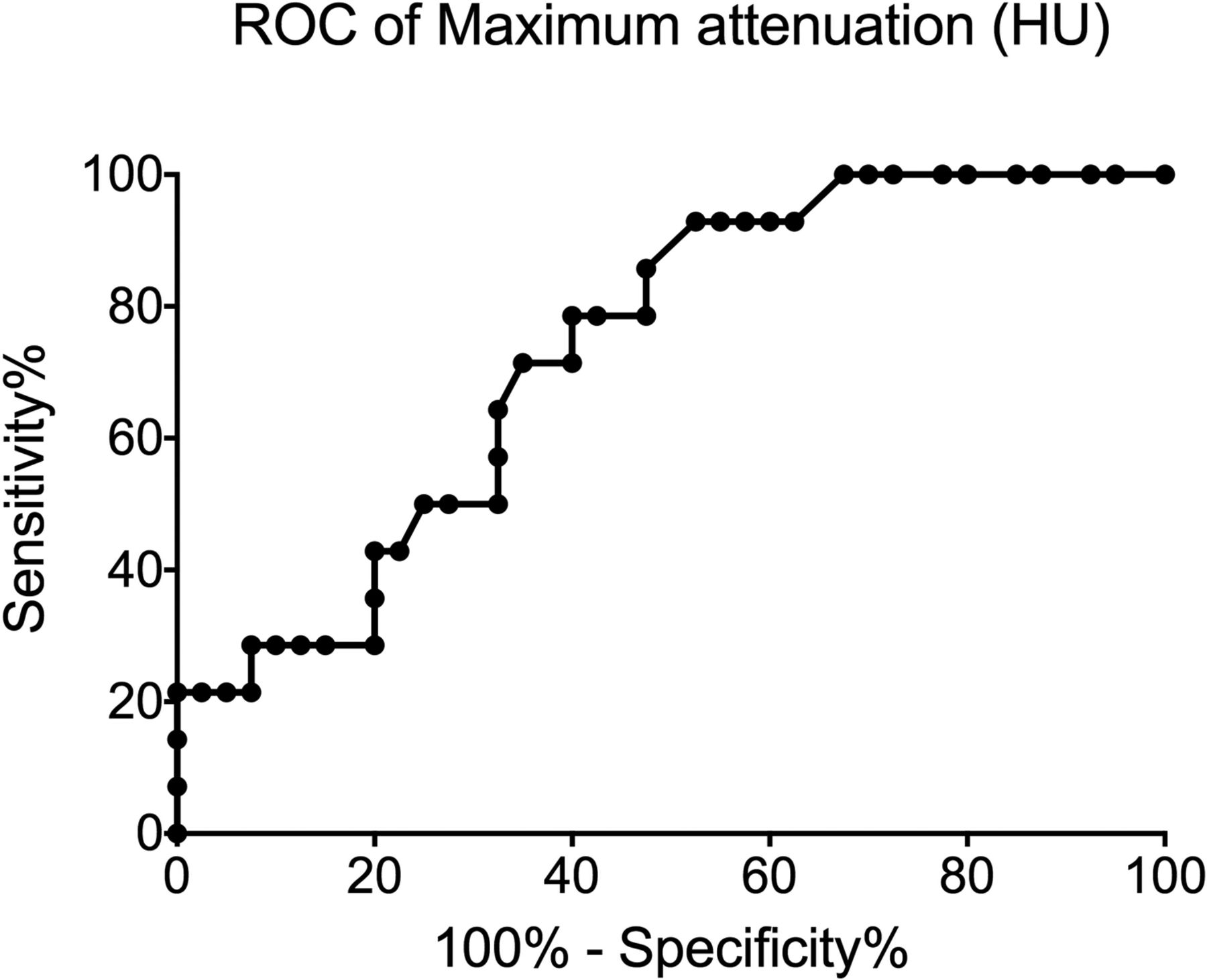
  • Fig 3.
    • Download figure
    • Open in new tab
    • Download powerpoint
    Fig 3.

    Receiver operator characteristic (ROC) curve showing sensitivity and specificity of maximum hyperdensity attenuation in identifying patients developing ICH.
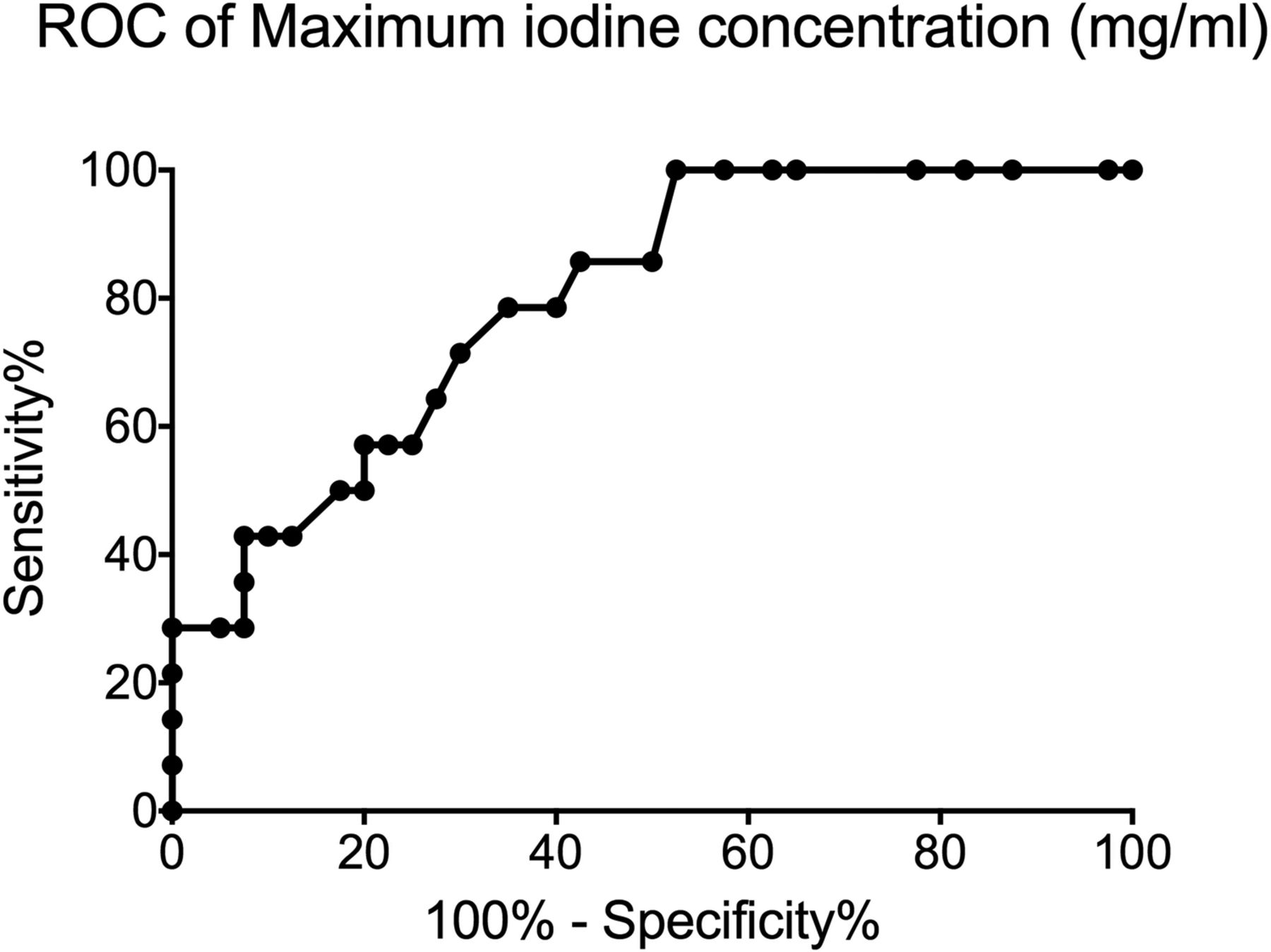
  • Fig 4.
    • Download figure
    • Open in new tab
    • Download powerpoint
    Fig 4.

    Receiver operator characteristic (ROC) curve showing sensitivity and specificity of maximum iodine concentration in identifying patients developing ICH.
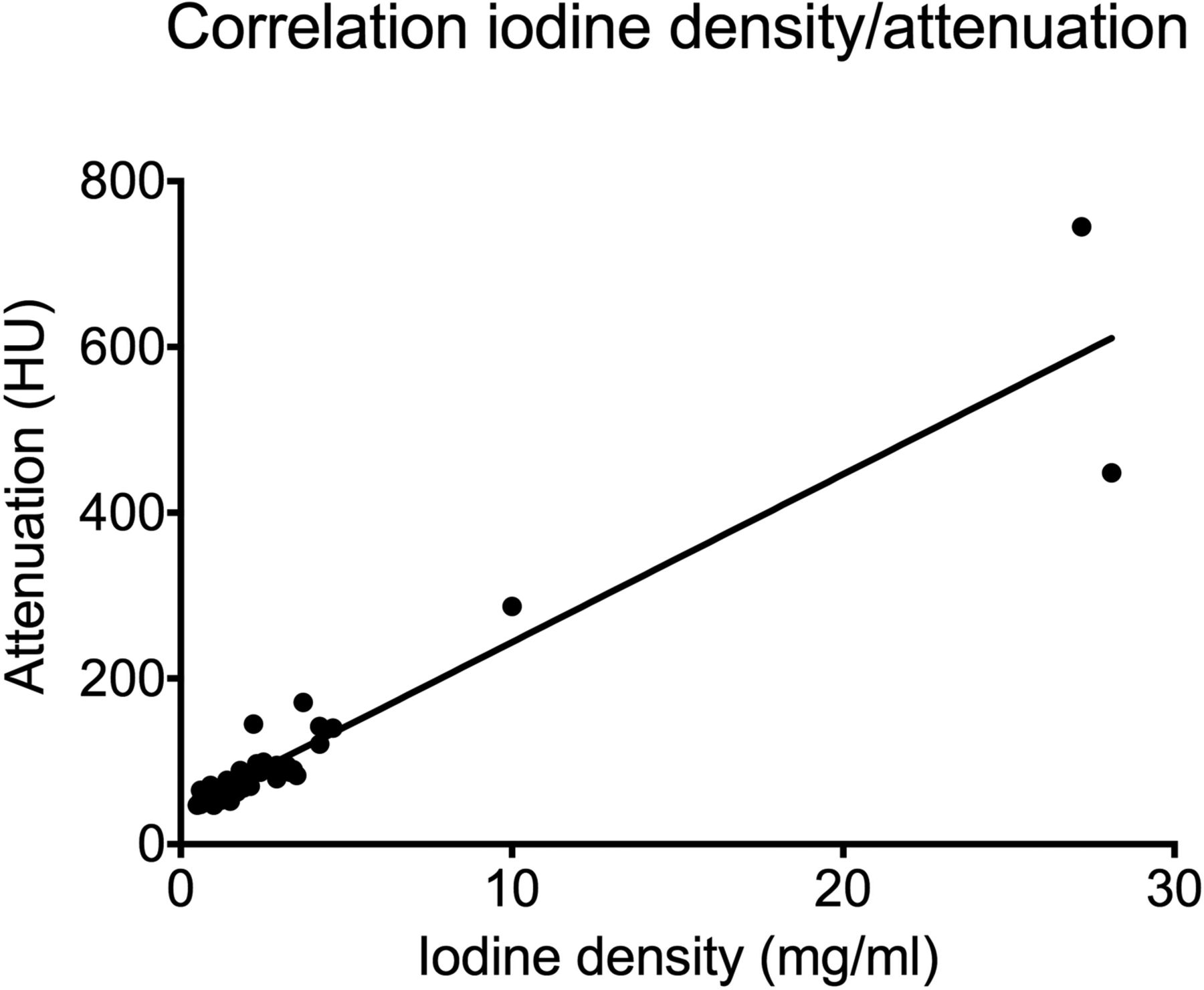
  • Fig 5.
    • Download figure
    • Open in new tab
    • Download powerpoint
    Fig 5.

    Graph showing the linear correlation existing between maximum hyperdensity attenuation and maximum iodine concentration.
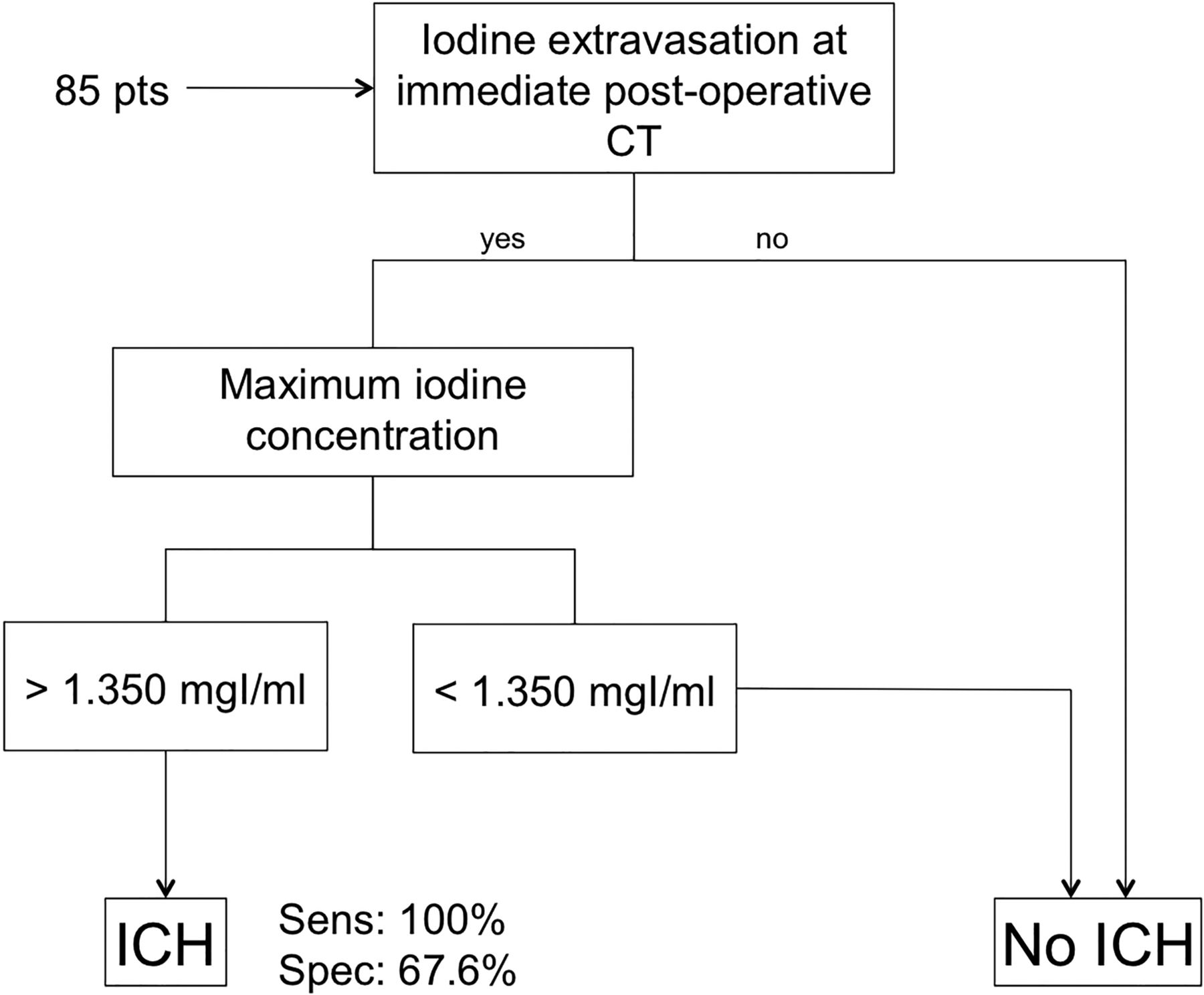
  • Fig 6.
    • Download figure
    • Open in new tab
    • Download powerpoint
    Fig 6.

    Diagnostic algorithm that enables identifying patents developing ICH with 100% sensitivity and 67.6% specificity.

Tables

  • Figures
    • View popup
    Table 1:

    Scanning parameters for postoperative unenhanced dual-energy brain CT and follow-up unenhanced brain CT

    DECTSECT
    Scanning techniqueSpiralSpiral
    Scan directionCaudocranialCaudocranial
    kVp80/140120
    mAs ref310/155390
    Collimation (mm)40 × 0.6128 × 0.6
    Rotation time (sec)0.51
    Pitch0.70.55
    CARE Dose 4DaOnOn
    CARE-kVaNot availableOff
    X-CAREaNot availableOn
    KernelD34fH40s
    • Note:—SECT indicates single-energy CT; ref, reference.

    • ↵a Siemens.

    • View popup
    Table 2:

    Results of postoperative DECT image analysis with interreader concordance and subsequent consensus

    Reader 1Reader 2Interreader ConcordanceConsensus
    Presence of parenchymal hyperdensity
        Yes53/85 (62.4%)53/85 (62.4%)κ = 0.95; 95% CI, 0.882–1.00054/85 (63.5%)
        No32/85 (37.6%)32/85 (37.6%)31/85 (36.5%)
    Hyperdensity location
        DWM26/53 (49.1%)31/53 (58.5%)κ = 0.745; 95% CI, 0.588–0.90329/54 (53.7%)
        DWM+cortical/subcortical21/53 (39.6%)16/53 (30.2%)18/54 (33.3%)
        Cortical/subcortical6/53 (11.3%)6/53 (11.3%)7/54 (13.0%)
    Hyperdensity homogeneity
        Homogeneous29/53 (54.7%)31/53 (58.5%)κ = 0.799; 95% CI, 0.632–0.96630/54 (55.6%)
        Inhomogeneous24/53 (45.3%)22/53 (41.5%)24/54 (44.4%)
    Mean maximum attenuation (HU)100 (median 77, range 45–701)103 (median 76, range 47–789)P = .99974 (range, 47–745)
    Presence of iodine extravasation
        Yes53/85 (62.4%)53/85 (62.4%)κ = 0.976; 95% CI, 0.929–1.00054/85 (63.5%)
        No32/85 (37.6%)32/85 (37.6%)31/85 (36.5%)
    Mean maximum iodine concentration (mg/dL)3.0 (median,1.8; range 0.5–28.4)2.9 (median, 1.6; range 0.6–27.7)P = .9261.8 (range, 0.5–28.1)
    Hemorrhage on VNC images
        Yes5/85 (5.9%)5/85 (5.9%)κ = 1.000; 95% CI, 1.000–1.0005/85 (5.9%)
        No80/85 (94.1%)80/85 (94.1%)80/85 (94.1%)
    • Note:—DWM indicates deep white matter.

    • View popup
    Table 3:

    Univariate analysis—association between postoperative dual-energy CT parameters and subsequent intracerebral hemorrhage developmenta

    Non-ICH Group (n = 71)ICH Group (n = 14)P Value
    No. (%)No. (%)
    Hyperdensity on simulated 120-kV images.002b
        Yes40/71 (56.3%)14/14 (100%)
        No31/71 (43.7%)0/14 (0%)
    Hyperdensity site.342
        DWM23/40 (57.5%)6/14 (42.9%)
        DWM + cortical11/40 (27.5%)7/14 (50.0%)
        Cortical6/40 (15.0%)1/14 (7.1%)
    Hyperdensity homogeneity.028b
        Homogeneous26/40 (65.0%)4/14 (28.6%)
        Inhomogeneous14/40 (35.0%)10/14 (71.4%)
    Hyperdensity maximum attenuation.010b
        Median (HU)6885
    Iodine extravasation on iodine map images.002b
        Yes40/71 (56.3%)14/14 (100%)
        No31/71 (43.7%)0/14 (0%)
    Maximum iodine concentration<.001b
        Median (mg/mL)1.402.63
    Hemorrhage on VNC images<.001b
        Yes0/71 (0%)5/14 (35.7%)
        No71/71 (100%)9/14 (64.3%)
    • Note:—DWM indicates deep white matter.

    • ↵a The strength of the association is quantified by the P value (significant if P < .05).

    • ↵b Significant.

Next
Back to top
Advertisement
Print
Download PDF
Email Article
Cite this article
0 Responses
Respond to this article
Share
Bookmark this article
Iodine Extravasation Quantification on Dual-Energy CT of the Brain Performed after Mechanical Thrombectomy for Acute Ischemic Stroke Can Predict Hemorrhagic Complications
M. Bonatti, F. Lombardo, G.A. Zamboni, F. Vittadello, R. Currò Dossi, B. Bonetti, R. Pozzi Mucelli, G. Bonatti
American Journal of Neuroradiology Jan 2018, DOI: 10.3174/ajnr.A5513
del.icio.us logo Twitter logo Facebook logo Mendeley logo
  • Tweet Widget
Purchase

Jump to section

  • Article
    • Abstract
    • ABBREVIATIONS:
    • Materials and Methods
    • Results
    • Discussion
    • Conclusions
    • Footnotes
    • References
  • Figures & Data
  • Info & Metrics
  • Responses
  • References
  • PDF

Related Articles

  • No related articles found.
  • PubMed
  • Google Scholar

Cited By...

  • Determinants and Clinical Relevance of Iodine Contrast Extravasation after Endovascular Thrombectomy: A Dual-Energy CT Study
  • Quantitative Intracerebral Iodine Extravasation in Risk Stratification for Intracranial Hemorrhage in Patients with Acute Ischemic Stroke
  • Prediction of Hemorrhage after Successful Recanalization in Patients with Acute Ischemic Stroke: Improved Risk Stratification Using Dual-Energy CT Parenchymal Iodine Concentration Ratio Relative to the Superior Sagittal Sinus
  • Quantification of Iodine Leakage on Dual-Energy CT as a Marker of Blood-Brain Barrier Permeability in Traumatic Hemorrhagic Contusions: Prediction of Surgical Intervention for Intracranial Pressure Management
  • Dual energy CT after stroke thrombectomy alters assessment of hemorrhagic complications
  • Crossref
  • Google Scholar

This article has not yet been cited by articles in journals that are participating in Crossref Cited-by Linking.

More in this TOC Section

Adult Brain

  • Diagnostic Neuroradiology of Monoclonal Antibodies
  • Clinical Outcomes After Chiari I Decompression
  • Segmentation of Brain Metastases with BLAST
Show more Adult Brain

Neurointervention

  • Cavernous dural AVF treated by transfacial route
  • A Retrospective Study in Tentorial DAVFs
  • Proximal Protection Devices for Carotid Stenting
Show more Neurointervention

Similar Articles

Advertisement

Indexed Content

  • Current Issue
  • Accepted Manuscripts
  • Article Preview
  • Past Issues
  • Editorials
  • Editor's Choice
  • Fellows' Journal Club
  • Letters to the Editor
  • Video Articles

Cases

  • Case Collection
  • Archive - Case of the Week
  • Archive - Case of the Month
  • Archive - Classic Case

More from AJNR

  • Trainee Corner
  • Imaging Protocols
  • MRI Safety Corner
  • Book Reviews

Multimedia

  • AJNR Podcasts
  • AJNR Scantastics

Resources

  • Turnaround Time
  • Submit a Manuscript
  • Submit a Video Article
  • Submit an eLetter to the Editor/Response
  • Manuscript Submission Guidelines
  • Statistical Tips
  • Fast Publishing of Accepted Manuscripts
  • Graphical Abstract Preparation
  • Imaging Protocol Submission
  • Evidence-Based Medicine Level Guide
  • Publishing Checklists
  • Author Policies
  • Become a Reviewer/Academy of Reviewers
  • News and Updates

About Us

  • About AJNR
  • Editorial Board
  • Editorial Board Alumni
  • Alerts
  • Permissions
  • Not an AJNR Subscriber? Join Now
  • Advertise with Us
  • Librarian Resources
  • Feedback
  • Terms and Conditions
  • AJNR Editorial Board Alumni

American Society of Neuroradiology

  • Not an ASNR Member? Join Now

© 2025 by the American Society of Neuroradiology All rights, including for text and data mining, AI training, and similar technologies, are reserved.
Print ISSN: 0195-6108 Online ISSN: 1936-959X

Powered by HighWire
Email this Article

Thank you for your interest in spreading the word on American Journal of Neuroradiology.

NOTE: We only request your email address so that the person you are recommending the page to knows that you wanted them to see it, and that it is not junk mail. We do not capture any email address.

Enter multiple addresses on separate lines or separate them with commas.
Iodine Extravasation Quantification on Dual-Energy CT of the Brain Performed after Mechanical Thrombectomy for Acute Ischemic Stroke Can Predict Hemorrhagic Complications
(Your Name) has sent you a message from American Journal of Neuroradiology
(Your Name) thought you would like to see the American Journal of Neuroradiology web site.
CAPTCHA
This question is for testing whether or not you are a human visitor and to prevent automated spam submissions.
Cite this article
M. Bonatti, F. Lombardo, G.A. Zamboni, F. Vittadello, R. Currò Dossi, B. Bonetti, R. Pozzi Mucelli, G. Bonatti
Iodine Extravasation Quantification on Dual-Energy CT of the Brain Performed after Mechanical Thrombectomy for Acute Ischemic Stroke Can Predict Hemorrhagic Complications
American Journal of Neuroradiology Jan 2018, DOI: 10.3174/ajnr.A5513

Citation Manager Formats

  • BibTeX
  • Bookends
  • EasyBib
  • EndNote (tagged)
  • EndNote 8 (xml)
  • Medlars
  • Mendeley
  • Papers
  • RefWorks Tagged
  • Ref Manager
  • RIS
  • Zotero

We use cookies on this site to enhance your user experience. By clicking any link on this page you are giving your consent for us to set cookies.