Case of the Week
Section Editors: Matylda Machnowska1 and Anvita Pauranik2
1University of Toronto, Toronto, Ontario, Canada
2BC Children's Hospital, University of British Columbia, Vancouver, British Columbia, Canada
Sign up to receive an email alert when a new Case of the Week is posted.
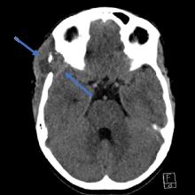
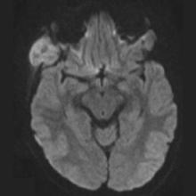
Initial axial CT imaging bone windows (A) demonstrate a sharply marginated, lytic skull base defect along the lateral margin of the right sphenoid ridge (red arrow). CT soft-tissue windows (B) demonstrate a heterogeneously dense soft-tissue process or mass spanning the lytic defect (blue arrows). Follow-up MRI study demonstrates a well-defined, T2 heterogeneously hyperintense mass (C, axial T2), with central homogeneous diffusion restriction (D: DWI, E: ADC), and with homogeneous enhancement on postcontrast imaging (F, axial postcontrast T1).


