
This article requires a subscription to view the full text. If you have a subscription you may use the login form below to view the article. Access to this article can also be purchased.
Graphical Abstract

Abstract
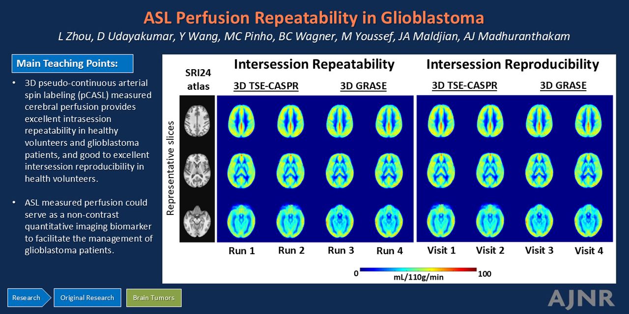
BACKGROUND AND PURPOSE: Arterial spin-labeling (ASL) MRI has gained recognition as a quantitative perfusion imaging method for managing patients with brain tumors. Limited studies have so far investigated the reproducibility of ASL-derived perfusion in these patients. This study aimed to evaluate intrasession repeatability and intersession reproducibility of perfusion measurements using 3D pseudocontinuous ASL (pCASL) with TSE Cartesian acquisition with spiral profile reordering (TSE-CASPR) in healthy volunteers (HV) and patients with glioblastoma (GBM) at 3T and to compare them against 3D pCASL with gradient and spin echo (GRASE).
MATERIALS AND METHODS: This prospective study (NCT03922984) was approved by the institutional review board, and written informed consent was obtained from all subjects. HV underwent repeat pCASL evaluations 2–4 weeks apart between November 2021 and October 2022. Patients with GBM were recruited for longitudinal MRI from September 2019 to February 2023. Intrasession repeatability (HV and GBM) and intersession reproducibility (HV only) of pCASL were assessed using linear regression, Bland–Altman analyses, the intraclass correlation coefficient (ICC) with 95% CI, and within-subject coefficients of variation (wsCV).
RESULTS: Twenty HV (9 men; mean age, 25.1 [SD, 1.7] years; range, 23–30 years) and 21 patients with GBM (15 men; mean age, 59.8 [SD, 14.3] years; range, 28–81 years) were enrolled. In imaging sessions, 3D pCASL-measured perfusion with TSE-CASPR and GRASE, respectively, achieved high R2 values (0.88–0.95; 0.93–0.96), minimal biases (−0.46−0.81; −0.08−0.35 mL/100 g/min), high ICCs [95% CI], 0.96–0.98 [0.94–0.98]; 0.96–0.98 [0.92–0.99]), and low wsCV (6.64%−9.07%; 5.20%−8.16%) in HV (n = 20) and patients with GBM (n = 21). Across imaging sessions, 3D pCASL in HV (n = 20) achieved high R2 values (0.71; 0.82), minimal biases (−1.2; −0.90 mL/100 g/min), high ICC [95% CI] values (0.85 [0.81–0.89]; 0.90 [0.87–0.93]), and low wsCV values (13.82%; 9.98%).
CONCLUSIONS: Our study demonstrated excellent intrasession repeatability of 3D pCASL-measured cerebral perfusion in HV and patients with GBM and good-to-excellent intersession reproducibility in HV. 3D pCASL with GRASE performed slightly better than 3D pCASL with TSE-CASPR in HV; however, in patients with GBM, 3D pCASL with TSE-CASPR showed better performance in tumor regions with a nearly 2-fold higher SNR. ASL-measured perfusion could serve as a noncontrast quantitative imaging biomarker to facilitate the management of patients with GBM.
ABBREVIATIONS:
- ASL
- arterial spin-labeling
- CASPR
- Cartesian acquisition with spiral profile reordering
- GBM
- glioblastoma
- GRASE
- gradient and spin echo
- HV
- healthy volunteers
- ICC
- intraclass correlation coefficient
- M0
- proton-density-weighted image
- NSA
- number of signals averaged
- pCASL
- pseudocontinuous arterial spin-labeling
- PLD
- postlabel delay
- QI
- quantitative imaging
- wsCV
- within-subject coefficients of variation
- © 2025 by American Journal of Neuroradiology
Log in using your username and password
Log in through your institution


Jump to section
Related Articles
Cited By...
- No citing articles found.