Advertisement

Main menu

  • Home
  • Content
    • Current Issue
    • Accepted Manuscripts
    • Article Preview
    • Past Issue Archive
    • Video Articles
    • AJNR Case Collection
    • Case of the Week Archive
    • Case of the Month Archive
    • Classic Case Archive
  • Special Collections
    • AJNR Awards
    • Low-Field MRI
    • Alzheimer Disease
    • ASNR Foundation Special Collection
    • Photon-Counting CT
    • View All
  • Multimedia
    • AJNR Podcasts
    • AJNR SCANtastic
    • Trainee Corner
    • MRI Safety Corner
    • Imaging Protocols
  • For Authors
    • Submit a Manuscript
    • Submit a Video Article
    • Submit an eLetter to the Editor/Response
    • Manuscript Submission Guidelines
    • Statistical Tips
    • Fast Publishing of Accepted Manuscripts
    • Graphical Abstract Preparation
    • Imaging Protocol Submission
    • Author Policies
  • About Us
    • About AJNR
    • Editorial Board
    • Editorial Board Alumni
  • More
    • Become a Reviewer/Academy of Reviewers
    • Subscribers
    • Permissions
    • Alerts
    • Feedback
    • Advertisers
    • ASNR Home

User menu

  • Alerts
  • Log in

Search

  • Advanced search
American Journal of Neuroradiology

American Journal of Neuroradiology

ASHNR American Society of Functional Neuroradiology ASHNR American Society of Pediatric Neuroradiology ASSR
  • Alerts
  • Log in

Advanced Search

  • Home
  • Content
    • Current Issue
    • Accepted Manuscripts
    • Article Preview
    • Past Issue Archive
    • Video Articles
    • AJNR Case Collection
    • Case of the Week Archive
    • Case of the Month Archive
    • Classic Case Archive
  • Special Collections
    • AJNR Awards
    • Low-Field MRI
    • Alzheimer Disease
    • ASNR Foundation Special Collection
    • Photon-Counting CT
    • View All
  • Multimedia
    • AJNR Podcasts
    • AJNR SCANtastic
    • Trainee Corner
    • MRI Safety Corner
    • Imaging Protocols
  • For Authors
    • Submit a Manuscript
    • Submit a Video Article
    • Submit an eLetter to the Editor/Response
    • Manuscript Submission Guidelines
    • Statistical Tips
    • Fast Publishing of Accepted Manuscripts
    • Graphical Abstract Preparation
    • Imaging Protocol Submission
    • Author Policies
  • About Us
    • About AJNR
    • Editorial Board
    • Editorial Board Alumni
  • More
    • Become a Reviewer/Academy of Reviewers
    • Subscribers
    • Permissions
    • Alerts
    • Feedback
    • Advertisers
    • ASNR Home
  • Follow AJNR on Twitter
  • Visit AJNR on Facebook
  • Follow AJNR on Instagram
  • Join AJNR on LinkedIn
  • RSS Feeds

AJNR Awards, New Junior Editors, and more. Read the latest AJNR updates

Research ArticleNeurodegenerative Disorder Imaging

Automated Detection of Normal Pressure Hydrocephalus Using CT Imaging for Calculating the Ventricle-to-Subarachnoid Volume Ratio

Jacob J. Knittel, Justin L. Hoskin, Dylan J. Hoyt, Jonathan A. Abdo, Emily L. Foldes, Molly M. McElvogue, Clay M. Oliver, Daniel A. Keesler, Terry D. Fife, F. David Barranco, Kris A. Smith, J. Gordon McComb, Matthew T. Borzage and Kevin S. King
American Journal of Neuroradiology January 2025, 46 (1) 141-146; DOI: https://doi.org/10.3174/ajnr.A8451
Jacob J. Knittel
aFrom the Creighton University School of Medicine (J.J.K., J.A.A., C.M.O.), Phoenix, Arizona
  • Find this author on Google Scholar
  • Find this author on PubMed
  • Search for this author on this site
Justin L. Hoskin
bDepartment of Neurology (J.L.H., T.D.F.), Barrow Neurological Institute, St. Joseph’s Hospital and Medical Center, Phoenix, Arizona
  • Find this author on Google Scholar
  • Find this author on PubMed
  • Search for this author on this site
Dylan J. Hoyt
cDepartment of Radiology (D.J.H., D.A.K.), St. Joseph’s Hospital and Medical Center, Phoenix, Arizona
  • Find this author on Google Scholar
  • Find this author on PubMed
  • Search for this author on this site
Jonathan A. Abdo
aFrom the Creighton University School of Medicine (J.J.K., J.A.A., C.M.O.), Phoenix, Arizona
  • Find this author on Google Scholar
  • Find this author on PubMed
  • Search for this author on this site
Emily L. Foldes
dDepartment of Neuroradiology (E.L.F., M.M.M., K.S.K.), Barrow Neurological Institute, St. Joseph’s Hospital and Medical Center, Phoenix, Arizona
  • Find this author on Google Scholar
  • Find this author on PubMed
  • Search for this author on this site
  • ORCID record for Emily L. Foldes
Molly M. McElvogue
dDepartment of Neuroradiology (E.L.F., M.M.M., K.S.K.), Barrow Neurological Institute, St. Joseph’s Hospital and Medical Center, Phoenix, Arizona
  • Find this author on Google Scholar
  • Find this author on PubMed
  • Search for this author on this site
  • ORCID record for Molly M. McElvogue
Clay M. Oliver
aFrom the Creighton University School of Medicine (J.J.K., J.A.A., C.M.O.), Phoenix, Arizona
  • Find this author on Google Scholar
  • Find this author on PubMed
  • Search for this author on this site
Daniel A. Keesler
bDepartment of Neurology (J.L.H., T.D.F.), Barrow Neurological Institute, St. Joseph’s Hospital and Medical Center, Phoenix, Arizona
  • Find this author on Google Scholar
  • Find this author on PubMed
  • Search for this author on this site
Terry D. Fife
bDepartment of Neurology (J.L.H., T.D.F.), Barrow Neurological Institute, St. Joseph’s Hospital and Medical Center, Phoenix, Arizona
  • Find this author on Google Scholar
  • Find this author on PubMed
  • Search for this author on this site
F. David Barranco
eDepartment of Neurosurgery (F.D.B., K.A.S.), Barrow Neurological Institute, St. Joseph’s Hospital and Medical Center, Phoenix, Arizona
  • Find this author on Google Scholar
  • Find this author on PubMed
  • Search for this author on this site
Kris A. Smith
eDepartment of Neurosurgery (F.D.B., K.A.S.), Barrow Neurological Institute, St. Joseph’s Hospital and Medical Center, Phoenix, Arizona
  • Find this author on Google Scholar
  • Find this author on PubMed
  • Search for this author on this site
  • ORCID record for Kris A. Smith
J. Gordon McComb
fDepartment of Neurosurgery (J.G.M.), Children’s Hospital of Los Angeles, Los Angeles, California
  • Find this author on Google Scholar
  • Find this author on PubMed
  • Search for this author on this site
  • ORCID record for J. Gordon McComb
Matthew T. Borzage
gDivision of Neonatology (M.T.B.), Children’s Hospital of Los Angeles, Los Angeles, California.
  • Find this author on Google Scholar
  • Find this author on PubMed
  • Search for this author on this site
  • ORCID record for Matthew T. Borzage
Kevin S. King
dDepartment of Neuroradiology (E.L.F., M.M.M., K.S.K.), Barrow Neurological Institute, St. Joseph’s Hospital and Medical Center, Phoenix, Arizona
  • Find this author on Google Scholar
  • Find this author on PubMed
  • Search for this author on this site
  • ORCID record for Kevin S. King
  • Article
  • Figures & Data
  • Supplemental
  • Info & Metrics
  • Responses
  • References
  • PDF
Loading

This article requires a subscription to view the full text. If you have a subscription you may use the login form below to view the article. Access to this article can also be purchased.

Graphical Abstract

Figure
  • Download figure
  • Open in new tab
  • Download powerpoint

Abstract

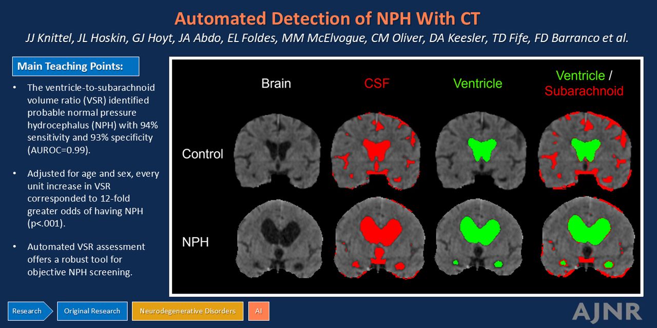
BACKGROUND AND PURPOSE: Normal pressure hydrocephalus (NPH) is a diagnostic challenge because its clinical symptoms and imaging appearance resemble normal aging and other forms of dementia. Identifying NPH is essential so that patients can receive timely treatment to improve gait distortion and quality of life. An automated marker of NPH was developed and evaluated on clinical CT images, and its utility was assessed in a large patient cohort.

MATERIALS AND METHODS: A retrospective review was conducted of CT images from 306 tap test–responsive patients with NPH between January 2015 and January 2022. Control CT images were obtained from patients in the emergency department who were evaluated for headache and had unremarkable CT findings between June 2021 and August 2022. The ventricle-to-subarachnoid volume ratio (VSR) was automatically calculated by the imaging software and used as a predictor of NPH in linear regression modeling with adjustment for age and sex. The correlations of VSR with age, sex, and the receiver operating characteristic were computed.

RESULTS: VSR was significantly greater in patients with NPH than controls (P < .001). Importantly, VSR was not significantly correlated with age (P = .56, R2 = 0.001). VSR identifies NPH with a sensitivity and specificity of 94.1% and 92.5%, respectively, with an area under the receiver operating characteristic curve of 0.99 (95% CI 0.975–0.995).

CONCLUSIONS: Automated assessment of the VSR on head CT images identified probable NPH with 93% accuracy. The assessment of a large cohort of patients with NPH supports the generalizability of clinical screening of CT images. Moreover, the results support the utility of ventricle-to-sulcal concordance often used by radiologists but not currently a part of the accepted guidelines for imaging markers of NPH.

ABBREVIATIONS:

AD
Alzheimer’s disease
AUC
area under the curve
AUROC
area under the receiver operating characteristic
ICV
intracranial volume
NIfTI
Neuroimaging Informatics Technology Initiative
NPH
normal pressure hydrocephalus
ROC
receiver operating characteristic
VP
ventriculoperitoneal
VSR
ventricle-to-subarachnoid volume ratio
  • © 2025 by American Journal of Neuroradiology
View Full Text

Log in using your username and password

Forgot your user name or password?

Log in through your institution

You may be able to gain access using your login credentials for your institution. Contact your library if you do not have a username and password.
PreviousNext
Back to top

In this issue

American Journal of Neuroradiology: 46 (1)
American Journal of Neuroradiology
Vol. 46, Issue 1
1 Jan 2025
  • Table of Contents
  • Index by author
  • Complete Issue (PDF)
Advertisement
Print
Download PDF
Email Article

Thank you for your interest in spreading the word on American Journal of Neuroradiology.

NOTE: We only request your email address so that the person you are recommending the page to knows that you wanted them to see it, and that it is not junk mail. We do not capture any email address.

Enter multiple addresses on separate lines or separate them with commas.
Automated Detection of Normal Pressure Hydrocephalus Using CT Imaging for Calculating the Ventricle-to-Subarachnoid Volume Ratio
(Your Name) has sent you a message from American Journal of Neuroradiology
(Your Name) thought you would like to see the American Journal of Neuroradiology web site.
CAPTCHA
This question is for testing whether or not you are a human visitor and to prevent automated spam submissions.
Cite this article
Jacob J. Knittel, Justin L. Hoskin, Dylan J. Hoyt, Jonathan A. Abdo, Emily L. Foldes, Molly M. McElvogue, Clay M. Oliver, Daniel A. Keesler, Terry D. Fife, F. David Barranco, Kris A. Smith, J. Gordon McComb, Matthew T. Borzage, Kevin S. King
Automated Detection of Normal Pressure Hydrocephalus Using CT Imaging for Calculating the Ventricle-to-Subarachnoid Volume Ratio
American Journal of Neuroradiology Jan 2025, 46 (1) 141-146; DOI: 10.3174/ajnr.A8451

Citation Manager Formats

  • BibTeX
  • Bookends
  • EasyBib
  • EndNote (tagged)
  • EndNote 8 (xml)
  • Medlars
  • Mendeley
  • Papers
  • RefWorks Tagged
  • Ref Manager
  • RIS
  • Zotero
0 Responses
Respond to this article
Share
Bookmark this article
Automated Hydrocephalus Detection via CT Imaging
Jacob J. Knittel, Justin L. Hoskin, Dylan J. Hoyt, Jonathan A. Abdo, Emily L. Foldes, Molly M. McElvogue, Clay M. Oliver, Daniel A. Keesler, Terry D. Fife, F. David Barranco, Kris A. Smith, J. Gordon McComb, Matthew T. Borzage, Kevin S. King
American Journal of Neuroradiology Jan 2025, 46 (1) 141-146; DOI: 10.3174/ajnr.A8451
del.icio.us logo Twitter logo Facebook logo Mendeley logo
  • Tweet Widget
  • Facebook Like
  • Google Plus One
Purchase

Jump to section

  • Article
    • Graphical Abstract
    • Abstract
    • ABBREVIATIONS:
    • MATERIALS AND METHODS
    • RESULTS
    • DISCUSSION
    • CONCLUSIONS
    • Acknowledgments
    • Footnotes
    • References
  • Figures & Data
  • Supplemental
  • Info & Metrics
  • Responses
  • References
  • PDF

Related Articles

  • PubMed
  • Google Scholar

Cited By...

  • No citing articles found.
  • Crossref
  • Google Scholar

This article has not yet been cited by articles in journals that are participating in Crossref Cited-by Linking.

More in this TOC Section

  • ICP Sign: New Imaging Marker for MSA-C vs SCA
  • Automatic quantification and classification in IPD
  • Vascular risk factors, CMB and WMH in cognition
Show more Neurodegenerative Disorder Imaging

Similar Articles

Advertisement

Indexed Content

  • Current Issue
  • Accepted Manuscripts
  • Article Preview
  • Past Issues
  • Editorials
  • Editor's Choice
  • Fellows' Journal Club
  • Letters to the Editor
  • Video Articles

Cases

  • Case Collection
  • Archive - Case of the Week
  • Archive - Case of the Month
  • Archive - Classic Case

More from AJNR

  • Trainee Corner
  • Imaging Protocols
  • MRI Safety Corner

Multimedia

  • AJNR Podcasts
  • AJNR Scantastics

Resources

  • Turnaround Time
  • Submit a Manuscript
  • Submit a Video Article
  • Submit an eLetter to the Editor/Response
  • Manuscript Submission Guidelines
  • Statistical Tips
  • Fast Publishing of Accepted Manuscripts
  • Graphical Abstract Preparation
  • Imaging Protocol Submission
  • Evidence-Based Medicine Level Guide
  • Publishing Checklists
  • Author Policies
  • Become a Reviewer/Academy of Reviewers
  • News and Updates

About Us

  • About AJNR
  • Editorial Board
  • Editorial Board Alumni
  • Alerts
  • Permissions
  • Not an AJNR Subscriber? Join Now
  • Advertise with Us
  • Librarian Resources
  • Feedback
  • Terms and Conditions
  • AJNR Editorial Board Alumni

American Society of Neuroradiology

  • Not an ASNR Member? Join Now

© 2025 by the American Society of Neuroradiology All rights, including for text and data mining, AI training, and similar technologies, are reserved.
Print ISSN: 0195-6108 Online ISSN: 1936-959X

Powered by HighWire