
This article requires a subscription to view the full text. If you have a subscription you may use the login form below to view the article. Access to this article can also be purchased.
Graphical Abstract

Abstract
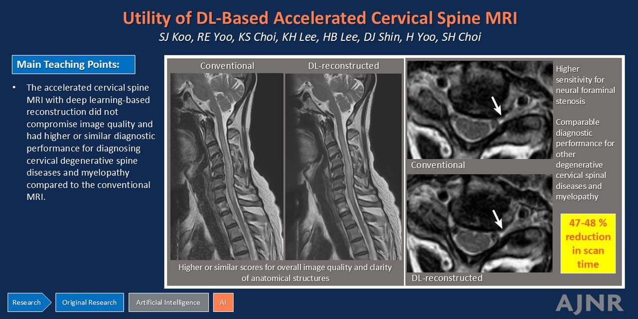
BACKGROUND AND PURPOSE: Deep learning (DL)-based reconstruction enables improving the quality of MR images acquired with a short scan time. We aimed to prospectively compare the image quality and diagnostic performance in evaluating cervical degenerative spine diseases and myelopathy between conventional cervical MRI and accelerated cervical MRI with a commercially available vendor-neutral DL-based reconstruction.
MATERIALS AND METHODS: Fifty patients with degenerative cervical spine disease or myelopathy underwent both conventional cervical MRI and accelerated cervical MRI by using a DL-based reconstruction operating within the DICOM domain. The images were evaluated both quantitatively, based on SNR and contrast-to-noise ratio (CNR), and qualitatively, by using a 5-point scoring system for the overall image quality and clarity of anatomic structures on sagittal T1WI, sagittal contrast-enhanced (CE) T1WI, and axial/sagittal T2WI. Four radiologists assessed the sensitivity and specificity of the 2 protocols for detecting degenerative diseases and myelopathy.
RESULTS: The DL-based protocol reduced MRI acquisition time by 47%–48% compared with the conventional protocol. DL-reconstructed images demonstrated a higher SNR on sagittal T1WI (P = .046) and a higher CNR on sagittal T2WI (P = .03) than conventional images. The SNR on sagittal T2WI and the CNR on sagittal T1WI did not significantly differ (P > .05). DL-reconstructed images had better overall image quality on sagittal T1WI (P < .001), sagittal T2WI (Dixon in-phase or TSE) (P < .001), and sagittal T2WI (Dixon water-only) (P = .013) and similar image quality on axial T2WI and sagittal CE T1WI (P > .05). DL-reconstructed images had better clarity of anatomic structures (P values were < .001 for all structures, except for the neural foramen [P = .024]). DL-reconstructed images had a higher sensitivity for detecting neural foraminal stenosis (P = .005) and similar sensitivities for diagnosing other degenerative spinal diseases and myelopathy (P > .05). The specificities for diagnosing degenerative spinal diseases and myelopathy did not differ between the 2 images (P > .05).
CONCLUSIONS: The accelerated cervical MRI reconstructed with a vendor-neutral DL-based reconstruction algorithm did not compromise image quality and had higher or similar diagnostic performance for diagnosing cervical degenerative spine diseases and myelopathy compared with the conventional protocol.
ABBREVIATIONS:
- CE
- contrast-enhanced
- CNR
- contrast-to-noise ratio
- DL
- deep learning
- HIVD
- herniated intervertebral disc
- © 2025 by American Journal of Neuroradiology
Log in using your username and password
Log in through your institution


Jump to section
Related Articles
Cited By...
- No citing articles found.