
Article Figures & Data
Figures
- FIG 1.
A and B, antero-posterior (AP) and lateral projections of a single-shot image of the stent on discharge. No deformity was observed. C and D, Single-shot images of the stent 14 days after the procedure, after the onset of symptoms. A noticeable narrowing and elongation of the distal part of the stent can be seen (orange arrow). This observation is concurrent with our observation of the carotid artery’s reaction to the implant. Because this case is the one in which our patient exhibited acute symptoms from the occurrence, we believe it is extremely illustrative for our point to come across. E, AP projection of the intraprocedural DSA performed prior to the implantation of the FD stent. A saccular aneurysm of the ophthalmic segment of the left ICA was the target of treatment. F and G, DWI MRI sequence, which was performed after the initial onset of right-sided weakness with which the patient presented. No acute ischemic changes in the brain parenchyma can be seen, which led us to believe that the changes in the cerebral blood flow were transient in nature.
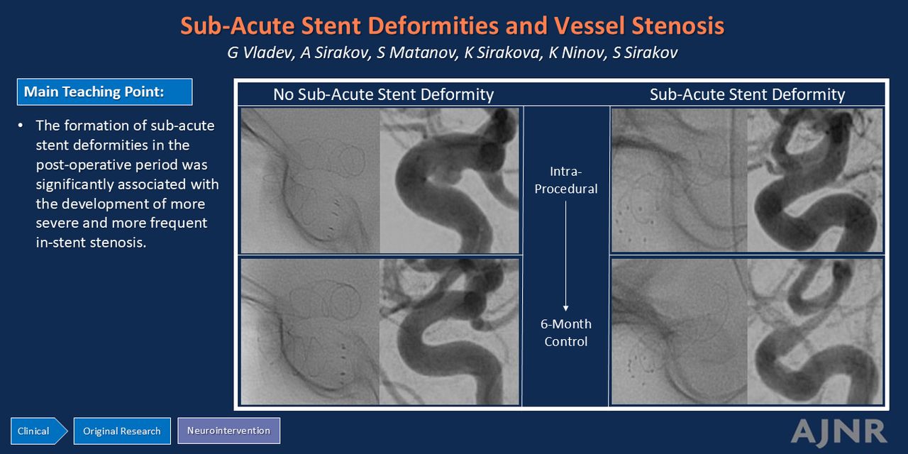
- FIG 2.
Six-month DSA control performed on the patient (anteroposterior projections). A severe narrowing of the distal left ICA in the segment of the placed FD and reduced blood flow in the left middle and anterior cerebral arteries can be seen (A). These findings correlate with the symptoms observed in the patient in the first month postprocedure. A compensatory development of leptomeningeal collaterals, collaterals from the posterior circulation and the right ICA, is the probable cause for the lack of vascular incidents during that time (B and C).
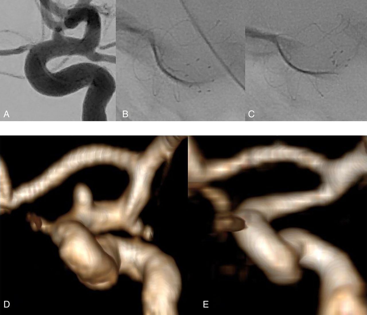
- FIG 3.
A, Intraprocedural DSA of the aneurysm on the left ICA. B and C, Fluoroscopy images of the stent, which illustrate the progressive narrowing and stenosis of the distal part of the implant. We can see a notable deformity on the 14-day single-shot (C), compared with the procedural image of the implant (B). The images of the deformity correlate well with the conducted follow-up MRI. We highlight a noticeable reduction of diameter of the distal ICA on follow-up (E), when compared with the pretreatment 3D-TOF reconstruction of the artery (D).


Tables
Characteristics n=48 Age (years) 31–71 (mean = 51.4 SD = 11,938) Female 40 (83%) Stent deformities in N of observed patients 28 (58%) Distal half stent deformities 26 (54%) Proximal half stent deformities 2 (4%) Proximal marker changes 18 (38%) Patients exhibiting clinically Notable symptoms 1 (2%) Aneurysms on the ophthalmic segment 34 (71%) Aneurysms on the terminal segment 14 (29%) Smoking 18 (38%) Hypertension 22 (46%) - Table 2:
Characteristics of patients with and without stent deformities with regards to the incidence of ISS and the de novo exhibition of a unilateral headache postprocedurally
ISS Incidence ISS Severity (6-Month Follow-Up) Novel Unilateral Headache at the 14-Day Follow-Up Mild Moderate Severe Patients with stent deformities at the 14-day follow-up (n=28) n=28 (100%) n=12/28 (43%) n=5/28 (18%) n=11/28 (39%) n=20/28 (71%) Patients without stent deformities at the 14-day follow-up (n=20) n=8 (40%) n=7/8 (87%) n=1/8 (13%) n=0 (0%) n=2/20 (10%)


Jump to section
Related Articles
Cited By...
- No citing articles found.