Advertisement

Main menu

  • Home
  • Content
    • Current Issue
    • Accepted Manuscripts
    • Article Preview
    • Past Issue Archive
    • Video Articles
    • AJNR Case Collection
    • Case of the Week Archive
    • Case of the Month Archive
    • Classic Case Archive
  • Special Collections
    • AJNR Awards
    • Low-Field MRI
    • Alzheimer Disease
    • ASNR Foundation Special Collection
    • Photon-Counting CT
    • View All
  • Multimedia
    • AJNR Podcasts
    • AJNR SCANtastic
    • Trainee Corner
    • MRI Safety Corner
    • Imaging Protocols
  • For Authors
    • Submit a Manuscript
    • Submit a Video Article
    • Submit an eLetter to the Editor/Response
    • Manuscript Submission Guidelines
    • Statistical Tips
    • Fast Publishing of Accepted Manuscripts
    • Graphical Abstract Preparation
    • Imaging Protocol Submission
    • Author Policies
  • About Us
    • About AJNR
    • Editorial Board
    • Editorial Board Alumni
  • More
    • Become a Reviewer/Academy of Reviewers
    • Subscribers
    • Permissions
    • Alerts
    • Feedback
    • Advertisers
    • ASNR Home

User menu

  • Alerts
  • Log in

Search

  • Advanced search
American Journal of Neuroradiology

American Journal of Neuroradiology

ASHNR American Society of Functional Neuroradiology ASHNR American Society of Pediatric Neuroradiology ASSR
  • Alerts
  • Log in

Advanced Search

  • Home
  • Content
    • Current Issue
    • Accepted Manuscripts
    • Article Preview
    • Past Issue Archive
    • Video Articles
    • AJNR Case Collection
    • Case of the Week Archive
    • Case of the Month Archive
    • Classic Case Archive
  • Special Collections
    • AJNR Awards
    • Low-Field MRI
    • Alzheimer Disease
    • ASNR Foundation Special Collection
    • Photon-Counting CT
    • View All
  • Multimedia
    • AJNR Podcasts
    • AJNR SCANtastic
    • Trainee Corner
    • MRI Safety Corner
    • Imaging Protocols
  • For Authors
    • Submit a Manuscript
    • Submit a Video Article
    • Submit an eLetter to the Editor/Response
    • Manuscript Submission Guidelines
    • Statistical Tips
    • Fast Publishing of Accepted Manuscripts
    • Graphical Abstract Preparation
    • Imaging Protocol Submission
    • Author Policies
  • About Us
    • About AJNR
    • Editorial Board
    • Editorial Board Alumni
  • More
    • Become a Reviewer/Academy of Reviewers
    • Subscribers
    • Permissions
    • Alerts
    • Feedback
    • Advertisers
    • ASNR Home
  • Follow AJNR on Twitter
  • Visit AJNR on Facebook
  • Follow AJNR on Instagram
  • Join AJNR on LinkedIn
  • RSS Feeds

AJNR Awards, New Junior Editors, and more. Read the latest AJNR updates

Research ArticleNeurodegenerative Disorder Imaging

Predicting Post-Operative Side Effects in VIM MRgFUS Based on THalamus Optimized Multi Atlas Segmentation (THOMAS) on White-Matter-Nulled MRI: A Retrospective Study

Sonoko Oshima, Asher Kim, Xiaonan R. Sun, Ziad Rifi, Katy A. Cross, Katherine A. Fu, Noriko Salamon, Benjamin M. Ellingson, Ausaf A. Bari and Jingwen Yao
American Journal of Neuroradiology February 2025, 46 (2) 330-340; DOI: https://doi.org/10.3174/ajnr.A8448
Sonoko Oshima
aFrom the UCLA Brain Tumor Imaging Laboratory (BTIL), Center for Computer Vision and Imaging Biomarkers (S.O., A.K., B.M.E., J.Y.), University of California, Los Angeles, Los Angeles, California
bDepartment of Radiological Sciences (S.O., N.S., B.M.E., J.Y.), David Geffen School of Medicine, University of California, Los Angeles, Los Angeles, California
  • Find this author on Google Scholar
  • Find this author on PubMed
  • Search for this author on this site
Asher Kim
aFrom the UCLA Brain Tumor Imaging Laboratory (BTIL), Center for Computer Vision and Imaging Biomarkers (S.O., A.K., B.M.E., J.Y.), University of California, Los Angeles, Los Angeles, California
cDepartment of Bioengineering (A.K., B.M.E., J.Y.), Henry Samueli School of Engineering and Applied Science, University of California, Los Angeles, Los Angeles, California
  • Find this author on Google Scholar
  • Find this author on PubMed
  • Search for this author on this site
  • ORCID record for Asher Kim
Xiaonan R. Sun
dDepartment of Neurosurgery (X.R.S., Z.R., B.M.E., A.A.B.), David Geffen School of Medicine, University of California, Los Angeles, Los Angeles, California
  • Find this author on Google Scholar
  • Find this author on PubMed
  • Search for this author on this site
Ziad Rifi
dDepartment of Neurosurgery (X.R.S., Z.R., B.M.E., A.A.B.), David Geffen School of Medicine, University of California, Los Angeles, Los Angeles, California
  • Find this author on Google Scholar
  • Find this author on PubMed
  • Search for this author on this site
  • ORCID record for Ziad Rifi
Katy A. Cross
eDepartment of Neurology (K.A.C., K.A.F.), David Geffen School of Medicine, University of California, Los Angeles, Los Angeles, California
  • Find this author on Google Scholar
  • Find this author on PubMed
  • Search for this author on this site
Katherine A. Fu
eDepartment of Neurology (K.A.C., K.A.F.), David Geffen School of Medicine, University of California, Los Angeles, Los Angeles, California
  • Find this author on Google Scholar
  • Find this author on PubMed
  • Search for this author on this site
  • ORCID record for Katherine A. Fu
Noriko Salamon
bDepartment of Radiological Sciences (S.O., N.S., B.M.E., J.Y.), David Geffen School of Medicine, University of California, Los Angeles, Los Angeles, California
  • Find this author on Google Scholar
  • Find this author on PubMed
  • Search for this author on this site
  • ORCID record for Noriko Salamon
Benjamin M. Ellingson
aFrom the UCLA Brain Tumor Imaging Laboratory (BTIL), Center for Computer Vision and Imaging Biomarkers (S.O., A.K., B.M.E., J.Y.), University of California, Los Angeles, Los Angeles, California
bDepartment of Radiological Sciences (S.O., N.S., B.M.E., J.Y.), David Geffen School of Medicine, University of California, Los Angeles, Los Angeles, California
cDepartment of Bioengineering (A.K., B.M.E., J.Y.), Henry Samueli School of Engineering and Applied Science, University of California, Los Angeles, Los Angeles, California
dDepartment of Neurosurgery (X.R.S., Z.R., B.M.E., A.A.B.), David Geffen School of Medicine, University of California, Los Angeles, Los Angeles, California
fDepartment of Psychiatry and Biobehavioral Sciences (B.M.E.), David Geffen School of Medicine, University of California Los Angeles, Los Angeles, CA.
  • Find this author on Google Scholar
  • Find this author on PubMed
  • Search for this author on this site
  • ORCID record for Benjamin M. Ellingson
Ausaf A. Bari
dDepartment of Neurosurgery (X.R.S., Z.R., B.M.E., A.A.B.), David Geffen School of Medicine, University of California, Los Angeles, Los Angeles, California
  • Find this author on Google Scholar
  • Find this author on PubMed
  • Search for this author on this site
Jingwen Yao
aFrom the UCLA Brain Tumor Imaging Laboratory (BTIL), Center for Computer Vision and Imaging Biomarkers (S.O., A.K., B.M.E., J.Y.), University of California, Los Angeles, Los Angeles, California
bDepartment of Radiological Sciences (S.O., N.S., B.M.E., J.Y.), David Geffen School of Medicine, University of California, Los Angeles, Los Angeles, California
cDepartment of Bioengineering (A.K., B.M.E., J.Y.), Henry Samueli School of Engineering and Applied Science, University of California, Los Angeles, Los Angeles, California
  • Find this author on Google Scholar
  • Find this author on PubMed
  • Search for this author on this site
  • ORCID record for Jingwen Yao
  • Article
  • Figures & Data
  • Supplemental
  • Info & Metrics
  • Responses
  • References
  • PDF
Loading

This article requires a subscription to view the full text. If you have a subscription you may use the login form below to view the article. Access to this article can also be purchased.

Graphical Abstract

Figure
  • Download figure
  • Open in new tab
  • Download powerpoint

Abstract

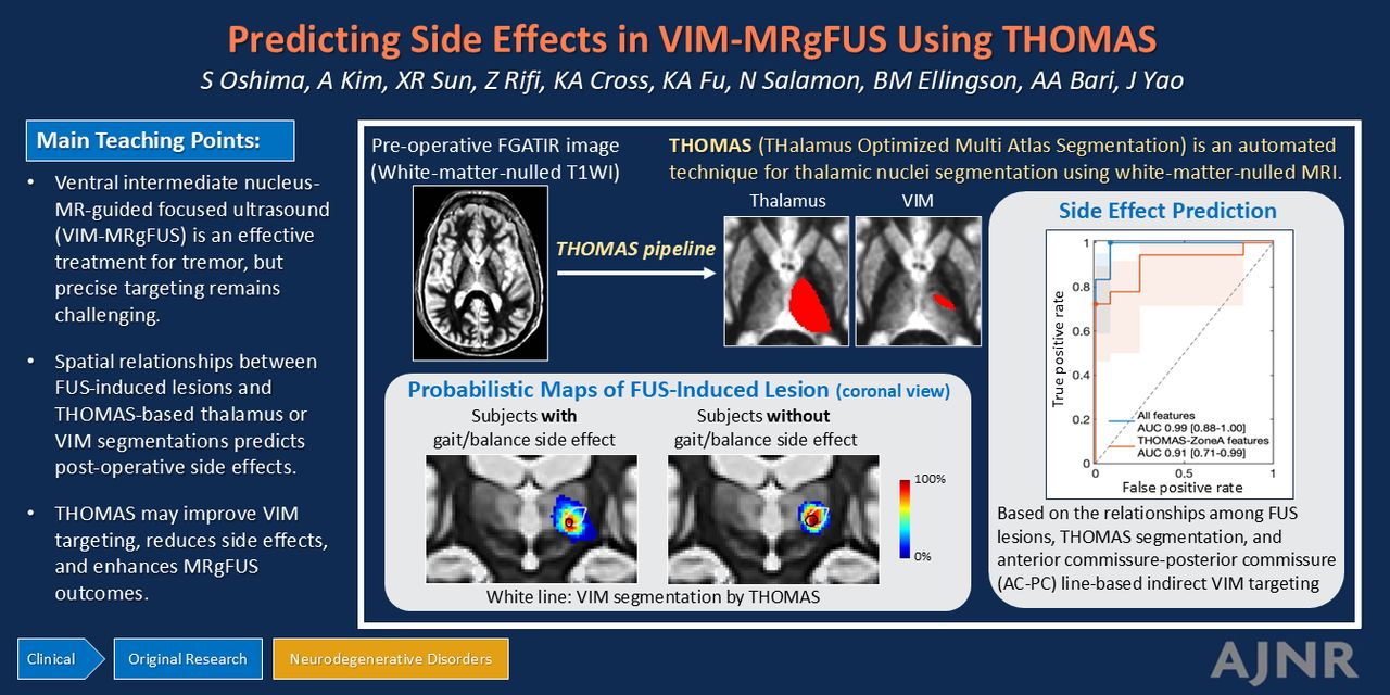
BACKGROUND AND PURPOSE: Precise and individualized targeting of the ventral intermediate thalamic nucleus for the MR-guided focused ultrasound is crucial for enhancing treatment efficacy and avoiding undesirable side effects. In this study, we tested the hypothesis that the spatial relationships between Thalamus Optimized Multi Atlas Segmentation derived segmentations and the post-focused ultrasound lesion can predict post-operative side effects in patients treated with MR-guided focused ultrasound.

MATERIALS AND METHODS: We retrospectively analyzed 30 patients (essential tremor, n = 26; tremor-dominant Parkinson’s disease, n = 4) who underwent unilateral ventral intermediate thalamic nucleus focused ultrasound treatment. We created ROIs of coordinate-based indirect treatment target, focused ultrasound-induced lesion, and thalamus and ventral intermediate thalamic nucleus segmentations. We extracted imaging features including 1) focused ultrasound-induced lesion volumes, 2) overlap between lesions and thalamus and ventral intermediate thalamic nucleus segmentations, 3) distance between lesions and ventral intermediate thalamic nucleus segmentation and 4) distance between lesions and the indirect standard target. These imaging features were compared between patients with and without post-operative gait/balance side effects using Wilcoxon rank-sum test. Multivariate prediction models of side effects based on the imaging features were evaluated using the receiver operating characteristic analyses.

RESULTS: Patients with self-reported gait/balance side effects had a significantly larger extent of focused ultrasound-induced edema, a smaller fraction of the lesion within the ventral intermediate thalamic nucleus segmentation, a larger fraction of the off-target lesion outside the thalamus segmentation, a more inferior centroid of the lesion from the ventral intermediate thalamic nucleus segmentation, and a larger distance between the centroid of the lesion and ventral intermediate thalamic nucleus segmentation (p < 0.05). Similar results were found for exam-based side effects. Multivariate regression models based on the imaging features achieved areas under the curve of 0.99 (95% CI: 0.88 to 1.00) and 0.96 (95% CI: 0.73 to 1.00) for predicting self-reported and exam-based side effects, respectively.

CONCLUSIONS: Thalamus Optimized Multi Atlas Segmentation-based patient-specific segmentation of the ventral intermediate thalamic nucleus can predict post-operative side effects, which has implications for aiding the direct targeting of MR-guided focused ultrasound and reducing side effects.

ABBREVIATIONS:

AC-PC
anterior commissure-posterior commissure
AUC
areas under the curve
ET
essential tremor
FGATIR
Fast Gray Matter Acquisition T1 Inversion Recovery
FTM
Fahn-Tolosa-Marín Clinical Rating Scale for Tremor
MRgFUS
MR-guided focused ultrasound
PD
Parkinson’s disease
THOMAS
THalamus Optimized Multi Atlas Segmentation
TH
thalamus
VIM
ventral intermediate nucleus
  • © 2025 by American Journal of Neuroradiology
View Full Text

Log in using your username and password

Forgot your user name or password?

Log in through your institution

You may be able to gain access using your login credentials for your institution. Contact your library if you do not have a username and password.
PreviousNext
Back to top

In this issue

American Journal of Neuroradiology: 46 (2)
American Journal of Neuroradiology
Vol. 46, Issue 2
1 Feb 2025
  • Table of Contents
  • Index by author
  • Complete Issue (PDF)
Advertisement
Print
Download PDF
Email Article

Thank you for your interest in spreading the word on American Journal of Neuroradiology.

NOTE: We only request your email address so that the person you are recommending the page to knows that you wanted them to see it, and that it is not junk mail. We do not capture any email address.

Enter multiple addresses on separate lines or separate them with commas.
Predicting Post-Operative Side Effects in VIM MRgFUS Based on THalamus Optimized Multi Atlas Segmentation (THOMAS) on White-Matter-Nulled MRI: A Retrospective Study
(Your Name) has sent you a message from American Journal of Neuroradiology
(Your Name) thought you would like to see the American Journal of Neuroradiology web site.
CAPTCHA
This question is for testing whether or not you are a human visitor and to prevent automated spam submissions.
Cite this article
Sonoko Oshima, Asher Kim, Xiaonan R. Sun, Ziad Rifi, Katy A. Cross, Katherine A. Fu, Noriko Salamon, Benjamin M. Ellingson, Ausaf A. Bari, Jingwen Yao
Predicting Post-Operative Side Effects in VIM MRgFUS Based on THalamus Optimized Multi Atlas Segmentation (THOMAS) on White-Matter-Nulled MRI: A Retrospective Study
American Journal of Neuroradiology Feb 2025, 46 (2) 330-340; DOI: 10.3174/ajnr.A8448

Citation Manager Formats

  • BibTeX
  • Bookends
  • EasyBib
  • EndNote (tagged)
  • EndNote 8 (xml)
  • Medlars
  • Mendeley
  • Papers
  • RefWorks Tagged
  • Ref Manager
  • RIS
  • Zotero
0 Responses
Respond to this article
Share
Bookmark this article
Predicting VIM MRgFUS Outcomes Using THOMAS
Sonoko Oshima, Asher Kim, Xiaonan R. Sun, Ziad Rifi, Katy A. Cross, Katherine A. Fu, Noriko Salamon, Benjamin M. Ellingson, Ausaf A. Bari, Jingwen Yao
American Journal of Neuroradiology Feb 2025, 46 (2) 330-340; DOI: 10.3174/ajnr.A8448
del.icio.us logo Twitter logo Facebook logo Mendeley logo
  • Tweet Widget
  • Facebook Like
  • Google Plus One
Purchase

Jump to section

  • Article
    • Graphical Abstract
    • Abstract
    • ABBREVIATIONS:
    • MATERIALS AND METHODS
    • RESULTS
    • DISCUSSION
    • CONCLUSIONS
    • ACKNOWLEDGMENTS
    • Footnotes
    • REFERENCES
  • Figures & Data
  • Supplemental
  • Info & Metrics
  • Responses
  • References
  • PDF

Related Articles

  • PubMed
  • Google Scholar

Cited By...

  • No citing articles found.
  • Crossref
  • Google Scholar

This article has not yet been cited by articles in journals that are participating in Crossref Cited-by Linking.

More in this TOC Section

  • ICP Sign: New Imaging Marker for MSA-C vs SCA
  • Automatic quantification and classification in IPD
  • Vascular risk factors, CMB and WMH in cognition
Show more Neurodegenerative Disorder Imaging

Similar Articles

Advertisement

Indexed Content

  • Current Issue
  • Accepted Manuscripts
  • Article Preview
  • Past Issues
  • Editorials
  • Editor's Choice
  • Fellows' Journal Club
  • Letters to the Editor
  • Video Articles

Cases

  • Case Collection
  • Archive - Case of the Week
  • Archive - Case of the Month
  • Archive - Classic Case

More from AJNR

  • Trainee Corner
  • Imaging Protocols
  • MRI Safety Corner

Multimedia

  • AJNR Podcasts
  • AJNR Scantastics

Resources

  • Turnaround Time
  • Submit a Manuscript
  • Submit a Video Article
  • Submit an eLetter to the Editor/Response
  • Manuscript Submission Guidelines
  • Statistical Tips
  • Fast Publishing of Accepted Manuscripts
  • Graphical Abstract Preparation
  • Imaging Protocol Submission
  • Evidence-Based Medicine Level Guide
  • Publishing Checklists
  • Author Policies
  • Become a Reviewer/Academy of Reviewers
  • News and Updates

About Us

  • About AJNR
  • Editorial Board
  • Editorial Board Alumni
  • Alerts
  • Permissions
  • Not an AJNR Subscriber? Join Now
  • Advertise with Us
  • Librarian Resources
  • Feedback
  • Terms and Conditions
  • AJNR Editorial Board Alumni

American Society of Neuroradiology

  • Not an ASNR Member? Join Now

© 2025 by the American Society of Neuroradiology All rights, including for text and data mining, AI training, and similar technologies, are reserved.
Print ISSN: 0195-6108 Online ISSN: 1936-959X

Powered by HighWire