Advertisement

Main menu

  • Home
  • Content
    • Current Issue
    • Accepted Manuscripts
    • Article Preview
    • Past Issue Archive
    • Video Articles
    • AJNR Case Collection
    • Case of the Week Archive
    • Case of the Month Archive
    • Classic Case Archive
  • Special Collections
    • AJNR Awards
    • Low-Field MRI
    • Alzheimer Disease
    • ASNR Foundation Special Collection
    • Photon-Counting CT
    • View All
  • Multimedia
    • AJNR Podcasts
    • AJNR SCANtastic
    • Trainee Corner
    • MRI Safety Corner
    • Imaging Protocols
  • For Authors
    • Submit a Manuscript
    • Submit a Video Article
    • Submit an eLetter to the Editor/Response
    • Manuscript Submission Guidelines
    • Statistical Tips
    • Fast Publishing of Accepted Manuscripts
    • Graphical Abstract Preparation
    • Imaging Protocol Submission
    • Author Policies
  • About Us
    • About AJNR
    • Editorial Board
    • Editorial Board Alumni
  • More
    • Become a Reviewer/Academy of Reviewers
    • Subscribers
    • Permissions
    • Alerts
    • Feedback
    • Advertisers
    • ASNR Home

User menu

  • Alerts
  • Log in

Search

  • Advanced search
American Journal of Neuroradiology

American Journal of Neuroradiology

ASHNR American Society of Functional Neuroradiology ASHNR American Society of Pediatric Neuroradiology ASSR
  • Alerts
  • Log in

Advanced Search

  • Home
  • Content
    • Current Issue
    • Accepted Manuscripts
    • Article Preview
    • Past Issue Archive
    • Video Articles
    • AJNR Case Collection
    • Case of the Week Archive
    • Case of the Month Archive
    • Classic Case Archive
  • Special Collections
    • AJNR Awards
    • Low-Field MRI
    • Alzheimer Disease
    • ASNR Foundation Special Collection
    • Photon-Counting CT
    • View All
  • Multimedia
    • AJNR Podcasts
    • AJNR SCANtastic
    • Trainee Corner
    • MRI Safety Corner
    • Imaging Protocols
  • For Authors
    • Submit a Manuscript
    • Submit a Video Article
    • Submit an eLetter to the Editor/Response
    • Manuscript Submission Guidelines
    • Statistical Tips
    • Fast Publishing of Accepted Manuscripts
    • Graphical Abstract Preparation
    • Imaging Protocol Submission
    • Author Policies
  • About Us
    • About AJNR
    • Editorial Board
    • Editorial Board Alumni
  • More
    • Become a Reviewer/Academy of Reviewers
    • Subscribers
    • Permissions
    • Alerts
    • Feedback
    • Advertisers
    • ASNR Home
  • Follow AJNR on Twitter
  • Visit AJNR on Facebook
  • Follow AJNR on Instagram
  • Join AJNR on LinkedIn
  • RSS Feeds

AJNR Awards, New Junior Editors, and more. Read the latest AJNR updates

Research ArticleBrain Tumor Imaging
Open Access

IDH Status in Brain Gliomas Can Be Predicted by the Spherical Mean MRI Technique

Vojtěch Sedlák, Milan Němý, Martin Májovský, Adéla Bubeníková, Love Engstrom Nordin, Tomáš Moravec, Jana Engelová, Dalibor Sila, Dora Konečná, Tomáš Belšan, Eric Westman and David Netuka
American Journal of Neuroradiology January 2025, 46 (1) 121-128; DOI: https://doi.org/10.3174/ajnr.A8432
Vojtěch Sedlák
aFrom the Department of Radiology (V.S., T.B.), Military University Hospital, Prague, Czech Republic
  • Find this author on Google Scholar
  • Find this author on PubMed
  • Search for this author on this site
Milan Němý
bDivision of Clinical Geriatrics (M.N., L.E.N., E.W.), Department of Neurobiology, Care Sciences and Society, Center for Alzheimer Research, Karolinska Institute, Stockholm, Sweden
cDepartment of Biomedical Engineering and Assistive Technology (M.N.), Czech Institute of Informatics, Robotics and Cybernetics, Czech Technical University in Prague, Prague, Czech Republic
  • Find this author on Google Scholar
  • Find this author on PubMed
  • Search for this author on this site
  • ORCID record for Milan Němý
Martin Májovský
dDepartment of Neurosurgery and Neurooncology (M.M., A.B., T.M., D.K., D.N.), First Faculty of Medicine, Charles University and Military University Hospital, Prague, Czech Republic
  • Find this author on Google Scholar
  • Find this author on PubMed
  • Search for this author on this site
  • ORCID record for Martin Májovský
Adéla Bubeníková
dDepartment of Neurosurgery and Neurooncology (M.M., A.B., T.M., D.K., D.N.), First Faculty of Medicine, Charles University and Military University Hospital, Prague, Czech Republic
  • Find this author on Google Scholar
  • Find this author on PubMed
  • Search for this author on this site
Love Engstrom Nordin
bDivision of Clinical Geriatrics (M.N., L.E.N., E.W.), Department of Neurobiology, Care Sciences and Society, Center for Alzheimer Research, Karolinska Institute, Stockholm, Sweden
eDepartment of Diagnostic Medical Physics (L.E.N.), Karolinska University Hospital Solna, Stockholm, Sweden
  • Find this author on Google Scholar
  • Find this author on PubMed
  • Search for this author on this site
  • ORCID record for Love Engstrom Nordin
Tomáš Moravec
dDepartment of Neurosurgery and Neurooncology (M.M., A.B., T.M., D.K., D.N.), First Faculty of Medicine, Charles University and Military University Hospital, Prague, Czech Republic
  • Find this author on Google Scholar
  • Find this author on PubMed
  • Search for this author on this site
Jana Engelová
fRadiodiagnostic Department (J.E.), Proton Therapy Center Czech Ltd, Prague, Czech Republic
  • Find this author on Google Scholar
  • Find this author on PubMed
  • Search for this author on this site
Dalibor Sila
gDepartment of Neurosurgery and Spine Surgery (D.S.), Arberlandklinik Viechtach, Germany
  • Find this author on Google Scholar
  • Find this author on PubMed
  • Search for this author on this site
Dora Konečná
dDepartment of Neurosurgery and Neurooncology (M.M., A.B., T.M., D.K., D.N.), First Faculty of Medicine, Charles University and Military University Hospital, Prague, Czech Republic
  • Find this author on Google Scholar
  • Find this author on PubMed
  • Search for this author on this site
Tomáš Belšan
aFrom the Department of Radiology (V.S., T.B.), Military University Hospital, Prague, Czech Republic
  • Find this author on Google Scholar
  • Find this author on PubMed
  • Search for this author on this site
Eric Westman
bDivision of Clinical Geriatrics (M.N., L.E.N., E.W.), Department of Neurobiology, Care Sciences and Society, Center for Alzheimer Research, Karolinska Institute, Stockholm, Sweden
hDepartment of Neuroimaging (E.W.), Centre for Neuroimaging Science, Institute of Psychiatry, Psychology, and Neuroscience, King’s College London, London, UK
  • Find this author on Google Scholar
  • Find this author on PubMed
  • Search for this author on this site
  • ORCID record for Eric Westman
David Netuka
dDepartment of Neurosurgery and Neurooncology (M.M., A.B., T.M., D.K., D.N.), First Faculty of Medicine, Charles University and Military University Hospital, Prague, Czech Republic
  • Find this author on Google Scholar
  • Find this author on PubMed
  • Search for this author on this site
  • Article
  • Figures & Data
  • Info & Metrics
  • Responses
  • References
  • PDF
Loading

Article Figures & Data

Figures

  • Tables
  • Figure
    • Download figure
    • Open in new tab
    • Download powerpoint
  • FIG 1.
    • Download figure
    • Open in new tab
    • Download powerpoint
    FIG 1.

    Diffusion data-processing pipeline for glioma assessment. Initial multishell DWI (A) is followed by Marchenko-Pastur principal component analysis (MP-PCA) denoising (B) to reduce noise. Subsequent steps include Gibbs ringing artifact removal (C), susceptibility-induced distortion (SID) correction (D), and N4 bias field correction (E) to improve image quality. Structural imaging (F) is used for tumor segmentation (G), which is then coregistered (H) with the diffusion data. Modeling of the diffusion data (J) enables extraction of tumor parameters (K), such as ADC, MK, and μFA. The I letter was intentionally omitted to improve readability.
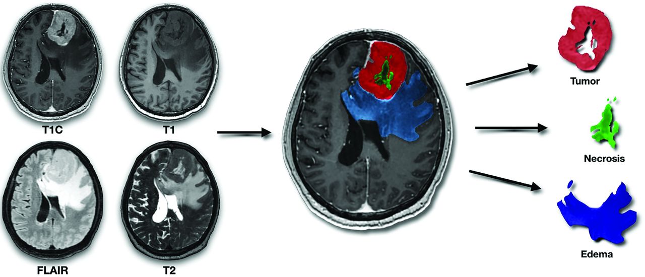
  • FIG 2.
    • Download figure
    • Open in new tab
    • Download powerpoint
    FIG 2.

    Illustration of tumor segmentation and generation of tumoral masks. The figure demonstrates the segmentation of gliomas using 4 image sequences (from left to right and top to bottom: contrast-enhanced T1-weighted, T1-weighted, FLAIR, and T2-weighted). These masks are subsequently coregistered with diffusion data sets.
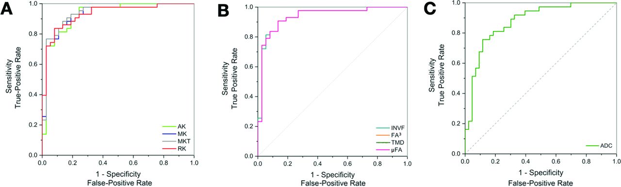
  • FIG 3.
    • Download figure
    • Open in new tab
    • Download powerpoint
    FIG 3.

    ROC curves for selected parameters illustrating the diagnostic accuracy in determining IDH status. The curves compare DKI (A), SMT parameters (B), and the standard clinically used ADC method (C), with an emphasis on parameters with AUC values of >0.90.

  • FIG 4.
    • Download figure
    • Open in new tab
    • Download powerpoint
    FIG 4.

    ROC curves for selected parameters illustrating the diagnostic accuracy in determining glioma grade.The curves compare DKI (A), and SMT (B), parameters with the standard clinically used ADC method (C), with an emphasis on parameters with AUC values of 0.90.

Tables

  • Figures
    • View popup
    Table 1:

    Patient characteristicsa

    CharacteristicResult
    No. of patients80
    Age, mean (yr)48 (SD, 16)
    HGG43 (54)
    IDHmt46 (58)
    Male sex49 (61)
    Postcontrast enhancement46 (58)
    Necrosis31 (39)
    T2 FLAIR mismatch6 (8)
    Hemorrhage24 (30)
    • Note:—HGG indicates high-grade glioma; IDHmt, IDH-mutant.

    • ↵a Unless otherwise noted, data represent the number of patients; data in parentheses are percentages.

    • View popup
    Table 2:

    Comparative diagnostic accuracy of investigated parameters differentiating IDH-mutant from IDH-wild-type gliomasa

    Models and ParametersAUC95% CIOptimal CutoffSensitivity (%)Specificity (%)P Value
    Nondiffusion
     Enhancementb0.770.66–0.880.5 ↓88.265.2<.001
     Necrosisb0.850.77–0.940.5 ↓79.491.3<.001
     T2LFMb0.570.44–0.700.5 ↑13.0100.03
     Hemorrhageb0.780.67–0.880.5 ↓61.893.5<.001
    ADC
     ADCc0.820.73–0.921.206 ↑67.491.2<.001
    DTI
     FA0.590.45–0.730.174 ↓50.082.6.050
     MDc0.830.75–0.920.945 ↑89.164.7<.001
    DKI
     AK0.900.83–0.970.583 ↓88.284.8<.001
     RK0.900.83–0.970.593 ↓94.178.3<.001
     MK0.900.83–0.970.567 ↓94.178.3<.001
     KFA0.550.42–0.680.807 ↑63.284.2.73
     MKT0.910.84–0.980.619 ↓88.284.8<.001
    SMT
     LMDc0.620.48–0.752.967 ↑73.958.8.009
     TMDc0.870.79–0.950.528 ↑78.391.2<.001
     µFA0.910.84–0.980.535 ↓91.284.8<.001
     µFA30.910.84–0.980.153 ↓91.284.8<.001
     MMDc0.850.77–0.941.369 ↑67.491.2<.001
     INVF0.910.84–0.980.339 ↓91.284.8<.001
     IDc0.720.61–0.832.006 ↑69.670.6<.001
     ETMDc0.770.66–0.871.301 ↑76.167.6<.001
     EMMDc0.650.53–0.781.502 ↑67.467.6.01
    • Note:—T2LFM indicates T2-FLAIR mismatch; FA, fractional anisotropy; MD, mean diffusivity; KFA, kurtosis fractional anisotropy; LMD, longitudinal microscopic diffusivity; µFA3, microscopic fractional anisotropy to the third power; MMD, microscopic mean diffusivity; ID, intrinsic diffusivity, ETMD, extra-neurite transverse microscopic diffusivity; EMMD, extra-neurite microscopic mean diffusivity.

    • ↵a Optimal cutoff levels to predict IDH type were assessed by the Youden index. Cutoffs were evaluated by sensitivity and specificity. An upward arrow (↑) indicates a positive correlation, in which values above the cutoff point predict an IDH-mutant glioma, whereas a downward arrow (↓) indicates a negative correlation, in which values below the cutoff point predict an IDH-mutant glioma. P values were computed by comparing the AUC against chance performance.

    • ↵b Binary variable, indicating the presence or absence of the feature.

    • ↵c Units in mm2/s × 10−3.

    • View popup
    Table 3:

    Comparative diagnostic accuracy of investigated parameters differentiating glioma gradesa

    Models and ParametersAUC95% CIOptimal CutoffSensitivity (%)Specificity (%)P Value
    Nondiffusion
     Enhancementb0.760.65–0.870.5 ↑81.470.2<.001
     Necrosisb0.810.71–0.910.5 ↑67.494.6<.001
     T2LFMb0.580.46–0.710.5 ↓16.2100.006
     Hemorrhageb0.730.61–0.840.5 ↑51.294.6<.001
    ADC
     ADCc0.880.80–0.961.217 ↓75.788.4<.001
    DTI
     FA0.640.52–0.770.174 ↑46.586.5.01
     MDc0.880.80–0.951.134 ↓83.876.7<.001
    DKI
     AK0.930.87–1.000.494 ↑97.775.7<.001
     RK0.930.88–1.000.627 ↑83.791.9<.001
     MK0.930.88–1.000.539 ↑93.081.1<.001
     KFA0.530.40–0.670.219 ↓78.444.2.85
     MKT0.940.88–0.990.564 ↑88.486.5<.001
    SMT
     LMDc0.660.53–0.792.967 ↓83.855.8<.001
     TMDc0.910.84–0.972.958 ↓91.979.1<.001
     µFA0.940.88–0.990.509 ↑90.786.5<.001
     µFA30.940.88–0.990.132 ↑90.786.5<.001
     MMDc0.900.83–0.971.311 ↓89.276.7<.001
     INVF0.940.88–0.990.319 ↑90.786.5<.001
     IDc0.770.67–0.881.946 ↓83.865.1<.001
     ETMDc0.820.72–0.911.208 ↓94.660.5<.001
     EMMDc0.720.60–0.841.502 ↓75.767.4<.001
    • Note:—T2LFM indicates T2 FLAIR mismatch; FA, fractional anisotropy; MD, mean diffusivity; KFA, kurtosis fractional anisotropy; LMD, longitudinal microscopic diffusivity; µFA3, microscopic fractional anisotropy to the third power; MMD, microscopic mean diffusivity; ID, intrinsic diffusivity; ETMD, extraneurite transverse microscopic diffusivity; EMMD, extraneurite microscopic mean diffusivity.

    • ↵a Optimal cutoff levels to predict glioma grade (low-grade versus high-grade) were assessed by the Youden index. Cutoffs were evaluated by sensitivity and specificity. An upward arrow (↑) indicates a positive correlation, in which values above the cutoff point predict a high-grade glioma, whereas a downward arrow (↓) indicates a negative correlation, in which values below the cutoff point predict a high-grade glioma. P values were computed by comparing the AUC against chance performance.

    • ↵b Binary variable, indicating the presence or absence of the feature.

    • ↵c Units mm2/s × 10−3.

PreviousNext
Back to top

In this issue

American Journal of Neuroradiology: 46 (1)
American Journal of Neuroradiology
Vol. 46, Issue 1
1 Jan 2025
  • Table of Contents
  • Index by author
  • Complete Issue (PDF)
Advertisement
Print
Download PDF
Email Article

Thank you for your interest in spreading the word on American Journal of Neuroradiology.

NOTE: We only request your email address so that the person you are recommending the page to knows that you wanted them to see it, and that it is not junk mail. We do not capture any email address.

Enter multiple addresses on separate lines or separate them with commas.
IDH Status in Brain Gliomas Can Be Predicted by the Spherical Mean MRI Technique
(Your Name) has sent you a message from American Journal of Neuroradiology
(Your Name) thought you would like to see the American Journal of Neuroradiology web site.
CAPTCHA
This question is for testing whether or not you are a human visitor and to prevent automated spam submissions.
Cite this article
Vojtěch Sedlák, Milan Němý, Martin Májovský, Adéla Bubeníková, Love Engstrom Nordin, Tomáš Moravec, Jana Engelová, Dalibor Sila, Dora Konečná, Tomáš Belšan, Eric Westman, David Netuka
IDH Status in Brain Gliomas Can Be Predicted by the Spherical Mean MRI Technique
American Journal of Neuroradiology Jan 2025, 46 (1) 121-128; DOI: 10.3174/ajnr.A8432

Citation Manager Formats

  • BibTeX
  • Bookends
  • EasyBib
  • EndNote (tagged)
  • EndNote 8 (xml)
  • Medlars
  • Mendeley
  • Papers
  • RefWorks Tagged
  • Ref Manager
  • RIS
  • Zotero
0 Responses
Respond to this article
Share
Bookmark this article
Predicting IDH Status in Brain Gliomas with MRI
Vojtěch Sedlák, Milan Němý, Martin Májovský, Adéla Bubeníková, Love Engstrom Nordin, Tomáš Moravec, Jana Engelová, Dalibor Sila, Dora Konečná, Tomáš Belšan, Eric Westman, David Netuka
American Journal of Neuroradiology Jan 2025, 46 (1) 121-128; DOI: 10.3174/ajnr.A8432
del.icio.us logo Twitter logo Facebook logo Mendeley logo
  • Tweet Widget
  • Facebook Like
  • Google Plus One
Purchase

Jump to section

  • Article
    • Graphical Abstract
    • Abstract
    • ABBREVIATIONS:
    • MATERIALS AND METHODS
    • RESULTS
    • DISCUSSION
    • Conclusions
    • Footnotes
    • References
  • Figures & Data
  • Info & Metrics
  • Responses
  • References
  • PDF

Related Articles

  • ErratumErratum
  • PubMed
  • Google Scholar

Cited By...

  • No citing articles found.
  • Crossref
  • Google Scholar

This article has not yet been cited by articles in journals that are participating in Crossref Cited-by Linking.

More in this TOC Section

  • Cognitive Control in Tumor Language Plasticity
  • Temporal Evolution of Vestibular schwannoma
  • CE MRI for Brain Metastasis Detection
Show more Brain Tumor Imaging

Similar Articles

Advertisement

Indexed Content

  • Current Issue
  • Accepted Manuscripts
  • Article Preview
  • Past Issues
  • Editorials
  • Editor's Choice
  • Fellows' Journal Club
  • Letters to the Editor
  • Video Articles

Cases

  • Case Collection
  • Archive - Case of the Week
  • Archive - Case of the Month
  • Archive - Classic Case

More from AJNR

  • Trainee Corner
  • Imaging Protocols
  • MRI Safety Corner

Multimedia

  • AJNR Podcasts
  • AJNR Scantastics

Resources

  • Turnaround Time
  • Submit a Manuscript
  • Submit a Video Article
  • Submit an eLetter to the Editor/Response
  • Manuscript Submission Guidelines
  • Statistical Tips
  • Fast Publishing of Accepted Manuscripts
  • Graphical Abstract Preparation
  • Imaging Protocol Submission
  • Evidence-Based Medicine Level Guide
  • Publishing Checklists
  • Author Policies
  • Become a Reviewer/Academy of Reviewers
  • News and Updates

About Us

  • About AJNR
  • Editorial Board
  • Editorial Board Alumni
  • Alerts
  • Permissions
  • Not an AJNR Subscriber? Join Now
  • Advertise with Us
  • Librarian Resources
  • Feedback
  • Terms and Conditions
  • AJNR Editorial Board Alumni

American Society of Neuroradiology

  • Not an ASNR Member? Join Now

© 2025 by the American Society of Neuroradiology All rights, including for text and data mining, AI training, and similar technologies, are reserved.
Print ISSN: 0195-6108 Online ISSN: 1936-959X

Powered by HighWire