Advertisement

Main menu

  • Home
  • Content
    • Current Issue
    • Accepted Manuscripts
    • Article Preview
    • Past Issue Archive
    • Video Articles
    • AJNR Case Collection
    • Case of the Week Archive
    • Case of the Month Archive
    • Classic Case Archive
  • Special Collections
    • AJNR Awards
    • Low-Field MRI
    • Alzheimer Disease
    • ASNR Foundation Special Collection
    • Photon-Counting CT
    • View All
  • Multimedia
    • AJNR Podcasts
    • AJNR SCANtastic
    • Trainee Corner
    • MRI Safety Corner
    • Imaging Protocols
  • For Authors
    • Submit a Manuscript
    • Submit a Video Article
    • Submit an eLetter to the Editor/Response
    • Manuscript Submission Guidelines
    • Statistical Tips
    • Fast Publishing of Accepted Manuscripts
    • Graphical Abstract Preparation
    • Imaging Protocol Submission
    • Author Policies
  • About Us
    • About AJNR
    • Editorial Board
    • Editorial Board Alumni
  • More
    • Become a Reviewer/Academy of Reviewers
    • Subscribers
    • Permissions
    • Alerts
    • Feedback
    • Advertisers
    • ASNR Home

User menu

  • Alerts
  • Log in

Search

  • Advanced search
American Journal of Neuroradiology

American Journal of Neuroradiology

ASHNR American Society of Functional Neuroradiology ASHNR American Society of Pediatric Neuroradiology ASSR
  • Alerts
  • Log in

Advanced Search

  • Home
  • Content
    • Current Issue
    • Accepted Manuscripts
    • Article Preview
    • Past Issue Archive
    • Video Articles
    • AJNR Case Collection
    • Case of the Week Archive
    • Case of the Month Archive
    • Classic Case Archive
  • Special Collections
    • AJNR Awards
    • Low-Field MRI
    • Alzheimer Disease
    • ASNR Foundation Special Collection
    • Photon-Counting CT
    • View All
  • Multimedia
    • AJNR Podcasts
    • AJNR SCANtastic
    • Trainee Corner
    • MRI Safety Corner
    • Imaging Protocols
  • For Authors
    • Submit a Manuscript
    • Submit a Video Article
    • Submit an eLetter to the Editor/Response
    • Manuscript Submission Guidelines
    • Statistical Tips
    • Fast Publishing of Accepted Manuscripts
    • Graphical Abstract Preparation
    • Imaging Protocol Submission
    • Author Policies
  • About Us
    • About AJNR
    • Editorial Board
    • Editorial Board Alumni
  • More
    • Become a Reviewer/Academy of Reviewers
    • Subscribers
    • Permissions
    • Alerts
    • Feedback
    • Advertisers
    • ASNR Home
  • Follow AJNR on Twitter
  • Visit AJNR on Facebook
  • Follow AJNR on Instagram
  • Join AJNR on LinkedIn
  • RSS Feeds

AJNR Awards, New Junior Editors, and more. Read the latest AJNR updates

OtherSpine Imaging and Spine image–Guided Interventions
Open Access

Perspectives from the Inaugural “Spinal CSF Leak: Bridging the Gap” Conference: A Convergence of Clinical and Patient Expertise

Andrew L. Callen, Samantha L. Pisani Petrucci, Peter Lennarson, Marius Birlea, Jennifer MacKenzie, Andrea J. Buchanan and the “Spinal CSF Leak: Bridging the Gap” Study Group
American Journal of Neuroradiology July 2024, 45 (7) 841-849; DOI: https://doi.org/10.3174/ajnr.A8181
Andrew L. Callen
aFrom the Department of Radiology (A.L.C., S.P.P.), Neuroradiology Section, University of Colorado Anschutz Medical Campus, Aurora, Colorado
  • Find this author on Google Scholar
  • Find this author on PubMed
  • Search for this author on this site
  • ORCID record for Andrew L. Callen
Samantha L. Pisani Petrucci
aFrom the Department of Radiology (A.L.C., S.P.P.), Neuroradiology Section, University of Colorado Anschutz Medical Campus, Aurora, Colorado
  • Find this author on Google Scholar
  • Find this author on PubMed
  • Search for this author on this site
Peter Lennarson
bDepartment of Neurosurgery (P.L.), University of Colorado Anschutz Medical Campus, Aurora, Colorado
  • Find this author on Google Scholar
  • Find this author on PubMed
  • Search for this author on this site
Marius Birlea
cDepartment of Neurology (M.B.), University of Colorado Anschutz Medical Campus, Aurora, Colorado
  • Find this author on Google Scholar
  • Find this author on PubMed
  • Search for this author on this site
  • ORCID record for Marius Birlea
Jennifer MacKenzie
dSpinal CSF Leak Foundation (J.M., A.B.), Spokane, Washington.
  • Find this author on Google Scholar
  • Find this author on PubMed
  • Search for this author on this site
  • ORCID record for Jennifer MacKenzie
Andrea J. Buchanan
dSpinal CSF Leak Foundation (J.M., A.B.), Spokane, Washington.
  • Find this author on Google Scholar
  • Find this author on PubMed
  • Search for this author on this site
  • ORCID record for Andrea J. Buchanan
  • Article
  • Figures & Data
  • Info & Metrics
  • Responses
  • References
  • PDF
Loading

Article Figures & Data

Figures

  • Tables
  • FIG 1.
    • Download figure
    • Open in new tab
    • Download powerpoint
    FIG 1.

    Normalization of MR imaging findings across time despite persistent spinal CSF leaks. A, Sagittal T1 noncontrast MR imaging in 2018 demonstrates sagging of posterior fossa structures (bracket), engorgement of the pituitary gland, and narrowed suprasellar distance (arrow). B, Axial FLAIR MR imaging in 2018 demonstrates a diffuse, thin subdural collection (arrows). Sagittal (C) and axial (D) T2 MR imaging of the cervical spine in 2018 shows a cervicothoracic ventral epidural fluid collection (arrows). The patient underwent a dorsal nontargeted epidural blood patch in 2018 with partial relief of symptoms. E, Sagittal T1 noncontrast MR imaging in 2023 demonstrates resolution of brain sag, pituitary engorgement, and narrowing of the suprasellar interval. F, Axial FLAIR MR imaging in 2023 with resolution of the subdural collection. Sagittal (G) and axial (H) T2 MR imaging of the cervical spine in 2023 shows persistence of the cervicothoracic ventral epidural fluid collection. I. Intraoperative photograph later in 2023, with a ventral dural defect identified at T2–T3 (arrows). After repair, the patient had substantial symptom improvement, with the Headache Impact Test score improving from 68 to 48 (Headache Impact Test: range, 36–78).
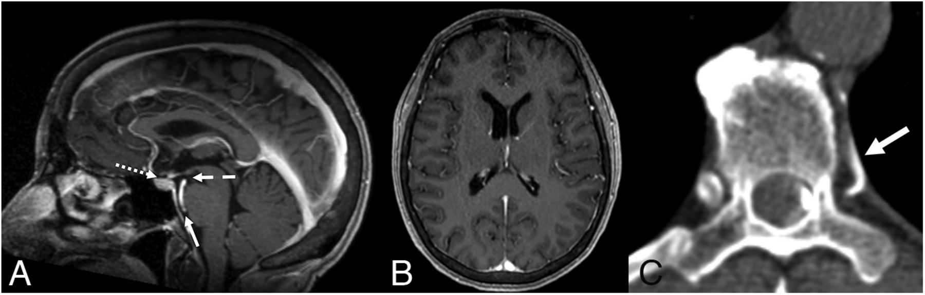
  • FIG 2.
    • Download figure
    • Open in new tab
    • Download powerpoint
    FIG 2.

    Brain MR imaging findings incorrectly reported as normal in a patient with spontaneous intracranial hypotension. A, Sagittal T1 postcontrast MR imaging demonstrates mild narrowing of the suprasellar (1 mm, dotted arrow), mamillopontine (4.8 mm, dashed arrow), and prepontine (3 mm, solid arrow) distances. B, No pachymeningeal thickening or subdural collection on axial T1 postcontrast MR imaging. C, Left-lateral decubitus dynamic CTM detected a CVF arising from the left T7–8 neural foramen (arrow).
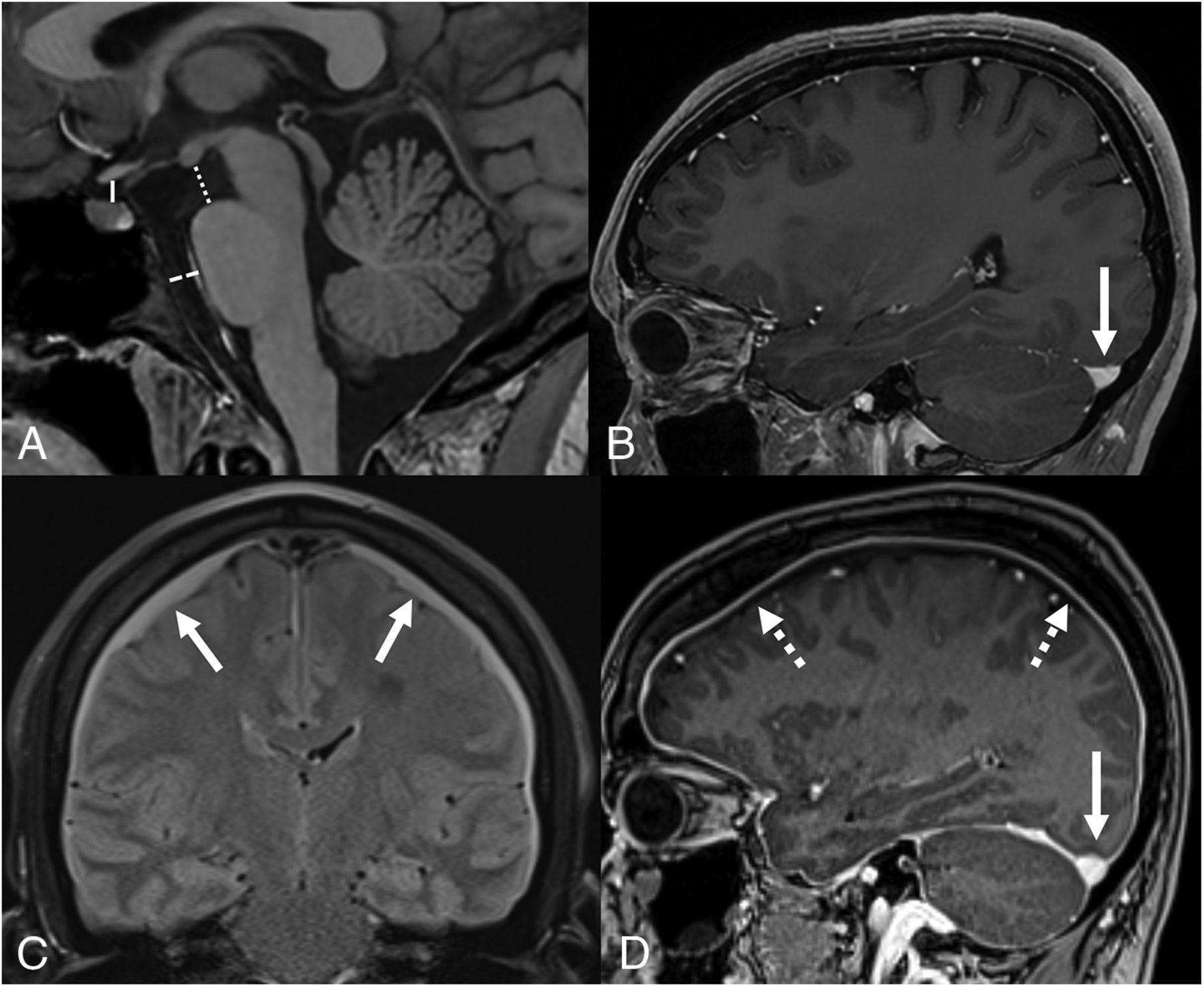
  • FIG 3.
    • Download figure
    • Open in new tab
    • Download powerpoint
    FIG 3.

    The Bern score. A, Sagittal T1 noncontrast MR imaging demonstrating the suprasellar interval (solid line, normal, >4 mm), mamillopontine interval (dotted line, normal, >6.5 mm), and prepontine interval (dashed line, normal, >5 mm). B, Sagittal T1 postcontrast MR imaging illustrates a normal flat appearance of the upward margin of the transverse sinus (arrow). C, Coronal FLAIR MR imaging with bilateral subdural fluid collections (arrows). D, Sagittal T1 postcontrast image demonstrates abnormal venous engorgement evidenced by an abnormal convex upward margin (solid arrow), as well as diffuse pachymeningeal enhancement (dotted arrows). The presence of a narrowed suprasellar interval, venous engorgement, or pachymeningeal enhancement are ascribed 2 points each, while a narrowed mamillopontine interval, prepontine interval, or the presence of subdural collections are ascribed 1 point each. A combined score of ≤2 equates to low probability, a score of 3 or 4 equates to moderate probability, and a score of ≥5 equates to high probability of localizing a CSF leak or venous fistula on subsequent myelography.20
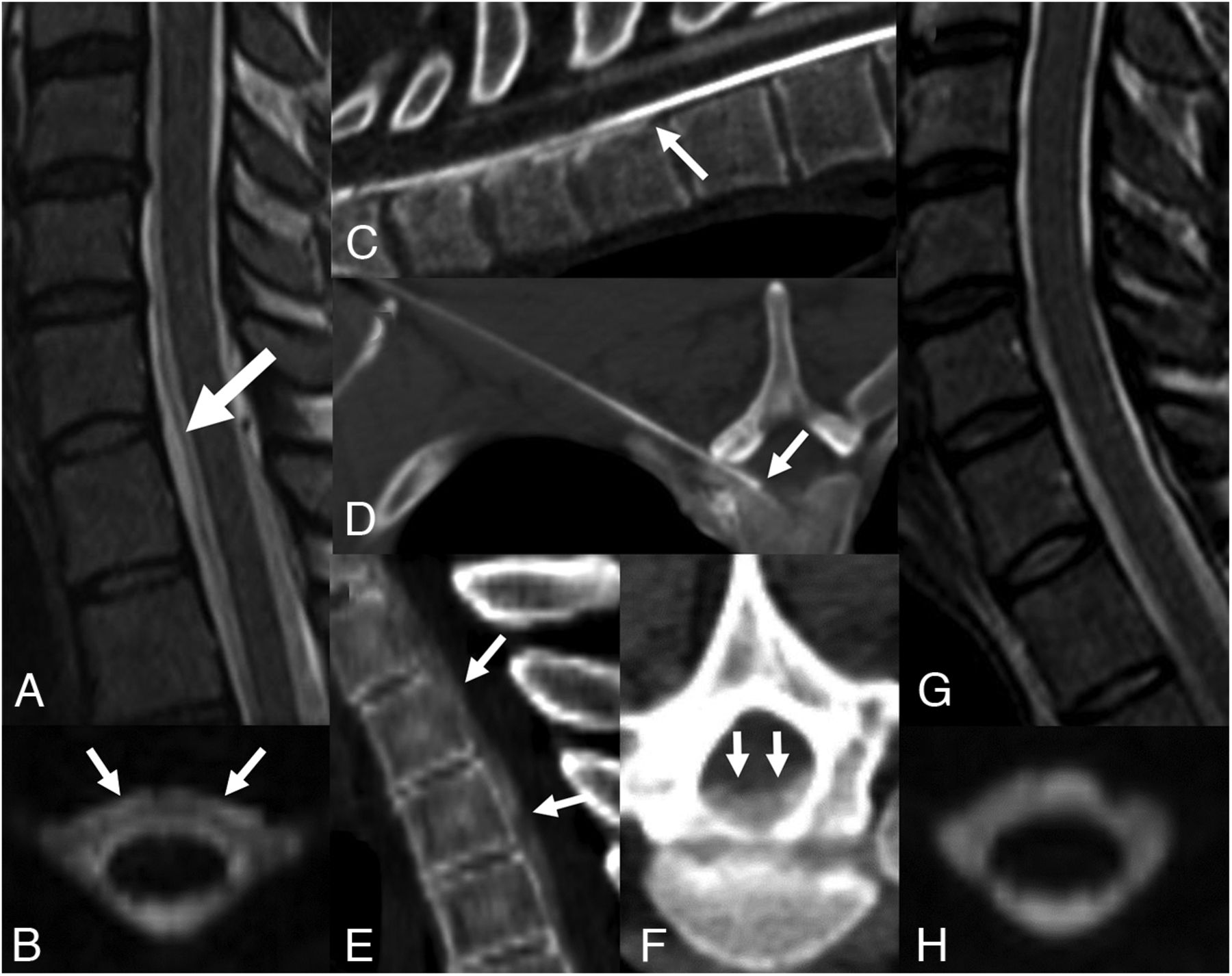
  • FIG 4.
    • Download figure
    • Open in new tab
    • Download powerpoint
    FIG 4.

    A 35-year-old man with spontaneous intracranial hypotension who underwent 2 prior nontargeted dorsal epidural blood patches. Sagittal (A) and axial (B) T2-weighted MR imaging sequences demonstrate a ventral epidural fluid collection (arrows). C, Prone dynamic CTM detected contrast extravasating from the subarachnoid into the ventral epidural space at T1-2, consistent with a ventral dural defect (arrow). D, Procedural image from a CT-guided epidural blood and fibrin patch using a 15-cm 22-ga spinal needle via a far-lateral transforaminal approach to target the ventral epidural space adjacent to the defect (arrow). Postinjection sagittal (E) and axial (F) images demonstrate spread of injected blood and fibrin glue along the ventral epidural space (arrows). Posttreatment sagittal T2 (G) and axial 3D T2 fat-saturated MR imaging (H) with resolution of the epidural fluid collection.
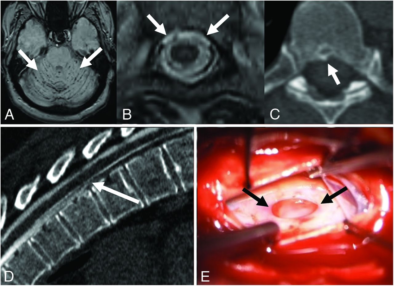
  • FIG 5.
    • Download figure
    • Open in new tab
    • Download powerpoint
    FIG 5.

    A 49-year-old man with a history of spontaneous intracranial hypotension, with persistent symptoms after 3 epidural blood patches. A, Axial SWI with hypointense signal along the cerebellar folia (arrows), consistent with superficial siderosis, a rare complication of chronic CSF leak. B, Axial T2-weighted MR imaging demonstrates a ventral epidural fluid collection (arrows). C, Axial noncontrast CT image shows a small osteophyte (arrows) along the ventral canal at T6–7. D, Prone dynamic CTM demonstrates extravasation of contrast from the subarachnoid space into the ventral epidural space at T6–7 (arrows), consistent with a ventral dural defect. E, Intraoperative photograph during repair of the ventral dural defect (arrows) at T6–7, accessed via hemilaminectomy, posterior durotomy, and lateral mobilization of the cord after dentate ligament resection .

Tables

  • Figures
  • Differential diagnoses in patients presenting with SCSFL

    Overlap with SCSFLKey Differences from SCSFL
    Chronic migraineHeadache as primary clinical presentation
    • Often evolves from less-frequent to more-frequent episodes

    • Less typically orthostatic

    • Often occur in the morning, rather than worsening throughout the day

    Postconcussion headacheBoth can be precipitated by trauma
    • Headache phenotype like migraine

    • Typically nonorthostatic

    • Associated cognitive issues

    Postural orthostatic tachycardia syndromeOrthostatic symptoms
    • Defined by changes in hemodynamic parameters on standing

    • If headache associated, more often migraine-like

    • Neck stiffness less common

    Chiari I malformationCerebellar tonsillar ectopia may be present in both conditions
    • Brain MR imaging should not demonstrate downward herniation of posterior fossa structures, pituitary/venous engorgement, or pachymeningeal enhancement

    • Orthostatic headache less common

PreviousNext
Back to top

In this issue

American Journal of Neuroradiology: 45 (7)
American Journal of Neuroradiology
Vol. 45, Issue 7
1 Jul 2024
  • Table of Contents
  • Index by author
  • Complete Issue (PDF)
Advertisement
Print
Download PDF
Email Article

Thank you for your interest in spreading the word on American Journal of Neuroradiology.

NOTE: We only request your email address so that the person you are recommending the page to knows that you wanted them to see it, and that it is not junk mail. We do not capture any email address.

Enter multiple addresses on separate lines or separate them with commas.
Perspectives from the Inaugural “Spinal CSF Leak: Bridging the Gap” Conference: A Convergence of Clinical and Patient Expertise
(Your Name) has sent you a message from American Journal of Neuroradiology
(Your Name) thought you would like to see the American Journal of Neuroradiology web site.
CAPTCHA
This question is for testing whether or not you are a human visitor and to prevent automated spam submissions.
Cite this article
Andrew L. Callen, Samantha L. Pisani Petrucci, Peter Lennarson, Marius Birlea, Jennifer MacKenzie, Andrea J. Buchanan, the “Spinal CSF Leak: Bridging the Gap” Study Group
Perspectives from the Inaugural “Spinal CSF Leak: Bridging the Gap” Conference: A Convergence of Clinical and Patient Expertise
American Journal of Neuroradiology Jul 2024, 45 (7) 841-849; DOI: 10.3174/ajnr.A8181

Citation Manager Formats

  • BibTeX
  • Bookends
  • EasyBib
  • EndNote (tagged)
  • EndNote 8 (xml)
  • Medlars
  • Mendeley
  • Papers
  • RefWorks Tagged
  • Ref Manager
  • RIS
  • Zotero
0 Responses
Respond to this article
Share
Bookmark this article
“Spinal CSF Leak: Bridging the Gap” Conference“Spinal CSF Leak: Bridging the Gap” Conference
Andrew L. Callen, Samantha L. Pisani Petrucci, Peter Lennarson, Marius Birlea, Jennifer MacKenzie, Andrea J. Buchanan, the “Spinal CSF Leak: Bridging the Gap” Study Group
American Journal of Neuroradiology Jul 2024, 45 (7) 841-849; DOI: 10.3174/ajnr.A8181
del.icio.us logo Twitter logo Facebook logo Mendeley logo
  • Tweet Widget
  • Facebook Like
  • Google Plus One
Purchase

Jump to section

  • Article
    • Abstract
    • ABBREVIATIONS:
    • MATERIALS AND METHODS
    • DISCUSSION
    • CONCLUSIONS
    • APPENDIX
    • Footnotes
    • References
  • Figures & Data
  • Info & Metrics
  • Responses
  • References
  • PDF

Related Articles

  • No related articles found.
  • PubMed
  • Google Scholar

Cited By...

  • Spinal CSF Leaks: The Neuroradiologist Transforming Care
  • Crossref (1)
  • Google Scholar

This article has been cited by the following articles in journals that are participating in Crossref Cited-by Linking.

  • Spinal CSF Leaks: The Neuroradiologist Transforming Care
    Mark D. Mamlouk, Andrew L. Callen, Ajay A. Madhavan, Niklas Lützen, Lalani Carlton Jones, Ian T. Mark, Waleed Brinjikji, John C. Benson, Jared T. Verdoorn, D.K. Kim, Timothy J. Amrhein, Linda Gray, William P. Dillon, Marcel M. Maya, Thien J. Huynh, Vinil N. Shah, Tomas Dobrocky, Eike I. Piechowiak, Joseph Levi Chazen, Michael D. Malinzak, Jessica L. Houk, Peter G. Kranz
    American Journal of Neuroradiology 2024 45 11

More in this TOC Section

  • Follow-up on Spinal Instrumentation Rescue
Show more Spine Imaging and Spine Image–guided Interventions

Similar Articles

Advertisement

Indexed Content

  • Current Issue
  • Accepted Manuscripts
  • Article Preview
  • Past Issues
  • Editorials
  • Editor's Choice
  • Fellows' Journal Club
  • Letters to the Editor
  • Video Articles

Cases

  • Case Collection
  • Archive - Case of the Week
  • Archive - Case of the Month
  • Archive - Classic Case

More from AJNR

  • Trainee Corner
  • Imaging Protocols
  • MRI Safety Corner
  • Book Reviews

Multimedia

  • AJNR Podcasts
  • AJNR Scantastics

Resources

  • Turnaround Time
  • Submit a Manuscript
  • Submit a Video Article
  • Submit an eLetter to the Editor/Response
  • Manuscript Submission Guidelines
  • Statistical Tips
  • Fast Publishing of Accepted Manuscripts
  • Graphical Abstract Preparation
  • Imaging Protocol Submission
  • Evidence-Based Medicine Level Guide
  • Publishing Checklists
  • Author Policies
  • Become a Reviewer/Academy of Reviewers
  • News and Updates

About Us

  • About AJNR
  • Editorial Board
  • Editorial Board Alumni
  • Alerts
  • Permissions
  • Not an AJNR Subscriber? Join Now
  • Advertise with Us
  • Librarian Resources
  • Feedback
  • Terms and Conditions
  • AJNR Editorial Board Alumni

American Society of Neuroradiology

  • Not an ASNR Member? Join Now

© 2025 by the American Society of Neuroradiology All rights, including for text and data mining, AI training, and similar technologies, are reserved.
Print ISSN: 0195-6108 Online ISSN: 1936-959X

Powered by HighWire