
Article Figures & Data
Figures
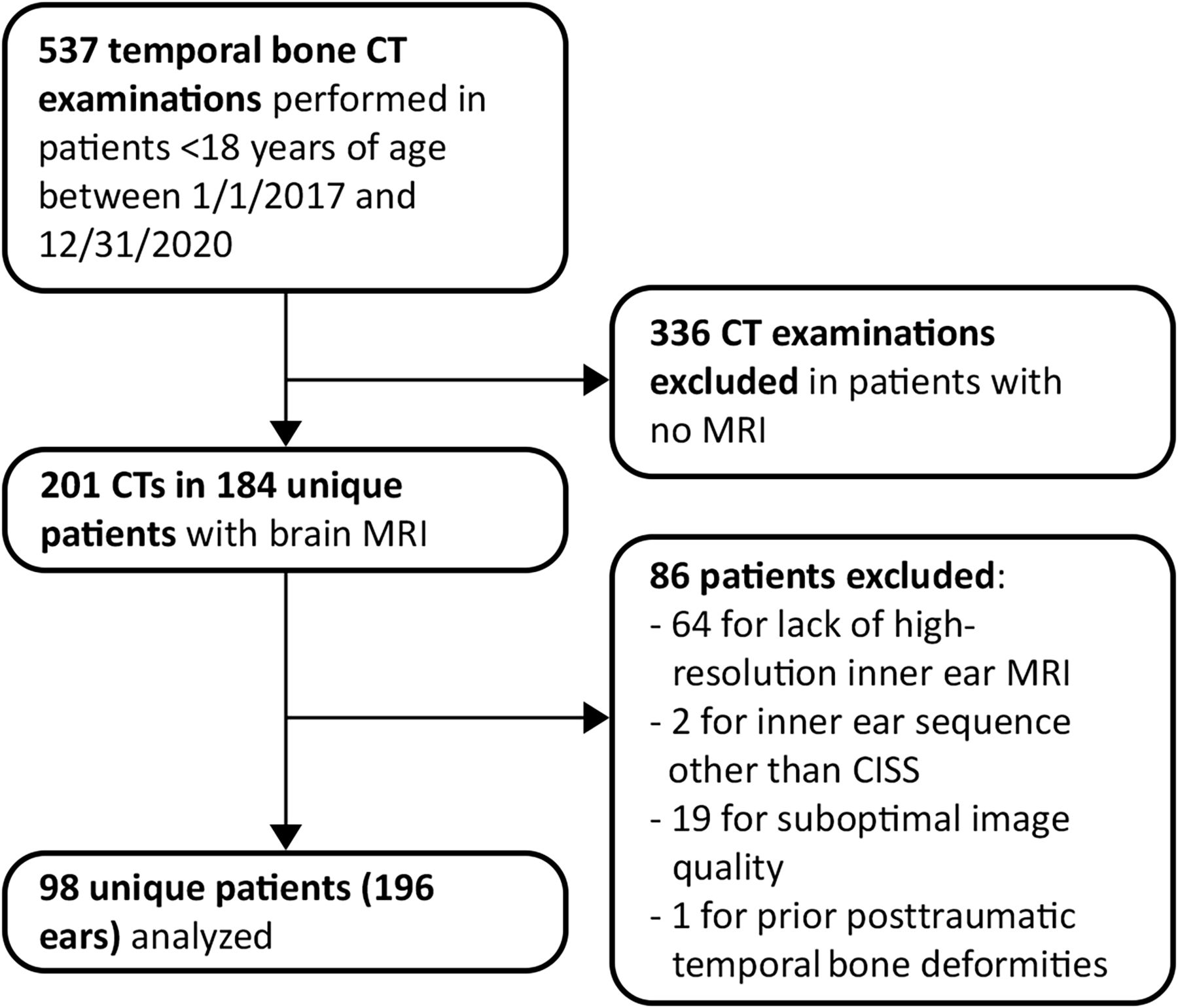
- FIG 1.
Flow chart summarizing identification, selection, and exclusion of subjects from this study.
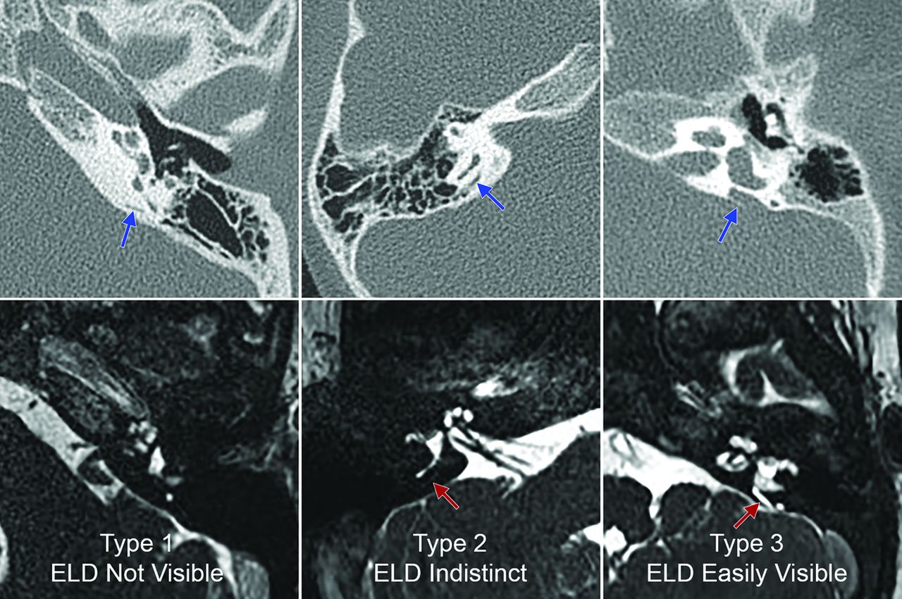
- FIG 2.
Examples of the 3 types of ELD visibility observed on MR imaging. Upper row, Representative axial-view non-contrast-enhanced temporal bone CT images of 3 different ears from different patients. Lower row, Axial temporal bone CISS MR images corresponding to the same ears as the CT images directly above demonstrate type 1 (lower left image), type 2 (lower middle image), and type 3 ELDs (lower right image). Blue arrows in the upper row indicate the vestibular aqueducts; red arrows in the lower row indicate the visible ELDs.
- FIG 3.
CISS MR images demonstrating additional examples of type 2 (faintly visible) ELDs (arrows) in 3 different patients: A, A 4-year-old girl with mild SNHL in the left ear. This patient demonstrated EVA on CT. B, A 1-year-old boy with profound SNHL in the right ear. This patient did not have VA enlargement on CT. C, An 8-year-old girl with profound SNHL in the left ear. This patient did not have VA enlargement on CT.
- FIG 4.
CISS MR images demonstrating additional examples of type 3 (easily visible) ELDs (arrows) in 3 different patients. A, A 7-month-old girl with severe SNHL in the right ear. B, A 6-year-old boy with severe SNHL in the right ear. C, A 4-year-old girl with mild SNHL in the left ear. All patients in these images demonstrated EVA on CT.


Tables
Patient-Level Characteristics Overall (n = 98 Patients) Demographic Age (mean) (SD) (yr) 6.2 (4.7) Female sex (No.) (%) 53 (54.1) Hearing loss laterality (No.) (%) Bilateral 66 (67.3) Unilateral 31 (31.6) Data not available 1 (1.0) MR imaging field strength (No.) (%) 1.5T (%) 38 (38.8) 3T (%) 60 (61.2) Characteristics by Evaluated Ears Overall (n = 196 Ears) Type of hearing loss (No.) (%) Normal 31 (15.8) Sensorineural 140 (71.4) Conductive 18 (9.2) Mixed 5 (2.5) Audiometry data not available 2 (1) Severity of hearing loss (No.) (%) Normal 31 (15.8) Mild 15 (7.7) Moderate 23 (11.7) Severe 42 (21.4) Profound 83 (42.3) Audiometry data not available 2 (1) ELD visualization (No.) (%) Type 1 145 (74.0) Type 2 29 (14.8) Type 3 22 (11.2) VA Status on CTa Type of ELD Visualization on MR Imaging Totals (Column %) Type 1, Not Visualized No. (Row %) Type 2, Faintly Visualized No. (Row %) Type 3, Easily Visualized No. (Row %) EVA – 136 (87.2) 20 (12.8) 0 (0) 156 (79.6) EVA + 9 (22.5) 9 (22.5) 22 (55.0) 40 (20.4) Totals (row %) 145 (74.0) 29 (14.8) 22 (11.2) 196 (100) ↵a There was evidence of an association between VA status and ELD visibility (P < .0008).
- Table 4:
Predicted probability of VA enlargement based on the type of MR Imaging visibility
Type of ELD Visualization on High-Resolution MR Imaging Predicted Probability of EVA 95% Confidence Level Type 1 0.053 (0.024–0.114) Type 2 0.288 (0.141–0.498) Type 3 0.997 (0.194–0.999) Type of ELD Visualization on High-Resolution MR Imaging VA Midpoint Width (Axial Plane) VA Opercular Width (Axial Plane) VA Midpoint Width (Pöschl Plane) Predicted Mean (mm) 95% CI Predicted Mean (mm) 95% CI Predicted Mean (mm) 95% CI Type 1 0.62 (0.55–0.69) 0.92 (0.80–1.03) 0.59 (0.53–0.64) Type 2 0.97 (0.83–1.11) 1.43 (1.20–1.67) 0.83 (0.72–0.94) Type 3 2.08 (1.92–2.25) 3.00 (2.73–3.28) 1.98 (1.85–2.11) ↵a All predicted means differed significantly from each other. For comparison of predicted mean VA opercular width between type 1 and type 2 ears, Tukey-adjusted P = .0004. For comparison of predicted mean VA midpoint width in the Pöschl plane between type 1 and type 2 ears, P = .0005. For all other comparisons, P < .0001.
Type of ELD Visualization on High-Resolution MR Imaging Reader 1 (Fellow and Senior Radiologist) Reader 2 (Junior Radiologist) Reader 3 (Neurotologist) Right Left Right Left Right Left Type 1 69 76 49 47 67 73 Type 2 17 12 34 38 20 16 Type 1 or 2 86 88 83 85 87 89 Type 3 12 10 15 13 11 9 Readers 3-Category Outcome: Fleiss κ (95% CI) 2-Category Outcome: Fleiss κ (95% CI) Right Left Right Left All 3 readers 0.691 (0.579–0.803) 0.531 (0.396–0.666) 0.849 (0.714–0.984) 0.825 (0.667–0.982) Pair-wise comparisons: Reader 1 vs 2 0.555 (0.402–0.709) 0.361 (0.186–0.537) 0.785 (0.604–0.966) 0.754 (0.547–0.960) Reader 1 vs 3 0.935 (0.863–1.007) 0.844 (0.724–0.963) 0.951 (0.885–1.047) 0.942 (0.828–1.055) Reader 2 vs 3 0.615 (0.468–0.762) 0.449 (0.279–0.619) 0.822 (0.654–0.991) 0.795 (0.601–0.989)


Jump to section
Related Articles
Cited By...
- No citing articles found.