Advertisement

Main menu

  • Home
  • Content
    • Current Issue
    • Accepted Manuscripts
    • Article Preview
    • Past Issue Archive
    • Video Articles
    • AJNR Case Collection
    • Case of the Week Archive
    • Case of the Month Archive
    • Classic Case Archive
  • Special Collections
    • AJNR Awards
    • Low-Field MRI
    • Alzheimer Disease
    • ASNR Foundation Special Collection
    • Photon-Counting CT
    • View All
  • Multimedia
    • AJNR Podcasts
    • AJNR SCANtastic
    • Trainee Corner
    • MRI Safety Corner
    • Imaging Protocols
  • For Authors
    • Submit a Manuscript
    • Submit a Video Article
    • Submit an eLetter to the Editor/Response
    • Manuscript Submission Guidelines
    • Statistical Tips
    • Fast Publishing of Accepted Manuscripts
    • Graphical Abstract Preparation
    • Imaging Protocol Submission
    • Author Policies
  • About Us
    • About AJNR
    • Editorial Board
    • Editorial Board Alumni
  • More
    • Become a Reviewer/Academy of Reviewers
    • Subscribers
    • Permissions
    • Alerts
    • Feedback
    • Advertisers
    • ASNR Home

User menu

  • Alerts
  • Log in

Search

  • Advanced search
American Journal of Neuroradiology

American Journal of Neuroradiology

ASHNR American Society of Functional Neuroradiology ASHNR American Society of Pediatric Neuroradiology ASSR
  • Alerts
  • Log in

Advanced Search

  • Home
  • Content
    • Current Issue
    • Accepted Manuscripts
    • Article Preview
    • Past Issue Archive
    • Video Articles
    • AJNR Case Collection
    • Case of the Week Archive
    • Case of the Month Archive
    • Classic Case Archive
  • Special Collections
    • AJNR Awards
    • Low-Field MRI
    • Alzheimer Disease
    • ASNR Foundation Special Collection
    • Photon-Counting CT
    • View All
  • Multimedia
    • AJNR Podcasts
    • AJNR SCANtastic
    • Trainee Corner
    • MRI Safety Corner
    • Imaging Protocols
  • For Authors
    • Submit a Manuscript
    • Submit a Video Article
    • Submit an eLetter to the Editor/Response
    • Manuscript Submission Guidelines
    • Statistical Tips
    • Fast Publishing of Accepted Manuscripts
    • Graphical Abstract Preparation
    • Imaging Protocol Submission
    • Author Policies
  • About Us
    • About AJNR
    • Editorial Board
    • Editorial Board Alumni
  • More
    • Become a Reviewer/Academy of Reviewers
    • Subscribers
    • Permissions
    • Alerts
    • Feedback
    • Advertisers
    • ASNR Home
  • Follow AJNR on Twitter
  • Visit AJNR on Facebook
  • Follow AJNR on Instagram
  • Join AJNR on LinkedIn
  • RSS Feeds

AJNR Awards, New Junior Editors, and more. Read the latest AJNR updates

Research ArticlePediatric Neuroimaging

Expanding the Imaging Spectrum of Polymorphous Low-Grade Neuroepithelial Tumor of the Young in Children

Carmen Cerron-Vela, Fabricio Guimarães Gonçalves, Angela N. Viaene, Luis Octavio Tierradentro-García and Arastoo Vossough
American Journal of Neuroradiology April 2024, 45 (4) 483-487; DOI: https://doi.org/10.3174/ajnr.A8145
Carmen Cerron-Vela
aFrom the Department of Radiology (C.C.-V., F.G.G., L.O.T.-G., A.V.), Children’s Hospital of Philadelphia, Philadelphia, Pennsylvania
  • Find this author on Google Scholar
  • Find this author on PubMed
  • Search for this author on this site
  • ORCID record for Carmen Cerron-Vela
Fabricio Guimarães Gonçalves
aFrom the Department of Radiology (C.C.-V., F.G.G., L.O.T.-G., A.V.), Children’s Hospital of Philadelphia, Philadelphia, Pennsylvania
  • Find this author on Google Scholar
  • Find this author on PubMed
  • Search for this author on this site
  • ORCID record for Fabricio Guimarães Gonçalves
Angela N. Viaene
bDepartment of Pathology and Laboratory Medicine (A.N.V.), Children's Hospital of Philadelphia, Philadelphia, Pennsylvania
  • Find this author on Google Scholar
  • Find this author on PubMed
  • Search for this author on this site
  • ORCID record for Angela N. Viaene
Luis Octavio Tierradentro-García
aFrom the Department of Radiology (C.C.-V., F.G.G., L.O.T.-G., A.V.), Children’s Hospital of Philadelphia, Philadelphia, Pennsylvania
  • Find this author on Google Scholar
  • Find this author on PubMed
  • Search for this author on this site
  • ORCID record for Luis Octavio Tierradentro-García
Arastoo Vossough
aFrom the Department of Radiology (C.C.-V., F.G.G., L.O.T.-G., A.V.), Children’s Hospital of Philadelphia, Philadelphia, Pennsylvania
  • Find this author on Google Scholar
  • Find this author on PubMed
  • Search for this author on this site
  • ORCID record for Arastoo Vossough
  • Article
  • Figures & Data
  • Supplemental
  • Info & Metrics
  • Responses
  • References
  • PDF
Loading

Article Figures & Data

Figures

  • Tables
  • FIGURE.
    • Download figure
    • Open in new tab
    • Download powerpoint
    FIGURE.

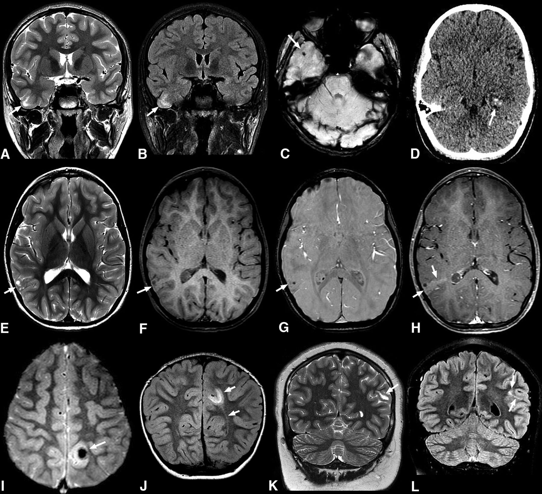
    Common imaging features of pediatric PLNTY tumors. A–C, A 9-year-old boy (case 5) with a 1-month history of headaches. Coronal T2WI (A) and FLAIR (B) sequences show a vague area of increased signal intensity in the cortical-subcortical region of the right temporal lobe (arrows in A and B). Axial SWI shows a small calcification (C). D, An 11-year-old girl with seizures (case 1). Nonenhanced axial CT shows a focal lesion in the left temporal lobe with intralesional calcifications exhibiting a punctate calcification pattern (arrow). E–H, A 6-year-old girl with complex focal seizures (case 3). Axial T2WI (E) image depicts a mixed cortical-subcortical lesion with solid and cystic components in the right parietal lobe. Pre- and postcontrast axial T1 TSE (F and H) reveals mild postcontrast enhancement (arrows in E and F). Axial SWI (G) demonstrates a punctate pattern of calcification (arrow). I and J, A 4-year-old girl with a 4-year history of seizures (case 6). Axial T2* gradient-echo image (I) reveals an ill-defined lesion in the left parietal lobe with a large central susceptibility in keeping with a chunky central calcification (arrow). Coronal FLAIR image (J) shows a related high-signal-intensity area in the subcortical white matter extending toward the left lateral ventricle, consistent with a TLS (arrows). K and L, A 14-year-old boy with new-onset seizures (case 8). Coronal T2WI (K) shows a hyperintense lesion in the left parietal region (arrow). Coronal FLAIR (L) shows an associated high signal area extending medially, in keeping with a TLS (arrows).

Tables

  • Figures
  • Distribution of imaging and pathologic features

    Percentage, Number
    Location
     Temporal lobe70%, n = 7
     Parietal lobe20%, n = 2
     Occipital lobe10%, n = 1
     Cortical-subcortical80%, n = 8
     Subcortical only20%, n = 2
    Morphology 
     Mixed solid and cystic80%, n = 8
     Solid10%, n = 1
     Cystic10%, n = 1
    Calcifications 
     Punctate50%, n = 5
     Chunky30%, n = 3
     No definite Ca2+ on imaging20%, n = 2
    Margins 
     Ill-defined90%, n = 9
     Well-defined10%, n = 1
    Contrast enhancement 
     Mild40%, n = 4
     None60%, n = 6
    TLS 
     Yes40%, n = 4
     None60%, n = 6
    Pathology proven cortical dysplasia30%, n = 3
    Pathology proven calcification70%, n = 7
    Mutation 
     FGFR2 alteration50%, n = 5
     BRAF mutation50%, n = 5
PreviousNext
Back to top

In this issue

American Journal of Neuroradiology: 45 (4)
American Journal of Neuroradiology
Vol. 45, Issue 4
1 Apr 2024
  • Table of Contents
  • Index by author
  • Complete Issue (PDF)
Advertisement
Print
Download PDF
Email Article

Thank you for your interest in spreading the word on American Journal of Neuroradiology.

NOTE: We only request your email address so that the person you are recommending the page to knows that you wanted them to see it, and that it is not junk mail. We do not capture any email address.

Enter multiple addresses on separate lines or separate them with commas.
Expanding the Imaging Spectrum of Polymorphous Low-Grade Neuroepithelial Tumor of the Young in Children
(Your Name) has sent you a message from American Journal of Neuroradiology
(Your Name) thought you would like to see the American Journal of Neuroradiology web site.
CAPTCHA
This question is for testing whether or not you are a human visitor and to prevent automated spam submissions.
Cite this article
Carmen Cerron-Vela, Fabricio Guimarães Gonçalves, Angela N. Viaene, Luis Octavio Tierradentro-García, Arastoo Vossough
Expanding the Imaging Spectrum of Polymorphous Low-Grade Neuroepithelial Tumor of the Young in Children
American Journal of Neuroradiology Apr 2024, 45 (4) 483-487; DOI: 10.3174/ajnr.A8145

Citation Manager Formats

  • BibTeX
  • Bookends
  • EasyBib
  • EndNote (tagged)
  • EndNote 8 (xml)
  • Medlars
  • Mendeley
  • Papers
  • RefWorks Tagged
  • Ref Manager
  • RIS
  • Zotero
0 Responses
Respond to this article
Share
Bookmark this article
Imaging of Low-Grade Neuroepithelial Tumors
Carmen Cerron-Vela, Fabricio Guimarães Gonçalves, Angela N. Viaene, Luis Octavio Tierradentro-García, Arastoo Vossough
American Journal of Neuroradiology Apr 2024, 45 (4) 483-487; DOI: 10.3174/ajnr.A8145
del.icio.us logo Twitter logo Facebook logo Mendeley logo
  • Tweet Widget
  • Facebook Like
  • Google Plus One
Purchase

Jump to section

  • Article
    • Abstract
    • ABBREVIATIONS:
    • CASE SERIES
    • DISCUSSION
    • CONCLUSIONS
    • Footnotes
    • References
  • Figures & Data
  • Supplemental
  • Info & Metrics
  • Responses
  • References
  • PDF

Related Articles

  • PubMed
  • Google Scholar

Cited By...

  • No citing articles found.
  • Crossref
  • Google Scholar

This article has not yet been cited by articles in journals that are participating in Crossref Cited-by Linking.

More in this TOC Section

  • Clinical SVR of Fetal Brain MRI
  • FRACTURE MR in Congenital Vertebral Anomalies
  • Comparing MRI Perfusion in Pediatric Brain Tumors
Show more Pediatric Neuroimaging

Similar Articles

Advertisement

Indexed Content

  • Current Issue
  • Accepted Manuscripts
  • Article Preview
  • Past Issues
  • Editorials
  • Editor's Choice
  • Fellows' Journal Club
  • Letters to the Editor
  • Video Articles

Cases

  • Case Collection
  • Archive - Case of the Week
  • Archive - Case of the Month
  • Archive - Classic Case

More from AJNR

  • Trainee Corner
  • Imaging Protocols
  • MRI Safety Corner
  • Book Reviews

Multimedia

  • AJNR Podcasts
  • AJNR Scantastics

Resources

  • Turnaround Time
  • Submit a Manuscript
  • Submit a Video Article
  • Submit an eLetter to the Editor/Response
  • Manuscript Submission Guidelines
  • Statistical Tips
  • Fast Publishing of Accepted Manuscripts
  • Graphical Abstract Preparation
  • Imaging Protocol Submission
  • Evidence-Based Medicine Level Guide
  • Publishing Checklists
  • Author Policies
  • Become a Reviewer/Academy of Reviewers
  • News and Updates

About Us

  • About AJNR
  • Editorial Board
  • Editorial Board Alumni
  • Alerts
  • Permissions
  • Not an AJNR Subscriber? Join Now
  • Advertise with Us
  • Librarian Resources
  • Feedback
  • Terms and Conditions
  • AJNR Editorial Board Alumni

American Society of Neuroradiology

  • Not an ASNR Member? Join Now

© 2025 by the American Society of Neuroradiology All rights, including for text and data mining, AI training, and similar technologies, are reserved.
Print ISSN: 0195-6108 Online ISSN: 1936-959X

Powered by HighWire