Advertisement

Main menu

  • Home
  • Content
    • Current Issue
    • Accepted Manuscripts
    • Article Preview
    • Past Issue Archive
    • Video Articles
    • AJNR Case Collection
    • Case of the Week Archive
    • Case of the Month Archive
    • Classic Case Archive
  • Special Collections
    • AJNR Awards
    • Low-Field MRI
    • Alzheimer Disease
    • ASNR Foundation Special Collection
    • Photon-Counting CT
    • View All
  • Multimedia
    • AJNR Podcasts
    • AJNR SCANtastic
    • Trainee Corner
    • MRI Safety Corner
    • Imaging Protocols
  • For Authors
    • Submit a Manuscript
    • Submit a Video Article
    • Submit an eLetter to the Editor/Response
    • Manuscript Submission Guidelines
    • Statistical Tips
    • Fast Publishing of Accepted Manuscripts
    • Graphical Abstract Preparation
    • Imaging Protocol Submission
    • Author Policies
  • About Us
    • About AJNR
    • Editorial Board
    • Editorial Board Alumni
  • More
    • Become a Reviewer/Academy of Reviewers
    • Subscribers
    • Permissions
    • Alerts
    • Feedback
    • Advertisers
    • ASNR Home

User menu

  • Alerts
  • Log in

Search

  • Advanced search
American Journal of Neuroradiology

American Journal of Neuroradiology

ASHNR American Society of Functional Neuroradiology ASHNR American Society of Pediatric Neuroradiology ASSR
  • Alerts
  • Log in

Advanced Search

  • Home
  • Content
    • Current Issue
    • Accepted Manuscripts
    • Article Preview
    • Past Issue Archive
    • Video Articles
    • AJNR Case Collection
    • Case of the Week Archive
    • Case of the Month Archive
    • Classic Case Archive
  • Special Collections
    • AJNR Awards
    • Low-Field MRI
    • Alzheimer Disease
    • ASNR Foundation Special Collection
    • Photon-Counting CT
    • View All
  • Multimedia
    • AJNR Podcasts
    • AJNR SCANtastic
    • Trainee Corner
    • MRI Safety Corner
    • Imaging Protocols
  • For Authors
    • Submit a Manuscript
    • Submit a Video Article
    • Submit an eLetter to the Editor/Response
    • Manuscript Submission Guidelines
    • Statistical Tips
    • Fast Publishing of Accepted Manuscripts
    • Graphical Abstract Preparation
    • Imaging Protocol Submission
    • Author Policies
  • About Us
    • About AJNR
    • Editorial Board
    • Editorial Board Alumni
  • More
    • Become a Reviewer/Academy of Reviewers
    • Subscribers
    • Permissions
    • Alerts
    • Feedback
    • Advertisers
    • ASNR Home
  • Follow AJNR on Twitter
  • Visit AJNR on Facebook
  • Follow AJNR on Instagram
  • Join AJNR on LinkedIn
  • RSS Feeds

AJNR Awards, New Junior Editors, and more. Read the latest AJNR updates

Research ArticleArtificial Intelligence

Identifying Patients with CSF-Venous Fistula Using Brain MRI: A Deep Learning Approach

Shahriar Faghani, Mana Moassefi, Ajay A. Madhavan, Ian T. Mark, Jared T. Verdoorn, Bradley J. Erickson and John C. Benson
American Journal of Neuroradiology April 2024, 45 (4) 439-443; DOI: https://doi.org/10.3174/ajnr.A8173
Shahriar Faghani
aFrom the Radiology Informatics Lab, Department of Radiology, Mayo Clinic, Rochester, Minnesota
  • Find this author on Google Scholar
  • Find this author on PubMed
  • Search for this author on this site
  • ORCID record for Shahriar Faghani
Mana Moassefi
aFrom the Radiology Informatics Lab, Department of Radiology, Mayo Clinic, Rochester, Minnesota
  • Find this author on Google Scholar
  • Find this author on PubMed
  • Search for this author on this site
  • ORCID record for Mana Moassefi
Ajay A. Madhavan
bDepartment of Radiology, Mayo Clinic, Rochester, Minnesota.
  • Find this author on Google Scholar
  • Find this author on PubMed
  • Search for this author on this site
  • ORCID record for Ajay A. Madhavan
Ian T. Mark
bDepartment of Radiology, Mayo Clinic, Rochester, Minnesota.
  • Find this author on Google Scholar
  • Find this author on PubMed
  • Search for this author on this site
  • ORCID record for Ian T. Mark
Jared T. Verdoorn
bDepartment of Radiology, Mayo Clinic, Rochester, Minnesota.
  • Find this author on Google Scholar
  • Find this author on PubMed
  • Search for this author on this site
  • ORCID record for Jared T. Verdoorn
Bradley J. Erickson
aFrom the Radiology Informatics Lab, Department of Radiology, Mayo Clinic, Rochester, Minnesota
  • Find this author on Google Scholar
  • Find this author on PubMed
  • Search for this author on this site
  • ORCID record for Bradley J. Erickson
John C. Benson
bDepartment of Radiology, Mayo Clinic, Rochester, Minnesota.
  • Find this author on Google Scholar
  • Find this author on PubMed
  • Search for this author on this site
  • ORCID record for John C. Benson
  • Article
  • Figures & Data
  • Info & Metrics
  • Responses
  • References
  • PDF
Loading

Article Figures & Data

Figures

  • Tables
  • FIG 1.
    • Download figure
    • Open in new tab
    • Download powerpoint
    FIG 1.

    Schematic illustration of the preprocessing, data augmentation, and model training pipeline. A, 3D contrast-enhanced T1 input volume. B, Preprocessed and augmented CET1 to mitigate overfitting. C, 3D-DenseNet model. D, Prediction of absence or presence of CSF leak.
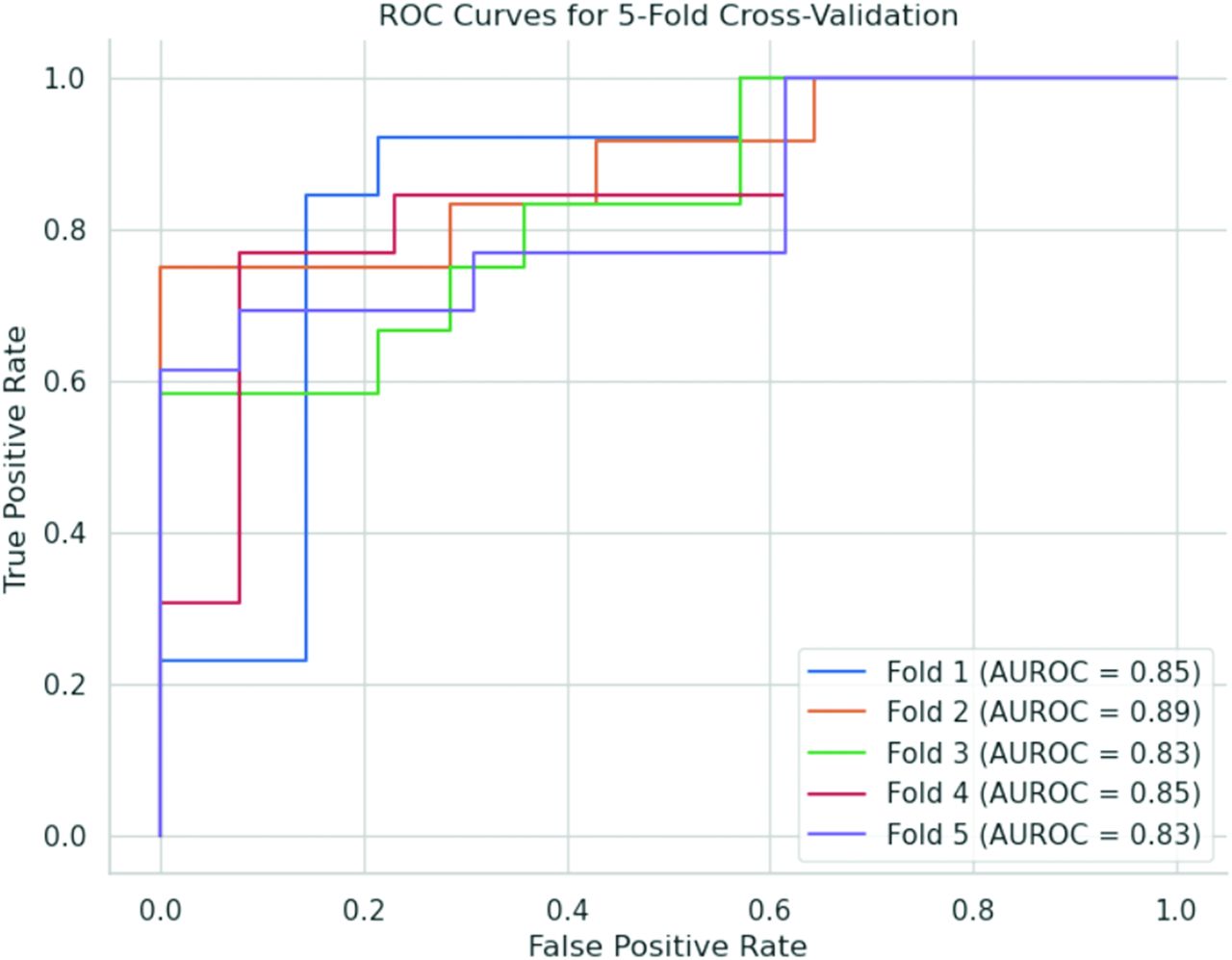
  • FIG 2.
    • Download figure
    • Open in new tab
    • Download powerpoint
    FIG 2.

    Receiver operating characteristic (ROC) curves for 5-fold cross-validation. Each curve represents the performance of the model on a distinct validation fold. The curves demonstrate the model’s ability to distinguish between the absence and presence of CSF leaks from brain MR imaging scans.

  • FIG 3.
    • Download figure
    • Open in new tab
    • Download powerpoint
    FIG 3.

    Three-part representation of the regions that are crucial to the model’s decision-making process in detecting CSF leaks. A, Sagittal view of contrast-enhanced T1 brain MR imaging. B, Occlusion mask overlaid on the original contrast-enhanced T1 image, highlighting the regions that significantly influence the model’s predictions. C, Occlusion mask generated to identify regions of interest.

Tables

  • Figures
    • View popup
    Table 1:

    Participant characteristics and Bern score status

    Subject CharacteristicsAll Subjects (n = 129)
    Median age in years (interquartile range)54 (20)
    Age range in years23–87
    Female84 (65.12%)
    Male45 (34.48%)
    Bern score
     Low risk (Bern score 0–2)47 (36.43%)
     Intermediate risk (Bern score 3–4)26 (20.15%)
     High risk (Bern score >4)56 (43.42%)
    • View popup
    Table 2:

    Summary of classifier performance per validation fold

    Fold NumberAUROC
    10.8988
    20.8284
    30.8580
    40.8580
    50.8910
PreviousNext
Back to top

In this issue

American Journal of Neuroradiology: 45 (4)
American Journal of Neuroradiology
Vol. 45, Issue 4
1 Apr 2024
  • Table of Contents
  • Index by author
  • Complete Issue (PDF)
Advertisement
Print
Download PDF
Email Article

Thank you for your interest in spreading the word on American Journal of Neuroradiology.

NOTE: We only request your email address so that the person you are recommending the page to knows that you wanted them to see it, and that it is not junk mail. We do not capture any email address.

Enter multiple addresses on separate lines or separate them with commas.
Identifying Patients with CSF-Venous Fistula Using Brain MRI: A Deep Learning Approach
(Your Name) has sent you a message from American Journal of Neuroradiology
(Your Name) thought you would like to see the American Journal of Neuroradiology web site.
CAPTCHA
This question is for testing whether or not you are a human visitor and to prevent automated spam submissions.
Cite this article
Shahriar Faghani, Mana Moassefi, Ajay A. Madhavan, Ian T. Mark, Jared T. Verdoorn, Bradley J. Erickson, John C. Benson
Identifying Patients with CSF-Venous Fistula Using Brain MRI: A Deep Learning Approach
American Journal of Neuroradiology Apr 2024, 45 (4) 439-443; DOI: 10.3174/ajnr.A8173

Citation Manager Formats

  • BibTeX
  • Bookends
  • EasyBib
  • EndNote (tagged)
  • EndNote 8 (xml)
  • Medlars
  • Mendeley
  • Papers
  • RefWorks Tagged
  • Ref Manager
  • RIS
  • Zotero
0 Responses
Respond to this article
Share
Bookmark this article
CSF-Venous Fistula Detection with Deep Learning
Shahriar Faghani, Mana Moassefi, Ajay A. Madhavan, Ian T. Mark, Jared T. Verdoorn, Bradley J. Erickson, John C. Benson
American Journal of Neuroradiology Apr 2024, 45 (4) 439-443; DOI: 10.3174/ajnr.A8173
del.icio.us logo Twitter logo Facebook logo Mendeley logo
  • Tweet Widget
  • Facebook Like
  • Google Plus One
Purchase

Jump to section

  • Article
    • Abstract
    • ABBREVIATIONS:
    • MATERIALS AND METHODS
    • RESULTS
    • DISCUSSION
    • CONCLUSIONS
    • Footnotes
    • References
  • Figures & Data
  • Info & Metrics
  • Responses
  • References
  • PDF

Related Articles

  • No related articles found.
  • PubMed
  • Google Scholar

Cited By...

  • Spinal CSF Leaks: The Neuroradiologist Transforming Care
  • Crossref (3)
  • Google Scholar

This article has been cited by the following articles in journals that are participating in Crossref Cited-by Linking.

  • Spinal CSF Leaks: The Neuroradiologist Transforming Care
    Mark D. Mamlouk, Andrew L. Callen, Ajay A. Madhavan, Niklas Lützen, Lalani Carlton Jones, Ian T. Mark, Waleed Brinjikji, John C. Benson, Jared T. Verdoorn, D.K. Kim, Timothy J. Amrhein, Linda Gray, William P. Dillon, Marcel M. Maya, Thien J. Huynh, Vinil N. Shah, Tomas Dobrocky, Eike I. Piechowiak, Joseph Levi Chazen, Michael D. Malinzak, Jessica L. Houk, Peter G. Kranz
    American Journal of Neuroradiology 2024 45 11
  • Expanded AI learning: AI as a Tool for Human Learning
    Shahriar Faghani, Christin A. Tiegs-Heiden, Mana Moassefi, Garret M. Powell, Michael D. Ringler, Bradley J. Erickson, Nicholas G. Rhodes
    Academic Radiology 2025 32 6
  • Advancing endometriosis detection in daily practice: a deep learning-enhanced multi-sequence MRI analytical model
    Mana Moassefi, Shahriar Faghani, Ceylan Colak, Shannon P. Sheedy, Pamela L. Causa Andrieu, Sherry S. Wang, Rachel L. McPhedran, Kristina T. Flicek, Garima Suman, Hiroaki Takahashi, Candice A. Bookwalter, Tatnai L. Burnett, Bradley J. Erickson, Wendaline M. VanBuren
    Abdominal Radiology 2025

More in this TOC Section

  • AI-Enhanced Photon-Counting CT of Temporal Bone
  • An AI De-identification Method for Pediatric MRIs
  • Aneurysm Segmentation on MRI-TOF with AI
Show more Artificial Intelligence

Similar Articles

Advertisement

Indexed Content

  • Current Issue
  • Accepted Manuscripts
  • Article Preview
  • Past Issues
  • Editorials
  • Editor's Choice
  • Fellows' Journal Club
  • Letters to the Editor
  • Video Articles

Cases

  • Case Collection
  • Archive - Case of the Week
  • Archive - Case of the Month
  • Archive - Classic Case

More from AJNR

  • Trainee Corner
  • Imaging Protocols
  • MRI Safety Corner

Multimedia

  • AJNR Podcasts
  • AJNR Scantastics

Resources

  • Turnaround Time
  • Submit a Manuscript
  • Submit a Video Article
  • Submit an eLetter to the Editor/Response
  • Manuscript Submission Guidelines
  • Statistical Tips
  • Fast Publishing of Accepted Manuscripts
  • Graphical Abstract Preparation
  • Imaging Protocol Submission
  • Evidence-Based Medicine Level Guide
  • Publishing Checklists
  • Author Policies
  • Become a Reviewer/Academy of Reviewers
  • News and Updates

About Us

  • About AJNR
  • Editorial Board
  • Editorial Board Alumni
  • Alerts
  • Permissions
  • Not an AJNR Subscriber? Join Now
  • Advertise with Us
  • Librarian Resources
  • Feedback
  • Terms and Conditions
  • AJNR Editorial Board Alumni

American Society of Neuroradiology

  • Not an ASNR Member? Join Now

© 2025 by the American Society of Neuroradiology All rights, including for text and data mining, AI training, and similar technologies, are reserved.
Print ISSN: 0195-6108 Online ISSN: 1936-959X

Powered by HighWire