
Article Figures & Data
Figures
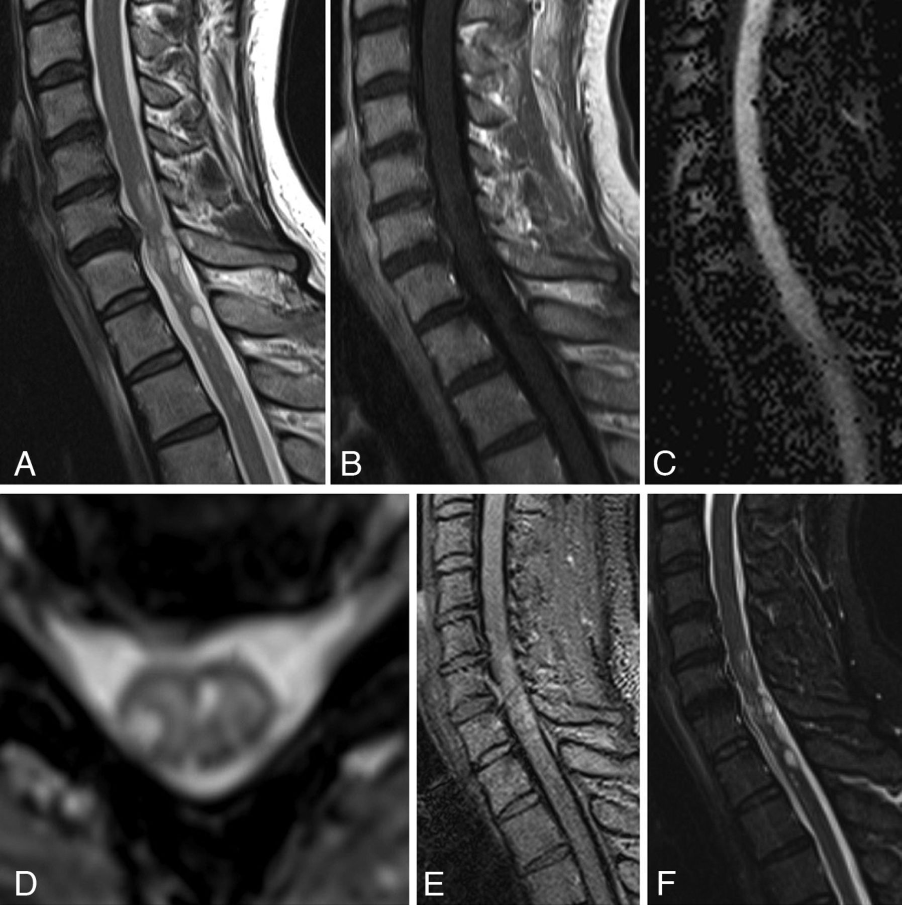
- FIG 1.
Case 1; 45-year-old woman A, Sagittal TSE T2-weighted image shows a multilocular intradural intramedullary lesion. There are multiple cystic-like changes within the spinal cord at different levels of the cervicothoracic junction. Note that there is neither syringomyelia nor T2-hypointense changes within the medulla. A cervical disk extrusion at C6–C7 can also be seen. B, Sagittal TSE T1-weighted image after the intravenous administration of gadolinium. No contrast enhancement can be seen. C, Sagittal echo-planar diffusion-weighted image shows no diffusion restriction within the lesion. D, Para-axial gradient recalled-echo T2-weighted image depicts the cystic-like intra-axial lesion. Note the sharp delineation of the lesions without perilesional edema. These lesions are located within the white matter of the spinal cord, without affecting the gray matter. E, Sagittal FLAIR T2-weighted image shows the T2-hyperintense lesion. Unlike perivascular spaces (also called Virchow-Robin spaces), there is no signal loss on FLAIR. F, Sagittal short tau inversion recovery T2-weighted image from a 12-month follow-up. No alterations in signal intensity or morphology were detected. Stable lesion.
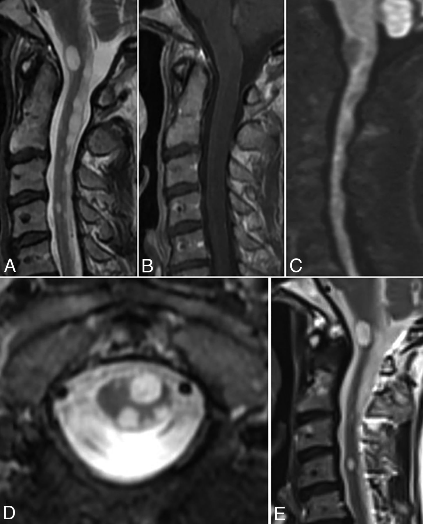
- FIG 2.
Case 2; 37-year-old woman A, Sagittal TSE T2-weighted image shows multilocular intradural intramedullary lesions. Similar to case 1, there was neither syringomyelia nor any T2-hypointense changes within the medulla. B, Sagittal T1-weighted image after the intravenous administration of gadolinium. No contrast enhancement can be seen. C, Sagittal echo-planar diffusion-weighted image shows no diffusion restriction within the lesions. D, Para-axial T2-weighted image depicts the cystic-like intra-axial lesions. Note the sharp delineation of the lesions. These lesions are located within the white matter of the spinal cord, without affecting the gray matter. E, Sagittal T2-weighted image from a 4-year follow-up MR-examination. No volume changes and/or signal intensity changes could be detected, allowing for differences in the field of view.


Jump to section
Related Articles
Cited By...
- No citing articles found.